CASP4 / Caspase-4 · IHC design guide

Design Immunohistochemistry for CASP4

Plan chromogenic CASP4 IHC in paraffin sections using tissue staining patterns (HPA tissue IHC) and catalog antibody conditions (datasheet A02941-1). Interpret staining with CASP4 processing in mind (UniProt).

Evidence assembled Oct 2026 · For research use; verify linked source records and product datasheet before use
Immunohistochemistry protocol sheet for CASP4 (IHC for CASP4): expected localisation Mostly cytoplasmic; gastro/lymphatic staining is membranous (HPA tissue IHC), antibody A02941-1, validated IHC image, and IHC protocol steps
Printable CASP4 IHC protocol sheet — expected localisation Mostly cytoplasmic; gastro/lymphatic staining is membranous (HPA tissue IHC), antibody A02941-1, controls and protocol steps. Open the full CASP4 IHC guide →

CASP4 Immunohistochemistry Experimental Design Guide

Expected localisation, validated protocols, controls and antibodies — the at-a-glance facts below, then the full design guide.

Must know before staining
Expected localisation Mostly cytoplasmic; gastro/lymphatic staining is membranous (HPA tissue IHC)
Staining pattern Mostly cytoplasmic; gastro/lymphatic cells show membrane staining (HPA tissue IHC)
Antigen retrieval Citrate pH 6 HIER, heat-mediated (datasheet A02941-1)
Positive control ⓘ Appendix+4 more · see all
Negative control ⓘ Adipose tissue+4 more · see all
Important caveats
Reasons your staining may differ from the expected pattern.
Fixation Keep fixation consistent across sections (standard IHC practice; not target-specific). Selected-image fixative and duration unreported (datasheet A02941-1); verify before use.
Caveat Activation-related cleavage may change epitope availability (UniProt)
Regulation Expression regulation is not specified (UniProt)
Isoform / epitope 5 isoforms; p20/p10 cleavage makes epitope placement relevant (UniProt)
Section 1

Recommended CASP4 IHC & IF Protocols

Compare the catalog antibody’s IHC-P protocol (datasheet A02941-1) with three published CASP4 tissue-staining protocols (PMC9148360; PMC9776729; PMC8943832).

Recommended immunohistochemistry (IHC-P) protocol parameters
SampleParaffin-embedded human colon cancer tissue; fixative not specified (datasheet A02941-1)
FixationImage fixative and duration unreported (datasheet A02941-1); verify before use.
Sectioning4–5 µm sections on charged slides (standard)
DeparaffinisationXylene, graded ethanol series to water (standard)
Antigen retrievalHeat retrieval: Citrate pH 6, 20 min (datasheet A02941-1)
Peroxidase block3% H2O2, 10 min, room temperature (standard)
Blocking10% goat serum (datasheet A02941-1)
Primary antibodyRabbit anti-CASP4, 0.5-1μg/ml (datasheet A02941-1)
Primary incubationOvernight at 4 °C (datasheet A02941-1)
DetectionStreptavidin-biotin complex (SABC), DAB chromogen (datasheet A02941-1)
CounterstainHematoxylin, blue, dehydrate and mount (standard)
Expected resultCASP4-positive staining in glandular cells of appendix (HPA tissue IHC: High). HPA tissue profile: Most tissues show cytoplasmic expression while gastro and lymphatic organs show high membranous expression. No signal in the no-primary control.
💡Decision noteStart with heat-mediated citrate retrieval at pH 6 (datasheet A02941-1). A published protocol also uses citrate (PMC9148360); boiling EDTA is another documented approach (PMC8943832).
Section 2

What Is the Expected CASP4 Staining Pattern?

CASP4 should stain mainly in the cytoplasm of glandular cells in several digestive organs and in non-germinal center cells of lymph nodes (HPA tissue IHC). Membranous staining can be prominent in gastrointestinal and lymphatic organs (HPA tissue IHC). CASP4 is predominantly ER associated but has no transmembrane segment (UniProt P49662 topology). HPA rates its tissue staining Approved, with medium consistency against RNA expression (HPA tissue IHC).

What am I looking at on my slide?
Cytoplasmic staining in appendix, duodenum or small-intestinal glandular cells.A plausible positive pattern: HPA reports high staining in these cells and cytoplasmic expression in most tissues (HPA tissue IHC). Compare the target cells with nearby tissue on the same section before scoring.
Distinct membranous staining in a gastrointestinal gland or lymphatic organ.This can fit the reported tissue pattern; HPA describes high membranous expression in gastrointestinal and lymphatic organs (HPA tissue IHC). CASP4's ER association does not make it an integral membrane protein (UniProt P49662 topology).
Predominantly nuclear staining, without the expected cytoplasmic or membranous pattern.Treat this as a questionable pattern and check controls and antibody specificity: the supported locations are cytosol and ER association, with no nuclear location listed (UniProt P49662 subcellular location; HPA subcellular ICC-IF).
Strong staining in adipocytes, cardiomyocytes or skeletal myocytes.These named cell populations are reported as not detected (HPA tissue IHC). Review cell identity and controls for nonspecific binding or endogenous chromogenic activity before calling the signal CASP4.
Broad haze across cells and stroma, or no staining in an expected positive gland.A uniform haze is difficult to assign to the reported cell pattern (HPA tissue IHC). Absence of signal in a high-staining gland warrants a control and procedure check; one failed section cannot establish absent CASP4 (general IHC practice).
💡Expected CASP4 appearanceCall a positive result when high cytoplasmic, sometimes membranous, staining identifies the reported glandular cells or lymph-node non-germinal center cells (HPA tissue IHC); diffuse haze or isolated nuclear staining is questionable against the supported locations (UniProt P49662 subcellular location; HPA subcellular ICC-IF).
How each factor affects the staining
Tissue and cell identityHPA reports high staining in appendix, breast, duodenum, gallbladder, prostate and small-intestinal glandular cells, and lymph-node non-germinal center cells; stomach glandular cells are medium (HPA tissue IHC). Score the named cell population, since a tissue name alone does not define every cell's expected result.
Compartment and topologyCASP4 is cytosolic and predominantly ER associated, without a transmembrane segment (UniProt P49662 subcellular location and topology). HPA nevertheless reports membranous staining in gastrointestinal and lymphatic tissues (HPA tissue IHC); assess compartment alongside tissue and cell identity.
Antibody evidenceThe listed antibodies HPA027588 and CAB037167 are each Approved for IHC, while the tissue profile has medium staining–RNA consistency (HPA antibodies; HPA tissue IHC). These ratings support pattern comparison but do not establish specificity for every cell or staining condition.
Processing and epitope interpretationCASP4 has a precursor and p20/p10 chains, plus 5 isoforms (UniProt P49662 processing and isoforms). No antibody epitope is supplied, so staining alone cannot distinguish precursor from processed CASP4 or identify which isoform is present.
IF/ICC relevance?HPA reports approved cytosolic localization with additional plasma-membrane localization in ICC-IF images (HPA subcellular ICC-IF). That finding can inform compartment interpretation; this section's primary readout remains chromogenic IHC in paraffin sections.
Why is my staining missing, weak or wrong?
SituationLikely causeNext action
No signal in an expected positive gland.A failed stain or an unsuitable assay condition is possible; HPA reports high glandular staining in several named tissues (HPA tissue IHC).Check a known-positive section, primary-antibody dilution and the detection reagents; review the catalog antibody's IHC-P procedure for its stated retrieval and detection conditions (general IHC practice).
Diffuse brown background obscures cell boundaries.Nonspecific binding or endogenous chromogenic activity can obscure localization (general IHC practice).Inspect a primary-omission control, blocking and washes; address endogenous activity appropriate to the chosen detection chemistry, then reassess the cell pattern (general IHC practice).
Nuclear staining dominates.The pattern conflicts with the supported cytosolic and ER-associated locations (UniProt P49662 subcellular location; HPA subcellular ICC-IF).Compare with an expected positive tissue and a primary-omission control; confirm that staining remains in the reported cell population before scoring it (HPA tissue IHC; general IHC practice).
Strong signal appears in a reported negative cell population.HPA reports no staining in adipocytes, cardiomyocytes and skeletal myocytes; cell misidentification or assay background is possible (HPA tissue IHC; general IHC practice).Verify the cell type on the counterstain and compare positive and omission controls; do not classify the signal as specific from intensity alone (general IHC practice).
Membrane staining seems unexpected.HPA describes high membranous expression in gastrointestinal and lymphatic organs, despite mainly cytoplasmic expression elsewhere (HPA tissue IHC).Check tissue and cell identity, then compare the signal with nearby cytoplasm and control sections before rejecting a membranous pattern (HPA tissue IHC; general IHC practice).
Brain staining seems to conflict with the record.UniProt says CASP4 is not detected in brain, whereas HPA lists low staining in specified cerebral cortex, cerebellar, hippocampal and caudate cell populations (UniProt P49662 tissue specificity; HPA tissue IHC).Record the exact brain region, cell type and signal strength; treat weak staining cautiously and use assay controls rather than a blanket brain-positive or brain-negative call (general IHC practice).

Sample controls for CASP4 IHC & IF

🧪Run appendix first and score its glandular cells, which show High CASP4 staining (HPA: appendix glandular cells, High). Use adipose tissue as the negative tissue, focusing on adipocytes (HPA: adipose adipocytes, Not detected); on the appendix slide, acellular areas should show no cellular stain, while other cell types should be scored by their observed staining rather than presumed negative (standard IHC practice).
Positive control tissue: Appendix (Glandular cells, HPA High)
Negative control tissue: Adipose tissue (HPA Not detected)
ICC-IF cell lines (HPA subcellular resource): HPA ICC-IF images show CASP4 in A-431, U-251MG, U2OS, with annotated localisation: Cytosol (approved) (HPA subcellular).
Technical controls: Include a no-primary, secondary-only control, a host-matched rabbit IgG isotype control, and a CASP4 knockout specimen as a biological negative (caption: rabbit primary antibody; standard IHC practice). Quench endogenous peroxidase for DAB detection, and block endogenous biotin if using the caption’s streptavidin–biotin system (caption: SABC/DAB; standard IHC practice).
⚠️Feasibility: A target-specific fixation window or fixation effect is unreported, and the selected A02941-1 paraffin-section caption does not state a fixative (caption: fixative not stated). That caption uses citrate retrieval at pH 6 for 20 minutes, but does not establish that retrieval is required for every specimen (caption: citrate retrieval). Paraffin IHC has a worked example, whereas the supplied evidence establishes neither an easier frozen-section approach nor an easier tissue IF approach; in appendix, peroxidase-positive inflammatory cells could complicate DAB interpretation, so interpret glandular staining against the controls (caption: paraffin IHC and DAB; HPA: appendix glandular cells, High; standard IHC practice).

HPA tissue IHC evidence for CASP4

Comprehensive Human Protein Atlas IHC scoring per tissue (reliability: Approved — Medium consistency between antibody staining and RNA expression data.). Rows are taken directly from the HPA tissue chart — click any row's HPA link to view the source.

Positive expression · recommended positive controls

TissueCell typeLevelEvidenceSource
Appendix Glandular cells High Protein (IHC) HPA →
Breast Glandular cells High Protein (IHC) HPA →
Duodenum Glandular cells High Protein (IHC) HPA →
Gallbladder Glandular cells High Protein (IHC) HPA →
Lymph node Non-germinal center cells High Protein (IHC) HPA →

Undetected expression · recommended negative controls

TissueCell typeLevelEvidenceSource
Adipose tissue Adipocytes Not detected Protein (IHC) HPA →
Heart muscle Cardiomyocytes Not detected Protein (IHC) HPA →
Ovary Ovarian stroma cells Not detected Protein (IHC) HPA →
Skeletal muscle Myocytes Not detected Protein (IHC) HPA →
Smooth muscle Smooth muscle cells Not detected Protein (IHC) HPA →
Section 3

Advanced CASP4 IHC Tips

Troubleshoot CASP4 staining in paraffin sections by checking retrieval, cell type, compartment and controls before interpreting chromogenic signal.

What retrieval should I try first if CASP4 staining is weak?
Start with heat-mediated citrate buffer at pH 6 for 20 minutes on paraffin sections (datasheet A02941-1). The selected image used 1 μg/mL catalog antibody overnight at 4°C, so match those conditions when assessing weak staining (datasheet A02941-1). Run a glandular-cell-rich positive control alongside the specimen; appendix and duodenum glandular cells show high staining (HPA: tissue IHC). If signal remains weak, compare a longer retrieval time or another buffer on adjacent sections while monitoring tissue damage (standard IHC practice). Keep detection and imaging conditions matched across sections so a retrieval change can be evaluated fairly (standard IHC practice).
Could fixation explain weak or uneven CASP4 staining?
The selected CASP4 image identifies a paraffin section but does not state its fixative, leaving target-specific fixation sensitivity unknown (datasheet A02941-1). Record the specimen's fixative and fixation duration before comparing staining between blocks, since these variables affect antigen accessibility in IHC (standard IHC practice). If staining varies, process adjacent sections with the same citrate pH 6 retrieval for 20 minutes (datasheet A02941-1). Include a consistently processed positive control to help separate a specimen preparation problem from an antibody or detection problem (standard IHC practice). Do not infer CASP4 fixation tolerance from its tissue distribution or subcellular location (HPA: tissue IHC; UniProt P49662: subcellular location).
Should CASP4 staining be cytoplasmic, membranous or nuclear?
Assess cytoplasmic staining first: most tissues show cytoplasmic expression, and cellular imaging places CASP4 mainly in the cytosol (HPA: tissue IHC; HPA: subcellular). Membranous staining can also be plausible in gastro and lymphatic organs, where tissue staining is high at membranes (HPA: tissue IHC). CASP4 associates with the ER membrane through TMEM214 despite lacking a transmembrane segment (UniProt P49662: subcellular location and topology). Compare the pattern with cell boundaries and morphology on the counterstained section before assigning a compartment (standard IHC practice). Treat isolated nuclear colour cautiously and check the no-primary control and adjacent tissue before calling it CASP4 (standard IHC practice; HPA: subcellular).
Can this stain distinguish CASP4 isoforms or activated CASP4?
CASP4 has five reported isoforms, but the supplied caption does not identify the catalog antibody's epitope or isoform coverage (UniProt P49662: isoforms; datasheet A02941-1). Its precursor contains a CARD region at residues 1–91 and is processed into p20 and p10 chains (UniProt P49662: domains and processing). Epitope position therefore matters when asking whether a stain represents precursor, processed protein or both (UniProt P49662: processing; standard IHC interpretation). Seek an epitope map and appropriate validation before making an isoform-specific or cleavage-specific claim (standard IHC practice). Report ordinary positive staining as CASP4 immunoreactivity, with scoring and compartment recorded, unless that additional validation is available (standard IHC practice).
How should I check a CASP4 pattern in multiplex IF?
Use IF as a separate application and confirm that the antibody works under the chosen IF fixation conditions before interpreting fluorescence (standard IF practice). Pair CASP4 with a marker identifying the expected cell population, then assess signal within those cells rather than across the whole field (standard IF practice). Include single-label and no-primary controls, and choose fluorophores whose emission is distinguishable from the specimen's autofluorescence (standard IF practice). CASP4 is mainly cytosolic but can associate with the ER membrane through TMEM214 (HPA: subcellular; UniProt P49662: subcellular location). For an intracellular epitope, optimise permeabilisation for access to the relevant side of that membrane, using the epitope map if available (standard IF practice; UniProt P49662: topology).
How can I reduce diffuse brown staining without losing CASP4 signal?
Check a no-primary section for staining from the detection system, and inspect tissue edges and damaged areas separately (standard IHC practice). The selected workflow used goat serum block, a biotinylated secondary, an avidin-biotin complex and DAB (datasheet A02941-1). Include a peroxidase block and evaluate whether the chosen biotin-based detection contributes background in the specimen (standard chromogenic IHC practice). If the control is clean but CASP4 staining remains diffuse, compare antibody concentration and incubation using adjacent sections (standard IHC practice). The caption's starting conditions are 10% goat serum and 1 μg/mL primary overnight at 4°C (datasheet A02941-1).
How should I quantify CASP4 staining across paraffin sections? ⚠ ANSWER MARKED FOR VERIFICATION
Define the cell population and anatomical region before scoring, then use the same criteria for every section (standard IHC quantification practice). For cellular staining, report the percentage of positive cells and an intensity-weighted H-score, with cytoplasmic and membranous patterns scored separately when visible (standard IHC quantification practice; HPA: tissue IHC). For discrete positive cells, density per mm² can supplement the percentage, provided the sampled area is stated (standard IHC quantification practice). Normalise counts to eligible cells or analysed tissue area, and exclude folds, edges and necrotic regions consistently (standard IHC quantification practice). Match retrieval, chromogen development and image acquisition across groups before comparing scores (standard IHC quantification practice).
When is a brown CASP4 signal likely to be artefactual?
Give greatest weight to staining in intact cells with a plausible cytoplasmic or membranous pattern (HPA: tissue IHC; HPA: subcellular). Glandular cells in appendix and duodenum show high staining, providing documented cell populations for comparison (HPA: tissue IHC). Be cautious with signal confined to section edges, necrotic material or tissue lacking clear cell boundaries (standard IHC interpretation). Brown colour in a no-primary control suggests a detection-system contribution; endogenous peroxidase can also produce chromogenic background (standard chromogenic IHC practice). CASP4 staining alone does not establish enzyme activation or pyroptosis, because its precursor is processed during activation and ordinary immunoreactivity does not identify the detected form (UniProt P49662: processing and function; standard IHC interpretation).
Boster reagents

Best CASP4 / Caspase-4 IHC Antibodies

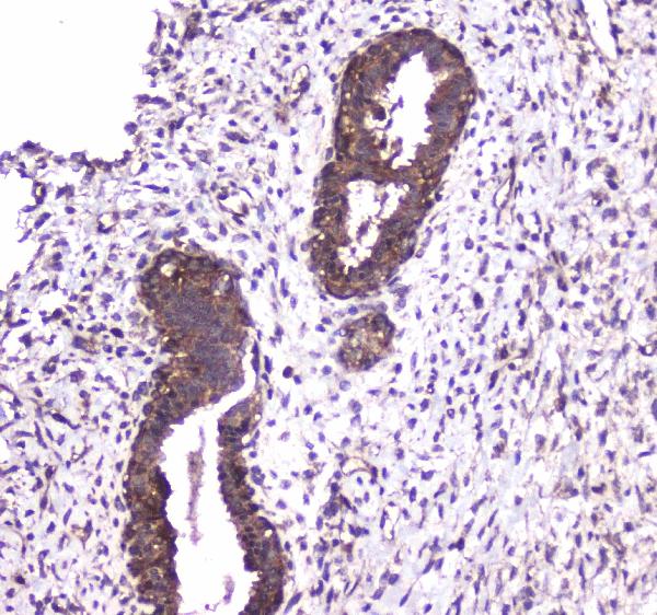
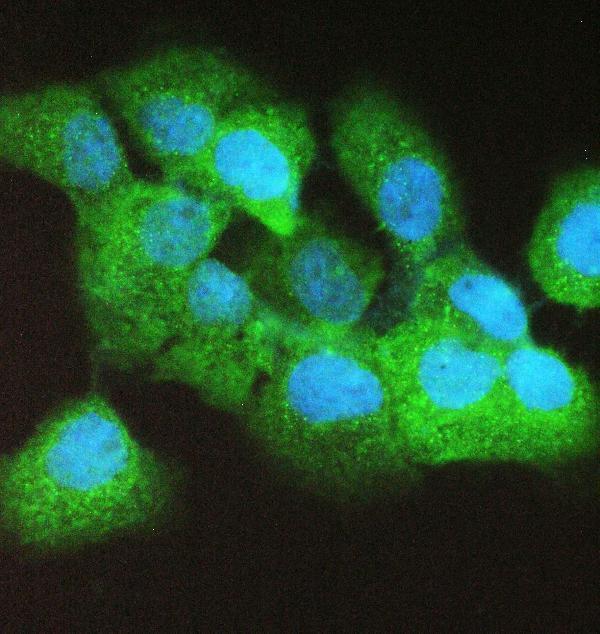
Human paraffin-section IHC images are provided for all three antibodies (catalog IHC captions); IF images show A02941-1 in A431 cells and human tonsil (A02941-1 IF captions).

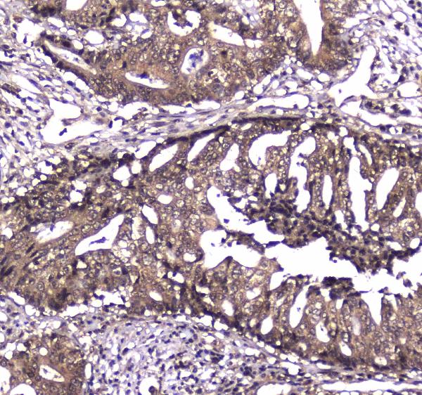
Real IHC data IHC analysis of Caspase 4 using anti-Caspase 4 antibody (A02941-1). Caspase 4 was detected in paraffin-embedded section of human colon cancer tissue. Heat mediated antigen retrieval was performed in citrate buffer (pH6, epitope retrieval solution) for 20 mins. The tissue section was blocked with 10% goat serum. The tissue section was then incubated with 1μg/ml rabbit anti-Caspase 4 Antibody (A02941-1) overnight at 4°C. Biotinylated goat anti-rabbit IgG was used as secondary antibody and incubated for 30 minutes at 37°C. The tissue section was developed using Strepavidin-Biotin-Complex (SABC)(Catalog # SA1022) with DAB as the chromogen.
Anti-Caspase 4/CASP4 Antibody ®
Cat # A02941-1
Real IHC data Immunohistochemistry analysis of Caspase-4 antibody in paraffin-embedded human lung carcinoma tissue.
Anti-Caspase-4 CASP4 Antibody
Cat # A02941
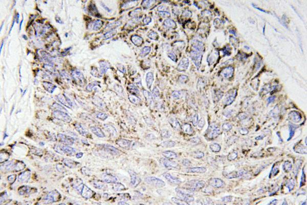
Real IHC data Anti-Caspase 4 antibody, PA1490, IHC(P) IHC(P): Human Mammary Cancer Tissue
Anti-Caspase 4/CASP4 Antibody ®
Cat # PA1490

A02941-1 shows human colon and mammary cancer paraffin-section IHC, plus A431-cell and human tonsil IF (A02941-1 image captions). A02941 shows human lung carcinoma paraffin-section IHC (A02941 IHC caption); PA1490 shows human mammary cancer IHC(P) (PA1490 IHC caption).

Which to pick: For tissue IHC, choose A02941-1 if a documented citrate pH 6 retrieval and 1 μg/ml incubation are useful (A02941-1 IHC captions); A02941 and PA1490 also have paraffin-section IHC images (A02941 and PA1490 IHC captions). For IF/ICC, A02941-1 lists both applications and has IF images, while A02941 lists both applications without an IF image (catalog applications and IF image lists). All three list Human only, so none has documented cross-species reactivity (catalog reactivity); the fixative is unreported in their IHC captions (catalog IHC captions).

Each figure is that product's own IHC / IF validation image from its datasheet.

References

  1. UniProt Consortium. UniProt entry P49662 (CASP4_HUMAN, Caspase-4).
  2. Human Protein Atlas. CASP4 tissue IHC expression (reliability: Approved).
  3. Human Protein Atlas. CASP4 subcellular location (ICC-IF): Mainly localized to the cytosol. In addition localized to the plasma membrane..
  4. Human Protein Atlas. CASP4 antibody validation summary (2 antibodies).
  5. Proteomic Profiling of Limited-Stage Follicular Lymphoma Reveals Differentially Expressed Proteins Linked to Disease Progression Post-Radiation Therapy. International journal of molecular sciences 2025 — PMC12525013.
  6. Pyroptosis-related genes regulate proliferation and invasion of pancreatic cancer and serve as the prognostic signature for modeling patient survival. Discover oncology 2022 — PMC9148360.
  7. Systematic Analysis of a Pyroptosis-Related Signature to Predict the Prognosis and Immune Microenvironment of Lower-Grade Glioma. Cells 2022 — PMC9776729.
  8. Identification of a Pyroptosis-Related Gene Signature and Effect of Silencing the CHMP4C and CASP4 in Pancreatic Adenocarcinoma. International journal of general medicine 2022 — PMC8943832.
  9. PubMed PMID:7743998 — UniProt-cited evidence.
  10. PubMed PMID:7797592 — UniProt-cited evidence.
  11. PubMed PMID:7797510 — UniProt-cited evidence.