This website uses cookies to ensure you get the best experience on our website.


- Table of Contents
Plan chromogenic CD81 IHC in paraffin sections using tonsil germinal centers as a high-staining reference (HPA tissue IHC). Start the IHC-P catalog antibody A01281 at 2 μg/mL and assess membrane and cytoplasmic staining by cell population (datasheet: A01281; HPA tissue IHC).

Expected localisation, validated protocols, controls and antibodies — the at-a-glance facts below, then the full design guide.
| Expected localisation | Stromal cytoplasm and membranes (HPA tissue IHC) | |
| Staining pattern | Mainly stromal staining; high in germinal center cells (HPA tissue IHC) | |
| Antigen retrieval | Citrate pH 6.0 HIER, 95–98 °C, 20 min (rule: cytoplasmic / membrane antigen) | |
| Positive control | Endometrium+4 more · see all | |
| Negative control | Adrenal gland+4 more · see all |
| Fixation | Keep fixation consistent across sections (standard IHC practice; not target-specific) | |
| Caveat | Stromal staining may complicate germinal center scoring (HPA tissue IHC) | |
| Regulation | No expression regulator specified (UniProt) | |
| Isoform / epitope | No isoforms annotated; extracellular versus cytoplasmic epitopes matter (UniProt) |
The catalog antibody protocol is followed by published CD81 staining methods for human breast tissue, human tonsil and lymphoma tissue, and mouse aorta (PMC6109364; PMC2813949; PMC4523685).
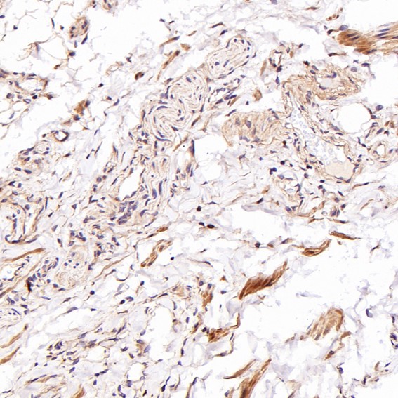
| Sample | Paraffin-embedded Human prostate cancer tissue; fixative not specified (datasheet M01281-1) |
| Fixation | Image fixative and duration unreported (datasheet M01281-1); verify before use. |
| Sectioning | 4–5 µm sections on charged slides (standard) |
| Deparaffinisation | Xylene, graded ethanol series to water (standard) |
| Antigen retrieval | Heat-induced epitope retrieval in citrate buffer, pH 6.0, 20 min at 95–98 °C (standard rule: cytoplasmic / membrane antigen) |
| Peroxidase block | 3% H2O2, 10 min, room temperature (standard) |
| Blocking | 10% normal serum of the secondary host, 30 min, room temperature (standard) |
| Primary antibody | Rabbit monoclonal (clone GAA-3) anti-CD81, 1:50-1:200 recommended; image 1:2000 (datasheet M01281-1) |
| Primary incubation | Overnight at 4 °C (standard) |
| Detection | HRP-polymer secondary, DAB chromogen 5–10 min (standard) |
| Counterstain | Hematoxylin, blue, dehydrate and mount (standard) |
| Expected result | CD81-positive staining in cells in endometrial stroma of endometrium (HPA tissue IHC: Medium). HPA tissue profile: Cytoplasmic and membranous expression in mainly in tissue stroma with additional high levels in seminiferous ducts and lymphoid germinal center cells. No signal in the no-primary control. |
CD81 is a four-pass membrane protein (UniProt P60033 topology); stroma and germinal centers stain (HPA IHC: Supported).
| Ovarian stromal cells stain strongly (HPA: High in ovarian stroma). | Membranous and cytoplasmic staining fits the observed pattern (HPA tissue IHC). |
| Nuclear-only staining (HPA tissue IHC: membrane and cytoplasm). | Suspect artefact; nuclear localisation is outside the reported pattern (HPA tissue IHC). |
| Strong bronchial epithelial staining (HPA: Not detected). | Check cross-reactivity or endogenous detection activity (standard IHC practice). |
| Diffuse staining across the section (standard IHC practice). | Suspect background from blocking, washing or detection (standard IHC practice). |
| No germinal center signal in tonsil (HPA: High). | The expected positive pattern is missing; assess the IHC run (HPA tissue IHC; standard IHC practice). |
| Protein topology (UniProt P60033). | Four transmembrane spans and two extracellular loops (UniProt P60033 topology). |
| IHC antibody validation (HPA antibodies). | HPA007234 and CAB002507 have Supported IHC status (HPA antibodies). |
| IF/ICC: where is CD81 seen? (HPA ICC-IF) | Mainly plasma membrane, with some cytosol (HPA ICC-IF: approved). |
| Situation | Likely cause | Next action |
|---|---|---|
| No tonsil germinal center signal (HPA: High). | An expected positive control failed (HPA tissue IHC). | Review the run and IHC-validated antibody (standard IHC practice). |
| Nuclear-only staining (HPA tissue IHC: membrane and cytoplasm). | Possible nonspecific staining (standard IHC practice). | Compare with a positive tissue and detection control (standard IHC practice). |
| Strong bronchial epithelial stain (HPA: Not detected). | Possible cross-reactivity or endogenous activity (standard IHC practice). | Check a detection-only control (standard IHC practice). |
| Diffuse section-wide haze (standard IHC practice). | Possible background from blocking or washing (standard IHC practice). | Review blocking and washes (standard IHC practice). |
| Signal only in cytoplasm (HPA tissue IHC: membrane and cytoplasm). | The expected membrane component is absent (HPA tissue IHC). | Compare with the membrane pattern in a positive control (standard IHC practice). |



Comprehensive Human Protein Atlas IHC scoring per tissue (reliability: Supported — Medium consistency between antibody staining and RNA expression data. Antibody staining in cells/structures not annotated, view images.). Rows are taken directly from the HPA tissue chart — click any row's HPA link to view the source.
| Tissue | Cell type | Level | Evidence | Source |
|---|---|---|---|---|
| Endometrium | Cells in endometrial stroma | Medium | Protein (IHC) | HPA → |
| Ovary | Ovarian stroma cells | High | Protein (IHC) | HPA → |
| Placenta | Decidual cells | High | Protein (IHC) | HPA → |
| Testis | Sertoli cells | High | Protein (IHC) | HPA → |
| Tonsil | Germinal center cells | High | Protein (IHC) | HPA → |
| Tissue | Cell type | Level | Evidence | Source |
|---|---|---|---|---|
| Adrenal gland | Glandular cells | Not detected | Protein (IHC) | HPA → |
| Breast | Adipocytes | Not detected | Protein (IHC) | HPA → |
| Bronchus | Respiratory epithelial cells | Not detected | Protein (IHC) | HPA → |
| Caudate | Glial cells | Not detected | Protein (IHC) | HPA → |
| Cerebellum | Cells in granular layer | Not detected | Protein (IHC) | HPA → |
Troubleshoot CD81 staining in paraffin sections by checking retrieval, cell identity, membrane localisation and assay controls before interpreting chromogenic signal.
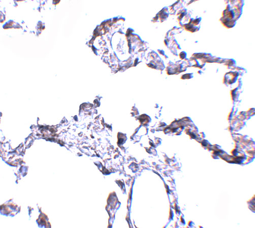
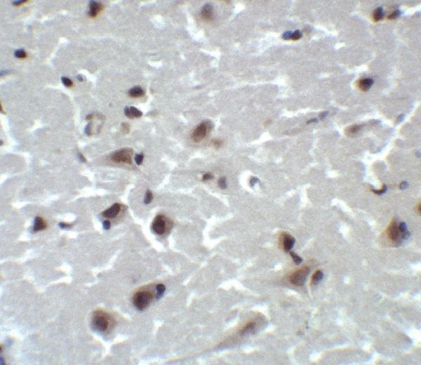
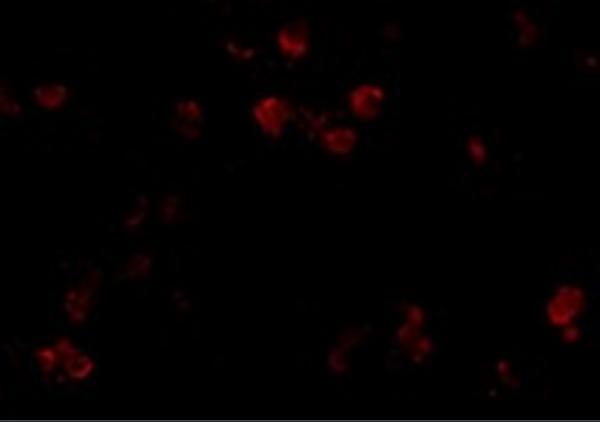
The catalog antibodies have IHC images from human tissues and IF images from A549 cells and human brain (catalog image captions). Both list human, mouse, and rat reactivity (catalog: A01281, M01281-1).
M01281-1 lists IHC and IF/ICC applications and has paraffin-section IHC images from human prostate cancer, testis cancer, Hodgkin’s lymphoma, and tonsil (catalog applications; M01281-1 image captions). A01281 lists IHC-P and IF applications, with IHC images from human lung and brain and IF images from A549 cells and human brain (catalog applications; A01281 image captions).
Which to pick: For paraffin-section tissue IHC, choose M01281-1 when its rabbit monoclonal format suits the experiment: its own caption documents a human prostate cancer paraffin section, but does not report the fixative (catalog: clone GAA-3; M01281-1 image caption). For IF, choose A01281 because its own captions show A549 cells and human brain; M01281-1 lists ICC/IF, but its supplied images document IHC only (A01281 image captions; catalog applications; M01281-1 image captions). For mouse or rat samples, either SKU lists reactivity, while the supplied IHC and IF images document human samples or A549 cells; confirm performance in the chosen species (catalog reactivity; catalog image captions).