CD82 / CD82 antigen · IHC design guide

Design Immunohistochemistry for CD82

Plan CD82 staining in paraffin sections with tissue controls and chromogenic detection. Expect membranous and cytoplasmic staining in lymphoid and glandular tissues (HPA tissue IHC), and assess each compartment separately.

Evidence assembled Oct 2026 · For research use; verify linked source records and product datasheet before use
Immunohistochemistry protocol sheet for CD82 (IHC for CD82): expected localisation Membranous and cytoplasmic tissue staining (HPA tissue IHC), antibody PB9170, validated IHC image, and IHC protocol steps
Printable CD82 IHC protocol sheet — expected localisation Membranous and cytoplasmic tissue staining (HPA tissue IHC), antibody PB9170, controls and protocol steps. Open the full CD82 IHC guide →

CD82 Immunohistochemistry Experimental Design Guide

Expected localisation, validated protocols, controls and antibodies — the at-a-glance facts below, then the full design guide.

Must know before staining
Expected localisation Membranous and cytoplasmic tissue staining (HPA tissue IHC)
Staining pattern Membranous and cytoplasmic staining in lymphoid and glandular cells (HPA tissue IHC)
Antigen retrieval EDTA pH 8.0 HIER, heat-mediated (datasheet PB9170)
Positive control ⓘ Placenta+4 more · see all
Negative control ⓘ Adipose tissue+4 more · see all
Important caveats
Reasons your staining may differ from the expected pattern.
Fixation Matched tissue-IHC evidence does not establish the fixation claim. Validate the specimen-specific method before use. (selected-SKU IHC image PB9170)
Caveat Lymphoid staining may complicate epithelial scoring (HPA tissue IHC)
Regulation No specific intensity regulator documented (UniProt)
Isoform / epitope 2 isoforms; epitope differences unknown; check loop vs tail (UniProt)
Section 1

Recommended CD82 IHC & IF Protocols

The catalog antibody’s IHC-P protocol is accompanied by published CD82 IHC workflows for frozen human gastric sections (PMC3107803) and paraffin renal carcinoma microarrays (PMC5584269).

Recommended immunohistochemistry (IHC-P) protocol parameters
SampleParaffin-embedded human tonsil tissue; fixative not specified (datasheet PB9170)
FixationImage fixative and duration unreported (datasheet PB9170); verify before use.
Sectioning4–5 µm sections on charged slides (standard)
DeparaffinisationXylene, graded ethanol series to water (standard)
Antigen retrievalHeat retrieval: EDTA pH 8.0 (datasheet PB9170); 20 min, 95–100 °C (standard)
Peroxidase block3% H2O2, 10 min, room temperature (standard)
Blocking10% goat serum (datasheet PB9170)
Primary antibodyRabbit anti-CD82, 2-5μg/ml (datasheet PB9170)
Primary incubationOvernight at 4 °C (datasheet PB9170)
DetectionHRP-conjugated secondary, DAB chromogen (datasheet PB9170)
CounterstainHematoxylin, blue, dehydrate and mount (standard)
Expected resultCD82-positive staining in decidual cells of placenta (HPA tissue IHC: High). HPA tissue profile: Cytoplasmic and membranous expression in several tissues, mainly in lymphoid tissues, glandular epithelia and islets of Langerhans. No signal in the no-primary control.
💡Decision noteStart with EDTA pH 8.0 heat retrieval for the catalog antibody (datasheet PB9170). The renal carcinoma study used 10 mM citrate, pH 6.0 (PMC5584269).
Section 2

What Is the Expected CD82 Staining Pattern?

CD82 is a four-pass membrane protein also found in cytoplasmic vesicles and phagosomes (UniProt P27701 topology and subcellular location). In paraffin-section IHC, expect membranous and cytoplasmic staining, chiefly in lymphoid cells, glandular epithelia, pancreatic endocrine cells and placental decidual cells (HPA tissue IHC). HPA rates its tissue pattern Supported, with medium consistency between antibody staining and RNA expression (HPA tissue IHC).

What am I looking at on my slide?
Decidual cells stain strongly; germinal center cells or pancreatic endocrine cells stain at moderate intensity (HPA tissue IHC).These cell types and relative levels match HPA observations: decidual cells are High, while germinal center and pancreatic endocrine cells are Medium (HPA tissue IHC). Assess signal in the named cells, since a tissue-wide impression can conceal which cells carry the stain (general IHC practice).
Signal outlines cells or forms cytoplasmic puncta in an expected positive cell type (HPA tissue IHC; UniProt P27701 subcellular location).Membranous and cytoplasmic staining fits the tissue profile; vesicle or phagosome localization offers a basis for puncta (HPA tissue IHC; UniProt P27701 subcellular location). Puncta alone do not identify a vesicle class or establish that every stained structure contains CD82 (general IHC interpretation).
The dominant signal is nuclear, with no convincing membrane or cytoplasmic staining.A predominantly nuclear pattern conflicts with the reported membrane and vesicle locations (UniProt P27701 subcellular location). Check morphology and controls before calling it CD82; an unsupported compartment can reflect nonspecific staining or a detection artefact (general IHC practice).
Strong signal appears in adipocytes or adrenal glandular cells, both reported Not detected (HPA tissue IHC).This conflicts with the named HPA cell-type observations, though it does not make every cell in those tissues a negative control (HPA tissue IHC). Consider cross-reactivity or endogenous chromogenic activity, and compare with a no-primary control (general IHC practice).
Diffuse colour covers cells and stroma, or a known-positive cell population has no signal.Widespread colour without cell or compartment definition is background; absent signal in a known-positive population makes a negative call elsewhere hard to interpret (general IHC practice). Compare a positive section and no-primary control processed in the same run before scoring (general IHC practice).
💡Expected CD82 appearanceCall a convincing positive when High decidual cells or Medium germinal center or pancreatic endocrine cells show defined membranous and/or cytoplasmic signal (HPA tissue IHC); dominant nuclear staining or uniform stromal colour is suspect (UniProt P27701 subcellular location; general IHC practice).
How each factor affects the staining
Cell type and reference intensity (HPA tissue IHC)HPA reports High decidual cells and Medium germinal center cells, pancreatic endocrine cells, collecting ducts and esophageal squamous cells (HPA tissue IHC). Its Not detected entries name specific cell types, such as adipocytes, rather than entire tissues (HPA tissue IHC).
Topology and epitope orientation (UniProt P27701 topology)CD82 has four transmembrane segments, cytoplasmic termini and two extracellular regions (UniProt P27701 topology). The supplied record gives no antibody epitope, so topology cannot predict this antibody's retrieval requirement or whether a particular membrane face will stain.
Antibody evidence (HPA antibody validation)HPA lists IHC as Supported for HPA028900 and CAB002508; it does not give an IHC status for HPA061418 (HPA antibody validation). Supported tissue staining is a useful reference pattern, but HPA reports only medium consistency with RNA data for CD82 (HPA tissue IHC).
IF/ICC Q: Should vesicular fluorescence replace the tissue IHC reference?A: No. HPA reports vesicles as an uncertain ICC-IF location, whereas its tissue IHC profile includes membranous and cytoplasmic staining (HPA subcellular; HPA tissue IHC). Interpret the assays against their respective evidence; the supplied ICC record establishes no paraffin-IHC protocol (HPA subcellular).
Why is my staining missing, weak or wrong?
SituationLikely causeNext action
No colour in decidual cells expected to stain High (HPA tissue IHC).The run may have failed at antibody binding, retrieval or detection; the supplied sources do not identify a CD82-specific fixation or retrieval failure (general IHC practice).Confirm the positive section and detection reagents worked, then review the validated antibody instructions and retrieval conditions for this run (general IHC practice).
Germinal center cells look faint beside strongly stained decidual cells (HPA tissue IHC).A real intensity difference is plausible: HPA calls germinal center cells Medium and decidual cells High (HPA tissue IHC).Judge each named cell population against its own HPA level and a concurrently stained positive section before changing conditions (HPA tissue IHC; general IHC practice).
The slide shows mostly nuclear colour.Nuclear localization is unsupported by the supplied CD82 locations; nonspecific binding or detection artefact is possible (UniProt P27701 subcellular location; general IHC practice).Check a no-primary control and review cell boundaries and counterstain before assigning the colour to CD82 (general IHC practice).
Colour is broad, hazy or present in the no-primary control.The pattern may arise from background or endogenous detection activity rather than specific CD82 binding (general chromogenic IHC practice).Review blocking, washes and the detection system; compare controls processed alongside the specimen (general chromogenic IHC practice).
An HPA Not detected cell type stains strongly, such as adipocytes (HPA tissue IHC).Cross-reactivity or endogenous activity is possible; the HPA designation applies to the listed cell type and observation (HPA tissue IHC; general IHC practice).Verify cell identity, inspect the no-primary control and compare staining with an expected positive cell population before scoring (general IHC practice).
A vesicular IF/ICC image appears different from a membrane-rich paraffin IHC section.The ICC-IF vesicle assignment is uncertain, while tissue IHC reports cytoplasmic and membranous expression (HPA subcellular; HPA tissue IHC).Use the tissue IHC pattern to assess this paraffin section; treat the ICC-IF image as separate, qualified localization evidence (HPA tissue IHC; HPA subcellular).

Sample controls for CD82 IHC & IF

🧪Run placenta first: decidual cells should stain (HPA: High in decidual cells). Use adipose tissue as a negative comparator (HPA: adipocytes Not detected); on the placenta slide, score non-decidual cells as internal negatives only if they show background staining without discernible membrane or vesicular signal (UniProt P27701 localization).
Positive control tissue: Placenta (Decidual cells, HPA High)
Negative control tissue: Adipose tissue (HPA Not detected)
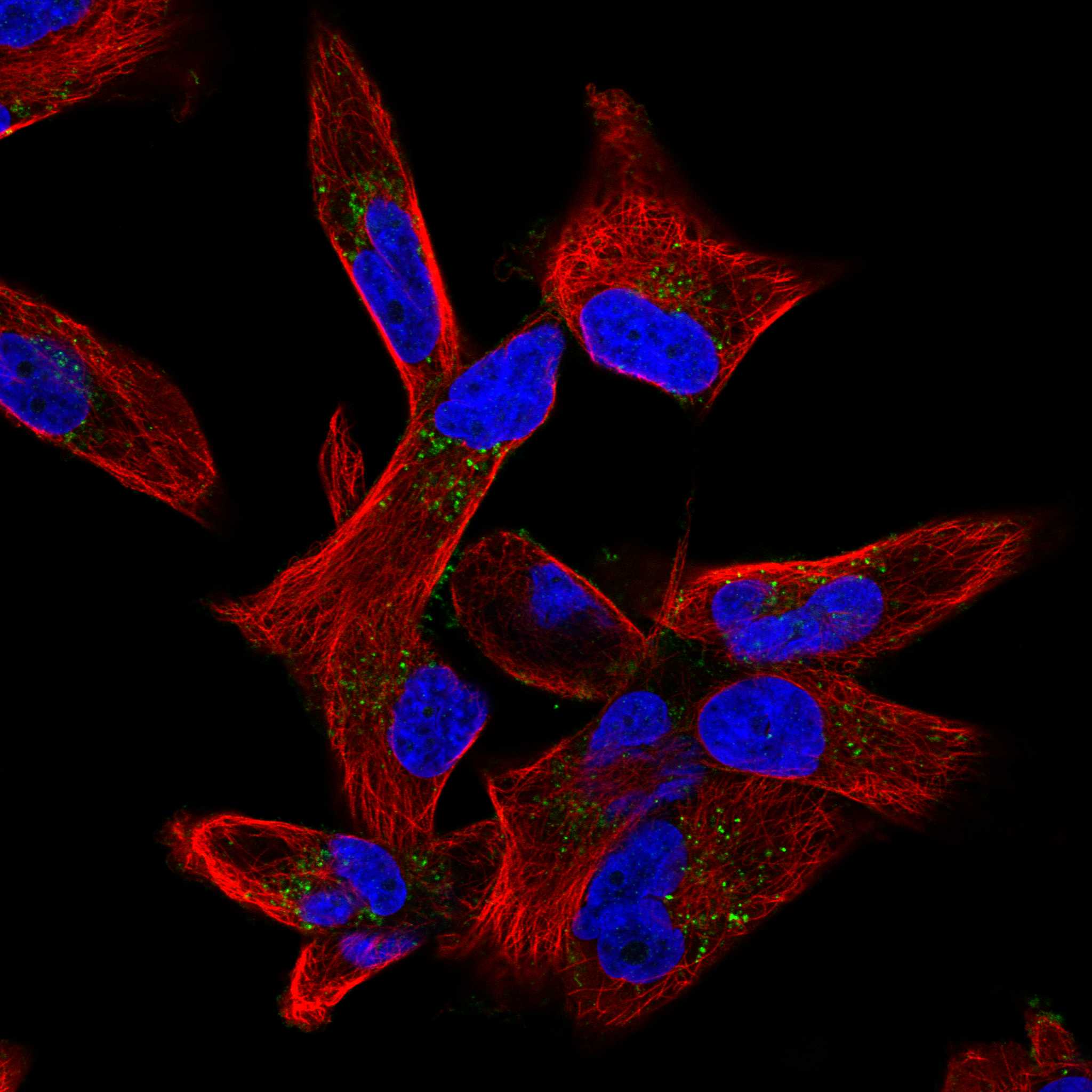
ICC-IF cell lines (HPA subcellular resource): HPA ICC-IF images show CD82 in Rh30, U-251MG, RPTEC/TERT1, hTERT-RPE1 (serum starved), with annotated localisation: Vesicles (uncertain) (HPA subcellular).
Technical controls: Include a no-primary, secondary-only control, matched rabbit IgG isotype control (caption: rabbit primary antibody), and CD82 knockout material as a biological negative. Quench endogenous peroxidase for DAB detection, especially in blood-rich placenta (caption: HRP/DAB detection; standard IHC practice).
⚠️Feasibility: No target-specific fixation window or fixation effect is reported in the supplied evidence, and the selected PB9170 paraffin-section caption does not state a fixative (caption: fixative unreported). The reported IHC procedure uses heat retrieval in EDTA at pH 8.0 (caption: EDTA retrieval); whether frozen sections or IF are easier is unreported. Placental blood can contribute endogenous peroxidase background in chromogenic IHC, so assess it with the no-primary control (standard IHC practice).

HPA tissue IHC evidence for CD82

Comprehensive Human Protein Atlas IHC scoring per tissue (reliability: Supported — Medium consistency between antibody staining and RNA expression data.). Rows are taken directly from the HPA tissue chart — click any row's HPA link to view the source.

Positive expression · recommended positive controls

TissueCell typeLevelEvidenceSource
Placenta Decidual cells High Protein (IHC) HPA →
Appendix Lymphoid tissue Medium Protein (IHC) HPA →
Esophagus Squamous epithelial cells Medium Protein (IHC) HPA →
Kidney Collecting ducts Medium Protein (IHC) HPA →
Lymph node Germinal center cells Medium Protein (IHC) HPA →

Undetected expression · recommended negative controls

TissueCell typeLevelEvidenceSource
Adipose tissue Adipocytes Not detected Protein (IHC) HPA →
Adrenal gland Glandular cells Not detected Protein (IHC) HPA →
Bone marrow Hematopoietic cells Not detected Protein (IHC) HPA →
Bronchus Basal cells Not detected Protein (IHC) HPA →
Caudate Glial cells Not detected Protein (IHC) HPA →
Section 3

Advanced CD82 IHC Tips

Troubleshoot CD82 staining in paraffin sections by checking retrieval, cell identity, subcellular pattern, and detection controls before comparing signal across samples.

What retrieval conditions should I try first for CD82 in paraffin sections?
Start with heat-mediated retrieval in EDTA at pH 8.0 (datasheet PB9170). The selected tonsil image used that condition before overnight incubation with 2 μg/ml primary antibody at 4°C (datasheet PB9170); use it as a reference when troubleshooting weak staining. Keep heating and cooling conditions consistent across sections, then compare signal in tonsil germinal center cells and background on the same run (HPA: Medium in tonsil germinal center cells). If signal remains weak, adjust retrieval duration in a small section series while keeping antibody and detection conditions fixed (standard IHC practice). Excessive retrieval can damage morphology or raise background, so judge both alongside signal (standard IHC practice).
How should I troubleshoot fixation-related loss of CD82 staining?
The selected paraffin-section caption does not state a fixative, so CD82 sensitivity to fixation under this protocol is unknown (datasheet PB9170: fixative not stated). Record fixative, time in fixative, processing history, and section age for each specimen before comparing staining (standard IHC practice). If signal varies between batches, stain sections together using the same EDTA pH 8.0 retrieval and detection run (datasheet PB9170; standard IHC practice). Include a tonsil section as a run control because germinal center cells show medium staining (HPA: Medium in tonsil germinal center cells). Do not assign a fixation effect from the presence or absence of membranous staining alone; evaluate morphology and controls together (standard IHC practice).
Should CD82 staining appear at the membrane, in the cytoplasm, or both?
Expect membranous staining with possible cytoplasmic puncta: CD82 is annotated at the cell membrane and in cytoplasmic vesicles and phagosomes (UniProt P27701: subcellular location). Tissue IHC also describes cytoplasmic and membranous expression, mainly in lymphoid tissues, glandular epithelia, and islets of Langerhans (HPA: tissue profile). Judge whether puncta sit within intact cells and whether membrane outlines follow cell borders; compare both patterns with an omission control (standard IHC practice). Diffuse stain over empty spaces or detached tissue lacks that cellular pattern and warrants a background check (standard IHC practice). Annotate membrane and cytoplasmic scores separately when the distinction matters to the experiment (standard IHC practice).
How can epitope location and isoforms affect CD82 IHC interpretation?
CD82 has 4 transmembrane segments and a large extracellular region spanning residues 111–228, with cytoplasmic termini and a short cytoplasmic loop (UniProt P27701: topology). It also has 3 annotated glycosylation sites at residues 129, 157, and 198, plus 2 isoforms (UniProt P27701: glycosylation and isoforms). The supplied caption does not identify the PB9170 epitope or establish isoform selectivity (datasheet PB9170: selected caption). Check the antibody's epitope documentation before interpreting a negative region or claiming isoform-specific staining (standard IHC practice). Compare matched sections under one retrieval condition; staining differences alone cannot identify which CD82 isoform is present (standard IHC practice).
How can I adapt the CD82 question to a multiplex IF experiment?
Use a marker for the expected cell population alongside CD82; tonsil germinal center cells offer a documented tissue reference (HPA: Medium in tonsil germinal center cells). Choose separated fluorophores and inspect unstained tissue in each channel before assigning faint signal, since tissue autofluorescence can complicate IF interpretation (standard IF practice). CD82 has extracellular and cytoplasmic regions, so the need for permeabilisation depends on the antibody's mapped epitope (UniProt P27701: topology; standard IF practice). If the epitope is cytoplasmic, compare gentle permeabilisation conditions; if it is extracellular, first assess staining without permeabilisation (standard IF practice). Include single-color controls and inspect both cell borders and puncta (standard IF practice; UniProt P27701: subcellular location).
What should I check when CD82 DAB staining is widespread?
Compare the stained section with a primary-antibody omission control to identify signal from secondary detection or tissue chemistry (standard IHC practice). The selected tonsil protocol used 10% goat serum blocking, a peroxidase-linked secondary antibody, and DAB development (datasheet PB9170). Use an endogenous peroxidase block and control DAB development time, especially when brown signal appears outside intact cells (standard chromogenic IHC practice). Check whether background tracks folds, damaged edges, or necrotic areas rather than cell borders and intracellular puncta (standard IHC practice; UniProt P27701: subcellular location). Reassess primary concentration against the documented 2 μg/ml starting condition before calling widespread stain CD82 expression (datasheet PB9170; standard IHC practice).
How should I quantify CD82 across paraffin sections? ⚠ ANSWER MARKED FOR VERIFICATION
Define the cell population and compartment before scoring, because tissue IHC reports both cytoplasmic and membranous CD82 patterns (HPA: tissue profile). For intact cells, record the percentage positive and an intensity-based H-score; report membrane and cytoplasmic results separately if both are analysed (standard IHC scoring practice). For spatial questions, count positive cells per mm² of viable, annotated tissue and normalise to the number of eligible cells or sampled area (standard IHC scoring practice). Keep retrieval, imaging, thresholds, and DAB development consistent across groups (standard IHC practice). Exclude folds and necrosis by predefined rules, then retain the same scoring rules for every section (standard IHC practice).
How do I distinguish convincing CD82 staining from artefact?
A convincing pattern follows cell membranes or intracellular vesicles in intact cells, consistent with CD82 localisation (UniProt P27701: subcellular location). Check cell identity: tonsil germinal center cells show medium staining, while adipocytes were not detected in the cited tissue survey (HPA: Medium in tonsil germinal center cells; HPA: Not detected in adipocytes). Compare any unexpected positive field with adjacent morphology and a primary-antibody omission control before assigning it to CD82 (standard IHC practice). Signal restricted to section edges, folds, necrosis, or areas with endogenous peroxidase activity needs separate evaluation (standard chromogenic IHC practice). Interpret a negative field cautiously if the run control or tissue preservation is poor (standard IHC practice).
Boster reagents

Best CD82 / CD82 antigen IHC Antibodies

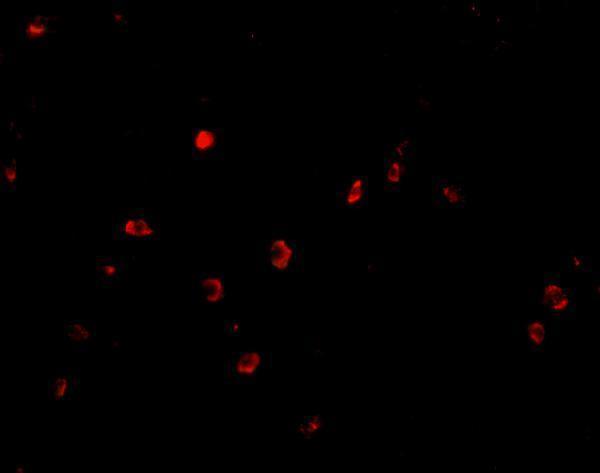
Anti-CD82 antibodies have pictured IHC results in human tonsil and colon tissue (PB9170 and A02300 image captions), plus IF in human colon cells (A02300 IF caption).

Real IHC data IHC analysis of CD82 using anti-CD82 antibody (PB9170). CD82 was detected in a paraffin-embedded section of human tonsil tissue. Heat mediated antigen retrieval was performed in EDTA buffer (pH 8.0, epitope retrieval solution). The tissue section was blocked with 10% goat serum. The tissue section was then incubated with 2 μg/ml rabbit anti-CD82 Antibody (PB9170) overnight at 4°C. Peroxidase Conjugated Goat Anti-rabbit IgG was used as secondary antibody and incubated for 30 minutes at 37°C. The tissue section was developed using HRP Conjugated Rabbit IgG Super Vision Assay Kit (Catalog # SV0002) with DAB as the chromogen.
Anti-CD82 Antibody ®
Cat # PB9170
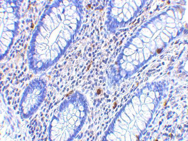
Real IHC data Immunohistochemistry of KAI1 in human colon tissue with KAI1 antibody at 2.5 μg/mL.
Anti-KAI1 CD82 Antibody
Cat # A02300

PB9170 has a paraffin-section IHC image from human tonsil and lists human reactivity (PB9170 IHC caption; catalog). A02300 has an IHC image from human colon tissue, an IF image from human colon cells, and listed human, mouse, and rat reactivity (A02300 image captions; catalog).

Which to pick: For tissue IHC matching the pictured method, choose PB9170: its human tonsil paraffin-section caption specifies EDTA retrieval at pH 8.0 and 2 μg/mL primary antibody overnight; the fixative is unreported (PB9170 IHC caption). For IF/ICC, choose A02300 because IF is listed and its human colon-cell IF image uses 20 μg/mL (A02300 catalog; IF caption). For mouse or rat work, A02300 lists those species and IHC-P and IF applications, but its pictured results are human; clonality is unspecified for both SKUs (A02300 catalog and image captions; PB9170 catalog).

Each figure is that product's own IHC / IF validation image from its datasheet.

References

  1. UniProt Consortium. UniProt entry P27701 (CD82_HUMAN, CD82 antigen).
  2. Human Protein Atlas. CD82 tissue IHC expression (reliability: Supported).
  3. Human Protein Atlas. CD82 subcellular location (ICC-IF): Localized to vesicles..
  4. Human Protein Atlas. CD82 antibody validation summary (3 antibodies).
  5. Clinical significance of KAI1/CD82 protein expression in nasopharyngeal carcinoma. Oncology letters 2015 — PMC4356316.
  6. Down-regulation of TM4SF is associated with the metastatic potential of gastric carcinoma TM4SF members in gastric carcinoma. World journal of surgical oncology 2011 — PMC3107803.
  7. KAI1/CD82 Genetically Engineered Endothelial Progenitor Cells Inhibit Metastasis of Human Nasopharyngeal Carcinoma in a Mouse Model. Medical science monitor : international medical journal of experimental and clinical research 2018 — PMC5975073.
  8. The metastasis suppressor CD82/KAI1 regulates cell migration and invasion via inhibiting TGF-β 1/Smad signaling in renal cell carcinoma. Oncotarget 2017 — PMC5584269.
  9. PubMed PMID:1842498 — UniProt-cited evidence.
  10. PubMed PMID:1401919 — UniProt-cited evidence.
  11. PubMed PMID:7754374 — UniProt-cited evidence.