This website uses cookies to ensure you get the best experience on our website.


- Table of Contents
Plan chromogenic CDH6 IHC in paraffin sections using kidney tubular cells as a high staining reference (HPA tissue IHC). Assess membrane and cytoplasmic signal against colon glandular cells, where CDH6 was not detected (HPA tissue IHC).

Expected localisation, validated protocols, controls and antibodies — the at-a-glance facts below, then the full design guide.
| Expected localisation | Membranous and cytoplasmic in tissues (HPA tissue IHC) | |
| Staining pattern | Kidney tubular cells show strong membrane and cytoplasmic staining (HPA tissue IHC) | |
| Antigen retrieval | EDTA pH 8.0 HIER, heat-mediated (datasheet A06353-1) | |
| Positive control | Gallbladder+4 more · see all | |
| Negative control | Adipose tissue+4 more · see all |
| Fixation | Keep fixation consistent across sections. (standard IHC practice; not target-specific) | |
| Caveat | Cytoplasmic signal can complicate membrane scoring (HPA tissue IHC) | |
| Regulation | Expression regulation not specified (UniProt) | |
| Isoform / epitope | 2 isoforms; check whether the epitope is extracellular or cytoplasmic (UniProt) |
The catalog antibody protocol uses EDTA pH 8.0 heat retrieval (datasheet A06353-1). The published options below report CDH6 staining conditions for lung cancer, thyroid cancer and reproductive tract sections (PMC13601855; PMC3772092; PMC5342521).
| Sample | Paraffin-embedded human ovarian cancer tissue; fixative not specified (datasheet A06353-1) |
| Fixation | Image fixative and duration unreported (datasheet A06353-1); verify before use. |
| Sectioning | 4–5 µm sections on charged slides (standard) |
| Deparaffinisation | Xylene, graded ethanol series to water (standard) |
| Antigen retrieval | Heat retrieval: EDTA pH 8.0 (datasheet A06353-1); 20 min, 95–100 °C (standard) |
| Peroxidase block | 3% H2O2, 10 min, room temperature (standard) |
| Blocking | 10% goat serum (datasheet A06353-1) |
| Primary antibody | Rabbit anti-CDH6, 2-5μg/ml (datasheet A06353-1) |
| Primary incubation | Overnight at 4 °C (datasheet A06353-1) |
| Detection | Streptavidin-biotin complex (SABC), DAB chromogen (datasheet A06353-1) |
| Counterstain | Hematoxylin, blue, dehydrate and mount (standard) |
| Expected result | CDH6-positive staining in glandular cells of gallbladder (HPA tissue IHC: High). HPA tissue profile: Membranous and cytoplasmic expression in several tissues, including kidney. No signal in the no-primary control. |
CDH6 is a cell-membrane protein with one transmembrane segment and an extracellular region (UniProt P55285 topology). In paraffin-section IHC, expect membranous staining, sometimes with cytoplasmic staining, in the cell types HPA reports: especially kidney tubule cells and gallbladder glandular cells, both High (HPA tissue IHC). Interpret weaker or absent staining by tissue and cell type; HPA rates its tissue profile Enhanced, with medium consistency between staining and RNA data (HPA tissue IHC).
| Kidney tubule cells or gallbladder glandular cells show clear membranous staining, with some cytoplasmic signal. | This fits HPA's High staining in those cells and its reported membranous and cytoplasmic tissue profile (HPA tissue IHC). Judge the relevant cells rather than the whole section: nearby cells need not share their intensity. Membrane localisation also agrees with CDH6 topology (UniProt P55285 topology). |
| The strongest signal is nuclear, or uniformly fills cells without a discernible membrane pattern. | That distribution does not match UniProt's cell-membrane annotation or HPA's tissue profile (UniProt P55285; HPA tissue IHC). Treat it as suspect rather than calling it CDH6 positive; review morphology, negative controls and chromogen background before scoring (general IHC practice). |
| Strong staining appears mainly in cells that HPA reports as Not detected, such as colon glandular cells. | This conflicts with that cell-level reference pattern (HPA: Not detected in colon glandular cells). Consider antibody cross-reactivity or endogenous detection activity, especially if signal also appears in a no-primary control (general IHC practice). An unexpected result warrants controls; HPA's reference profile is not proof that every specimen must be negative. |
| Color spreads across the section, including cell-free areas, and obscures cell borders. | Diffuse deposit is difficult to assign to CDH6-expressing cells and does not establish the reported membranous pattern (HPA tissue IHC). Check no-primary and detection controls, washing and blocking before interpreting intensity (general IHC practice). Avoid scoring diffuse background as cytoplasmic positivity merely because HPA also reports cytoplasmic staining. |
| Kidney tubule cells show no convincing signal while controls and tissue morphology are assessable. | This is unexpected against HPA's High kidney-tubule observation (HPA tissue IHC), but one negative section does not prove absent CDH6. Review antibody suitability, reagent performance and the chosen staining conditions with a positive control (general IHC practice). HPA reports medium staining–RNA consistency, so describe unresolved discrepancies rather than assigning a biological cause (HPA tissue IHC). |
| Compartment and epitope context | CDH6 has an extracellular region at residues 54–615, a transmembrane segment at 616–636 and a cytoplasmic region at 637–790 (UniProt P55285 topology). These positions support a membrane expectation. The supplied record gives no antibody epitope, so it cannot predict which region this antibody detects or specify retrieval conditions. |
| Cell-type context | HPA reports High staining in kidney tubule and gallbladder glandular cells; Medium staining in cerebellar Purkinje and cerebral-cortex neuronal cells (HPA tissue IHC). It reports Not detected in several other specified cell types, including colon glandular cells (HPA tissue IHC). Choose comparison cells by the annotated cell type, not the organ name alone. |
| Strength of the reference pattern | HPA labels the tissue IHC profile Enhanced while describing medium consistency between antibody staining and RNA expression (HPA tissue IHC). Its observed levels are useful expectations, not guaranteed intensities for every paraffin section. Record staining location and cell type alongside intensity when comparing a specimen with the reference (general IHC practice). |
| Antibody evidence | HPA lists Enhanced IHC status for HPA007047 and CAB025274, and Approved IHC status for HPA007456 (HPA antibodies). Those statuses belong to the listed antibodies; they do not validate an unspecified catalog antibody. Confirm the IHC status of the antibody actually used before transferring confidence to a result. |
| IF/ICC Q: What localisation is expected? | A: HPA reports CDH6 mainly at the plasma membrane and cell junctions in ICC-IF, with both locations supported (HPA subcellular). That finding provides a compartment comparison for fluorescence images; it is not a paraffin-IHC protocol or a reason to expect identical appearance across the two applications. |
| Situation | Likely cause | Next action |
|---|---|---|
| A kidney section is blank or barely stained. | The result conflicts with High staining in kidney tubule cells (HPA tissue IHC); the supplied sources do not identify why this section is negative. | Verify that tubule cells are present and assess a positive control, antibody suitability and reagent performance (general IHC practice). Record the discrepancy if controls pass; do not infer CDH6 absence from technical failure alone. |
| Signal is strongest in nuclei rather than at cell borders. | Nuclear dominance conflicts with the cell-membrane annotation and HPA's membranous tissue profile (UniProt P55285; HPA tissue IHC). | Inspect the no-primary control, detection background and tissue morphology (general IHC practice). Withhold a CDH6-positive call until an interpretable cell-associated pattern is present. |
| Unexpected cells stain strongly, including colon glandular cells. | HPA reports colon glandular cells as Not detected (HPA tissue IHC); cross-reactivity or endogenous detection activity is possible (general IHC practice). | Compare the unexpected cells with a no-primary control and a documented positive tissue; review antibody validation for the reagent used (general IHC practice; HPA antibodies). |
| Diffuse color obscures the expected membrane pattern. | Background can prevent cell-level localisation and scoring (general IHC practice); HPA's cytoplasmic profile does not establish that cell-free color is CDH6 (HPA tissue IHC). | Review blocking, washing and detection controls, then repeat or adjust the general IHC workflow as needed (general IHC practice). Score only interpretable cells. |
| Staining is present but weaker than in a kidney reference. | HPA reports different levels across cell types, including High kidney tubules and Medium Purkinje cells (HPA tissue IHC); intensity also needs a controlled comparison (general IHC practice). | First confirm the same annotated cell type and assess staining location, control performance and scoring consistency (HPA tissue IHC; general IHC practice). Do not treat every weaker signal as technical failure. |
| Fluorescence shows junctional signal that looks different from chromogenic IHC. | HPA supports plasma-membrane and cell-junction localisation in ICC-IF, while its tissue IHC profile includes membranous and cytoplasmic staining (HPA subcellular; HPA tissue IHC). | Interpret each application against its own HPA localisation evidence and controls (HPA subcellular; HPA tissue IHC; general IHC practice). Use the separate IF/ICC guide for its workflow. |


Comprehensive Human Protein Atlas IHC scoring per tissue (reliability: Enhanced — Medium consistency between antibody staining and RNA expression data.). Rows are taken directly from the HPA tissue chart — click any row's HPA link to view the source.
| Tissue | Cell type | Level | Evidence | Source |
|---|---|---|---|---|
| Gallbladder | Glandular cells | High | Protein (IHC) | HPA → |
| Kidney | Cells in tubules | High | Protein (IHC) | HPA → |
| Cerebellum | Purkinje cells | Medium | Protein (IHC) | HPA → |
| Cerebral cortex | Neuronal cells | Medium | Protein (IHC) | HPA → |
| Cervix | Squamous epithelial cells | Medium | Protein (IHC) | HPA → |
| Tissue | Cell type | Level | Evidence | Source |
|---|---|---|---|---|
| Adipose tissue | Adipocytes | Not detected | Protein (IHC) | HPA → |
| Appendix | Glandular cells | Not detected | Protein (IHC) | HPA → |
| Bone marrow | Hematopoietic cells | Not detected | Protein (IHC) | HPA → |
| Breast | Adipocytes | Not detected | Protein (IHC) | HPA → |
| Colon | Glandular cells | Not detected | Protein (IHC) | HPA → |
Troubleshoot CDH6 staining in paraffin sections by checking retrieval, compartment, cell type, and controls before comparing signal intensity (standard IHC practice).
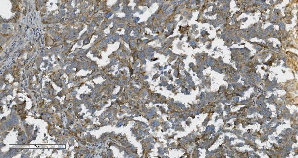
The IHC-validated anti-CDH6 antibody has an image from a human ovarian cancer paraffin section (A06353-1 image caption); catalog reactivity includes Human, Mouse, and Rat (catalog: A06353-1).
The rendered card is A06353-1, which lists IHC and Human reactivity (catalog: A06353-1). Its IHC image shows staining in a paraffin section of human ovarian cancer tissue (A06353-1 image caption).
Which to pick: For tissue IHC, choose A06353-1: its image documents a human ovarian cancer paraffin section, but does not report the fixative (A06353-1 image caption). For IF, consider A06353, a polyclonal antibody with IF listed as an application; ICC is not listed and no IF image is supplied (catalog: A06353). For cross-species planning, both SKUs list Human, Mouse, and Rat reactivity, while the supplied IHC image documents Human tissue only (catalog: A06353 and A06353-1; A06353-1 image caption).