This website uses cookies to ensure you get the best experience on our website.


- Table of Contents
Plan CERKL paraffin IHC using cytoplasmic tissue staining and high signal in lung alveolar cells as reference points (HPA tissue IHC). This guide covers the catalog antibody’s 1:100–1:300 IHC dilution (datasheet A09801-1) and the uncertain agreement between staining and RNA expression (HPA tissue IHC).

Expected localisation, validated protocols, controls and antibodies — the at-a-glance facts below, then the full design guide.
| Expected localisation | Cytoplasm (HPA tissue IHC); nucleolar enrichment is a molecular expectation (UniProt) | |
| Staining pattern | Cytoplasmic in most tissues; high in lung alveolar cells (HPA tissue IHC) | |
| Antigen retrieval | Tris-EDTA pH 8.0 HIER, heat-mediated (datasheet A09801-1) | |
| Positive control | Adrenal gland+4 more · see all | |
| Negative control | Heart muscle+4 more · see all |
| Fixation | Keep fixation conditions consistent across sections. (standard IHC practice; not target-specific) | |
| Caveat | Staining–RNA agreement is uncertain (HPA tissue IHC) | |
| Regulation | Isoform expression varies by tissue (UniProt) | |
| Isoform / epitope | 8 isoforms; epitope coverage is unknown (UniProt) |
The catalog antibody’s IHC-P protocol is accompanied by two published CERKL IHC methods (PMC8172666; PMC12645229).
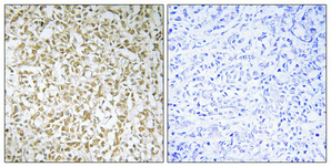
| Sample | Paraffin-embedded Human breast cancer tissue; fixative not specified (datasheet A09801-1) |
| Fixation | Image fixative and duration unreported (datasheet A09801-1); verify before use. |
| Sectioning | 4–5 µm sections on charged slides (standard) |
| Deparaffinisation | Xylene, graded ethanol series to water (standard) |
| Antigen retrieval | Heat retrieval: Tris-EDTA pH 8.0 (datasheet A09801-1); 20 min, 95–100 °C (standard) |
| Peroxidase block | 3% H2O2, 10 min, room temperature (standard) |
| Blocking | 10% normal serum of the secondary host, 30 min, room temperature (standard) |
| Primary antibody | Rabbit anti-CERKL, 1:100-1:300 (datasheet A09801-1) |
| Primary incubation | Overnight at 4 °C (standard) |
| Detection | HRP-polymer secondary, DAB chromogen 5–10 min (standard) |
| Counterstain | Hematoxylin, blue, dehydrate and mount (standard) |
| Expected result | CERKL-positive staining in glandular cells of adrenal gland (HPA tissue IHC: High). HPA tissue profile: Cytoplasmic expression in most tissues. No signal in the no-primary control. |
CERKL is mainly cytoplasmic in tissue IHC, with high staining reported in glandular cells, lung alveolar cells, and other listed cell populations (HPA: tissue IHC). Nuclear staining can also fit its annotated localisation (UniProt Q49MI3; HPA: approved ICC-IF nucleoplasm). CERKL has no transmembrane segment (UniProt Q49MI3 topology). Treat tissue intensity as provisional: HPA rates its IHC profile Uncertain because antibody staining and RNA expression have low consistency (HPA: tissue IHC).
| Cytoplasmic staining in pancreatic exocrine glandular cells, lung alveolar cells, or colonic glandular cells. | This matches cell populations scored High by tissue IHC (HPA: pancreas, lung, colon). Compare the cellular distribution with a negative control; high HPA staining is a reference pattern, not proof that an individual antibody signal is specific (HPA: IHC reliability Uncertain; standard IHC practice). |
| Nuclear staining accompanies cytoplasmic staining in otherwise plausible cells. | Nuclear signal can be biologically plausible: nucleoplasm and cytosol are approved ICC-IF locations (HPA: subcellular), and UniProt annotates nuclear and nucleolar localisation (UniProt Q49MI3). Tissue IHC is described chiefly as cytoplasmic, so assess nuclear detail and control staining before assigning it to CERKL (HPA: tissue IHC). |
| Signal is confined to cell membranes or extracellular material, with no convincing intracellular staining. | That pattern does not fit the reported cytoplasmic tissue profile or the annotated intracellular locations (HPA: tissue IHC; UniProt Q49MI3). Check the negative control and tissue morphology for nonspecific deposits or detection artefacts before interpreting the signal as CERKL (standard IHC practice). |
| Strong staining appears in cardiomyocytes or skeletal myocytes while expected comparison cells remain weak. | HPA reports CERKL as Not detected in those cell populations (HPA: heart muscle, skeletal muscle). This mismatch raises concern about cross-reactivity or endogenous detection activity; HPA's Uncertain IHC reliability means the discrepancy warrants controls rather than a definitive specificity judgment (HPA: tissue IHC; standard IHC practice). |
| Broad, even chromogen obscures cell borders and the negative control also stains. | A shared background pattern prevents reliable compartment or cell-type scoring (standard IHC practice). Review blocking, washing, and endogenous enzyme controls for the chosen detection system, then compare the corrected slide with HPA's predominantly cytoplasmic reference pattern (standard IHC practice; HPA: tissue IHC). |
| Tissue and cell selection | HPA reports High staining in pancreatic exocrine glandular cells and lung alveolar cells, but Not detected in cardiomyocytes and skeletal myocytes (HPA: tissue IHC). Compare named cell populations within intact tissue rather than treating every cell in a positive tissue as positive (standard IHC practice). |
| Compartment and isoform context | UniProt lists cytoplasm, nucleus, nucleolus, Golgi, and endoplasmic reticulum, with nucleolar enrichment except for isoform 5 (UniProt Q49MI3). HPA approves nucleoplasm and cytosol in ICC-IF, while tissue IHC is predominantly cytoplasmic (HPA: subcellular and tissue IHC). |
| Antibody evidence | HPA rates the overall tissue IHC profile Uncertain and lists HPA035444 as IHC Uncertain; neither status establishes a definitive positive or negative result for another antibody (HPA: tissue IHC; HPA: antibodies). Use the tested antibody's own controls when judging a section (standard IHC practice). |
| Chromogenic detection background | Endogenous enzyme activity can contribute color when an enzyme-based IHC detection system is used (standard IHC practice). A detection-only control helps separate that activity from primary-antibody staining; HPA's tissue pattern does not identify the source of background in a particular slide (standard IHC practice; HPA: tissue IHC). |
| Situation | Likely cause | Next action |
|---|---|---|
| No stain in pancreatic exocrine cells or lung alveolar cells. | Either the assay failed or this specimen differs from HPA's High reference pattern; HPA rates its IHC profile Uncertain (HPA: pancreas, lung, tissue IHC). | Confirm morphology and run a suitable positive control, then check primary-antibody dilution, retrieval, and detection steps using the antibody's validated IHC instructions (standard IHC practice). |
| Cardiomyocytes or skeletal myocytes stain strongly. | These cells are Not detected in HPA tissue IHC, so cross-reactivity or chromogenic background is possible (HPA: heart muscle, skeletal muscle; standard IHC practice). | Compare primary-omission and detection controls with the stained section; judge the suspect cells separately from adjacent tissue (standard IHC practice). |
| Only sharp membrane or extracellular staining is visible. | The distribution conflicts with CERKL's intracellular annotations and predominantly cytoplasmic tissue profile (UniProt Q49MI3; HPA: tissue IHC). | Inspect morphology and control slides for edge deposits or nonspecific signal; do not score that pattern alone as CERKL positivity (standard IHC practice). |
| Nuclear signal is present but cytoplasmic signal is weak. | Nuclear localisation is plausible, although HPA describes tissue IHC chiefly as cytoplasmic (UniProt Q49MI3; HPA: subcellular and tissue IHC). | Compare nuclear staining with the negative control and tissue cell types before scoring; report the observed compartment rather than forcing a cytoplasmic-only call (standard IHC practice). |
| Diffuse color makes intensity or localisation impossible to score. | Background may arise from incomplete blocking, inadequate washing, or endogenous detection activity in a chromogenic assay (standard IHC practice). | Review the detection-only control, blocking, and wash conditions; repeat scoring only where cell boundaries and compartments are discernible (standard IHC practice). |
| Two sections give different proportions of stained cells. | Cell composition may differ between sections, and HPA reports low consistency between antibody staining and RNA expression (standard IHC practice; HPA: tissue IHC). | Compare the same named cell population in both sections, document its compartment and intensity, and avoid treating HPA's High designation as a universal threshold (standard IHC practice; HPA: tissue IHC). |


![HPA ICC-IF BJ [Human fibroblast] — CERKL (HPA035443)](https://images.proteinatlas.org/35443/1527_F10_1_blue_red_green.jpg)
![HPA ICC-IF BJ [Human fibroblast] — CERKL (HPA035443)](https://images.proteinatlas.org/35443/1527_F10_2_blue_red_green.jpg)
Comprehensive Human Protein Atlas IHC scoring per tissue (reliability: Uncertain — Low consistency between antibody staining and RNA expression data.). Rows are taken directly from the HPA tissue chart — click any row's HPA link to view the source.
| Tissue | Cell type | Level | Evidence | Source |
|---|---|---|---|---|
| Adrenal gland | Glandular cells | High | Protein (IHC) | HPA → |
| Colon | Glandular cells | High | Protein (IHC) | HPA → |
| Duodenum | Glandular cells | High | Protein (IHC) | HPA → |
| Gallbladder | Glandular cells | High | Protein (IHC) | HPA → |
| Lung | Alveolar cells | High | Protein (IHC) | HPA → |
| Tissue | Cell type | Level | Evidence | Source |
|---|---|---|---|---|
| Heart muscle | Cardiomyocytes | Not detected | Protein (IHC) | HPA → |
| Ovary | Ovarian stroma cells | Not detected | Protein (IHC) | HPA → |
| Skeletal muscle | Myocytes | Not detected | Protein (IHC) | HPA → |
| Smooth muscle | Smooth muscle cells | Not detected | Protein (IHC) | HPA → |
| Soft tissue | Fibroblasts | Not detected | Protein (IHC) | HPA → |
Troubleshoot CERKL staining in paraffin sections by checking retrieval, controls, compartment, and cell type before comparing chromogenic IHC scores.
One anti-CERKL antibody has a real IHC image from human breast cancer paraffin sections; IF is listed, but no IF image is supplied (catalog applications; IHC image caption).
A09801-1 has an IHC image from human breast cancer paraffin sections (IHC image caption). A09801-1 lists human reactivity and IF among its applications, with no IF image supplied (catalog reactivity and applications; IF image list).
Which to pick: Choose A09801-1 for tissue IHC: its image shows human breast cancer paraffin sections, while the fixative is unreported (IHC image caption). For IF/ICC, A09801-1 lists IF but has no IF image or ICC listing; its host is rabbit and clonality is unreported (catalog applications, IF image list, host and clone). No cross-species option is documented: A09801-1 lists human reactivity only (catalog reactivity).