This website uses cookies to ensure you get the best experience on our website.


- Table of Contents
Plan CMKLR1 chromogenic IHC in paraffin sections using the observed cytoplasmic tissue pattern alongside its expected membrane location (HPA tissue IHC; UniProt). This guide covers controls, fixation, detection and scoring while accounting for uncertain agreement between tissue staining and RNA expression (HPA tissue IHC).

Expected localisation, validated protocols, controls and antibodies — the at-a-glance facts below, then the full design guide.
| Expected localisation | Cell membrane expected (UniProt); cytoplasm observed (HPA tissue IHC) | |
| Staining pattern | Cytoplasmic staining in most tissues, including parathyroid gland (HPA tissue IHC) | |
| Antigen retrieval | Tris-EDTA pH 9.0 HIER, heat-mediated (datasheet A02960) | |
| Positive control | Kidney+2 more · see all | |
| Negative control | Adipose tissue+4 more · see all |
| Fixation | Keep fixation conditions consistent across paraffin sections. (standard IHC practice; not target-specific) | |
| Caveat | Staining has low consistency with RNA expression (HPA tissue IHC) | |
| Regulation | High in differentiating adipocytes (UniProt) | |
| Isoform / epitope | 2 isoforms; check extracellular vs cytoplasmic epitope (UniProt) |
Start with the catalog antibody’s IHC-P protocol (datasheet A02960), then compare three published CMKLR1 IHC protocols (PMC10804379; PMC3670424; PMC10044805).
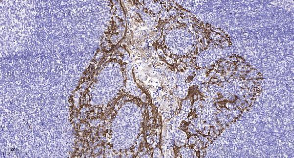
| Sample | Paraffin-embedded human tonsil tissue; fixative not specified (datasheet A02960) |
| Fixation | Image fixative and duration unreported (datasheet A02960); verify before use. |
| Sectioning | 4–5 µm sections on charged slides (standard) |
| Deparaffinisation | Xylene, graded ethanol series to water (standard) |
| Antigen retrieval | Heat retrieval: Tris-EDTA pH 9.0 (datasheet A02960); 20 min, 95–100 °C (standard) |
| Peroxidase block | 3% H2O2, 10 min, room temperature (standard) |
| Blocking | 10% normal serum of the secondary host, 30 min, room temperature (standard) |
| Primary antibody | Rabbit anti-CMKLR1, 1:100 recommended; image 1:200 (datasheet A02960) |
| Primary incubation | Overnight at 4 °C (standard) |
| Detection | HRP-polymer secondary, DAB chromogen 5–10 min (standard) |
| Counterstain | Hematoxylin, blue, dehydrate and mount (standard) |
| Expected result | CMKLR1-positive staining in cells in tubules of kidney (HPA tissue IHC: Medium). HPA tissue profile: Cytoplasmic expression in most tissues, including parathyroid gland and placenta. No signal in the no-primary control. |
CMKLR1 is a seven-transmembrane cell-surface receptor that can internalize after chemerin stimulation (UniProt Q99788 topology and subcellular annotation). Expression is reported in monocyte-derived macrophages, immature dendritic cells and blood monocytes (UniProt Q99788 tissue specificity). HPA reports mainly cytoplasmic tissue staining, including medium staining in kidney tubules, parathyroid glandular cells and Leydig cells, but rates its tissue IHC pattern Uncertain because staining and RNA expression have low consistency (HPA tissue IHC).
| Membrane-associated staining, with some intracellular staining, in cells of an appropriate population. | This fits a cell-surface receptor that internalizes in response to RARRES2 (UniProt Q99788 subcellular annotation). HPA also describes cytoplasmic staining across most tissues (HPA tissue IHC). Judge the cell identity and distribution together; a cytoplasmic component alone cannot distinguish internalized receptor from nonspecific signal (UniProt Q99788; HPA tissue IHC). |
| Strong, isolated nuclear staining without a convincing membrane or cytoplasmic component. | A nuclear-only pattern lacks support from the annotated membrane location and HPA's cytoplasmic tissue profile (UniProt Q99788 subcellular annotation; HPA tissue IHC). Treat it as suspect and check the primary-omission control, detection reagents and morphology before assigning CMKLR1 positivity (general IHC practice). |
| Staining dominates a cell type reported as undetected, such as appendix glandular cells or bone-marrow hematopoietic cells. | HPA lists these particular cell populations as Not detected, although its overall IHC reliability is Uncertain (HPA tissue IHC). Consider cross-reactivity or endogenous detection activity; verify cell identity and compare with an omission control before interpreting the signal (general IHC practice). |
| Diffuse color covers tissue, stroma or blank areas and obscures cell boundaries. | That distribution cannot establish receptor localization or a positive cell population (general IHC practice). Background may reflect nonspecific binding or detection chemistry; inspect a primary-omission control and assess whether signal tracks tissue structures rather than individual cells (general IHC practice). |
| No staining in kidney tubule cells, parathyroid glandular cells or testicular Leydig cells. | HPA reports Medium staining in these populations, so an absent signal calls for a run check (HPA tissue IHC). Their staining is not a definitive target-positive standard because HPA rates the pattern Uncertain; review control performance and morphology before calling a sample CMKLR1-negative (HPA tissue IHC; general IHC practice). |
| Receptor location and ligand response | CMKLR1 is annotated at the cell membrane and internalizes efficiently after RARRES2 stimulation (UniProt Q99788 subcellular annotation). Thus intracellular staining can be compatible with receptor biology, but a static IHC section cannot establish whether ligand-driven internalization occurred (UniProt Q99788; general IHC practice). |
| Epitope position | The receptor has 7 transmembrane segments and extracellular and cytoplasmic regions (UniProt Q99788 topology). The supplied record does not locate the antibody epitope, so it cannot predict which region the antibody recognizes or whether retrieval changes CMKLR1 detection (UniProt Q99788 topology; supplied antibody record). |
| Tissue and cell selection | UniProt reports expression in myeloid populations and multiple organs, while HPA's scored tissue examples vary by cell type and include undetected populations (UniProt Q99788 tissue specificity; HPA tissue IHC). Record the stained cell type, not just the organ name, when comparing sections (general IHC practice). |
| Evidence strength | HPA rates tissue IHC Uncertain owing to low consistency between antibody staining and RNA data; its listed antibody HPA047865 is also IHC Uncertain (HPA tissue IHC; HPA antibodies). A visually plausible pattern therefore needs controls and cautious interpretation (general IHC practice). |
| IF/ICC Q&A: Should the tissue IHC pattern be used as an IF/ICC protocol? | No protocol inference follows from these data: HPA summarizes the ICC-IF location as membrane but supplies no main-location assignment or imaged cell lines (HPA subcellular ICC-IF). Use that location as limited context; IF/ICC assay conditions belong in the separate guide (HPA subcellular ICC-IF). |
| Situation | Likely cause | Next action |
|---|---|---|
| No signal in a section chosen for expected staining. | The run may have failed, the sampled cell population may be absent, or expression may vary; HPA's medium-positive examples have Uncertain overall reliability (HPA tissue IHC; general IHC practice). | Confirm the expected cell type in the section and check a concurrently processed control. Review the validated antibody instructions and retrieval and detection steps before interpreting the sample as negative (HPA tissue IHC; general IHC practice). |
| Only nuclei appear positive. | Neither UniProt's membrane annotation nor HPA's cytoplasmic tissue profile supports a nuclear-only pattern (UniProt Q99788 subcellular annotation; HPA tissue IHC). | Compare primary-omission and stained sections, inspect precipitate or pigment, and reassess whether any cell-boundary or cytoplasmic pattern remains (general IHC practice). |
| Color persists broadly when the primary antibody is omitted. | A primary-independent signal points toward endogenous detection activity or another detection-system background source (general IHC practice). | Check the omission control beside the test section; optimize the relevant endogenous-activity block and detection reagents according to the assay chemistry (general IHC practice). |
| Color appears across many unrelated cells or extracellular areas. | Nonspecific binding or excess detection signal can obscure cell-specific staining (general IHC practice). HPA's Uncertain reliability makes appearance alone weak evidence of specificity (HPA tissue IHC). | Inspect tissue morphology and background controls, then optimize blocking, primary concentration and detection development within the validated IHC workflow (general IHC practice). |
| A reportedly undetected HPA cell population stains strongly. | HPA reports several cell-specific negative findings, but its tissue IHC pattern is Uncertain; cross-reactivity, endogenous activity or biological variation remain possible (HPA tissue IHC; general IHC practice). | Verify the exact cell population and compare matched controls. Describe the observation without converting an HPA negative listing into a definitive exclusion rule (HPA tissue IHC; general IHC practice). |
| Cytoplasmic staining is present with little visible membrane outline. | HPA reports cytoplasmic tissue staining, and UniProt notes receptor internalization after RARRES2 stimulation; neither establishes the mechanism in this section (HPA tissue IHC; UniProt Q99788 subcellular annotation). | Score the cell type and intracellular distribution, check background controls, and report the pattern as observed without assigning ligand exposure or internalization from IHC alone (general IHC practice). |


Comprehensive Human Protein Atlas IHC scoring per tissue (reliability: Uncertain — Low consistency between antibody staining and RNA expression data.). Rows are taken directly from the HPA tissue chart — click any row's HPA link to view the source.
| Tissue | Cell type | Level | Evidence | Source |
|---|---|---|---|---|
| Kidney | Cells in tubules | Medium | Protein (IHC) | HPA → |
| Parathyroid gland | Glandular cells | Medium | Protein (IHC) | HPA → |
| Testis | Leydig cells | Medium | Protein (IHC) | HPA → |
| Tissue | Cell type | Level | Evidence | Source |
|---|---|---|---|---|
| Adipose tissue | Adipocytes | Not detected | Protein (IHC) | HPA → |
| Appendix | Glandular cells | Not detected | Protein (IHC) | HPA → |
| Bone marrow | Hematopoietic cells | Not detected | Protein (IHC) | HPA → |
| Breast | Adipocytes | Not detected | Protein (IHC) | HPA → |
| Bronchus | Respiratory epithelial cells | Not detected | Protein (IHC) | HPA → |
These questions address paraffin-section chromogenic IHC for CMKLR1, with one entry on adapting the analysis to IF.
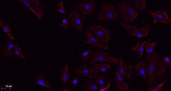
Two anti-CMKLR1 antibodies have IHC images from paraffin-embedded human tissue; one also has an IF image from A549 cells (catalog image captions). Both list human, mouse and rat reactivity (catalog).
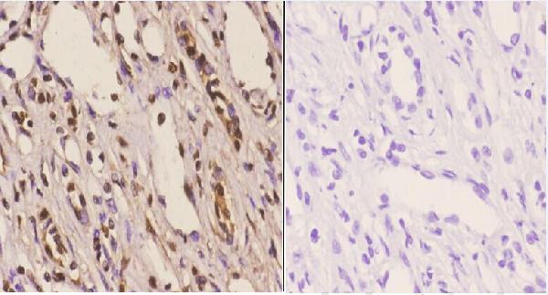
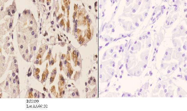
A02960 will render with human tonsil IHC; its catalog also shows A549 IF (A02960 image captions). A02960-1 will render with human kidney carcinoma IHC; its catalog also shows human stomach carcinoma IHC (A02960-1 image captions).
Which to pick: For paraffin-section IHC, choose A02960 for the tonsil example or A02960-1 for the kidney carcinoma example; both captions report paraffin embedding, but neither reports the fixative (IHC image captions). For IF/ICC, start with A02960 because it has an A549 IF image; A02960-1 lists IF as an application but has no IF image in the payload (catalog). For mouse or rat samples, either SKU lists reactivity, though the supplied IHC images show human tissue only (catalog reactivity; IHC image captions).