This website uses cookies to ensure you get the best experience on our website.


- Table of Contents
Plan COMT staining in paraffin sections using the catalog antibody’s IHC conditions and cell-specific tissue controls. The guide covers expected cytoplasmic staining, membrane staining in Bergmann glia, and how to interpret variable intensity (HPA tissue IHC).

Expected localisation, validated protocols, controls and antibodies — the at-a-glance facts below, then the full design guide.
| Expected localisation | Variable cytoplasmic staining; membrane staining in Bergmann glia (HPA tissue IHC) | |
| Staining pattern | Variable cytoplasmic staining across tissue cell types (HPA tissue IHC) | |
| Antigen retrieval | Citrate pH 6 HIER, heat-mediated (datasheet M00464) | |
| Positive control | Cerebellum+4 more · see all | |
| Negative control | Adipose tissue+3 more · see all |
| Fixation | Matched tissue-IHC evidence does not establish the fixation claim. Validate the specimen-specific method before use. (selected-SKU IHC image M00464) | |
| Caveat | Cell-type variation can complicate tissue-level scoring (HPA tissue IHC) | |
| Regulation | Variable expression across tissues (HPA tissue IHC) | |
| Isoform / epitope | 2 isoforms; epitope location may affect detection (UniProt) |
The catalog antibody’s IHC-P protocol (datasheet: M00464) appears alongside published COMT staining methods for prostate specimens (PMC8480839) and oral and pharyngeal tissues (PMC8268107).
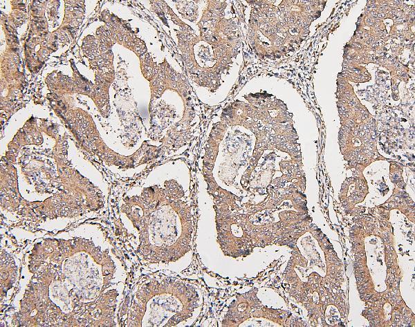
| Sample | Paraffin-embedded human intestinal cancer tissues; fixative not specified (datasheet M00464) |
| Fixation | Image fixative and duration unreported (datasheet M00464); verify before use. |
| Sectioning | 4–5 µm sections on charged slides (standard) |
| Deparaffinisation | Xylene, graded ethanol series to water (standard) |
| Antigen retrieval | Heat retrieval: Citrate pH 6, 20 min (datasheet M00464) |
| Peroxidase block | 3% H2O2, 10 min, room temperature (standard) |
| Blocking | 10% goat serum (datasheet M00464) |
| Primary antibody | Mouse monoclonal (clone 15C10) anti-COMT, 0.5-1μg/ml (datasheet M00464) |
| Primary incubation | Overnight at 4 °C (datasheet M00464) |
| Detection | Streptavidin-biotin complex (SABC), DAB chromogen (datasheet M00464) |
| Counterstain | Hematoxylin, blue, dehydrate and mount (standard) |
| Expected result | COMT-positive staining in bergmann glia - cytoplasm/membrane of cerebellum (HPA tissue IHC: High). HPA tissue profile: Cytoplasmic expression at variable levels in all tissues. No signal in the no-primary control. |
COMT is reported in cytoplasm and at the cell membrane (UniProt P21964 localization). Its membrane-bound isoform has a transmembrane segment at residues 7–26 (UniProt P21964 topology). In tissue IHC, expect variable cytoplasmic staining, including strong staining in Bergmann glia, kidney collecting ducts and placental cytotrophoblasts (HPA tissue IHC). HPA rates the tissue profile Enhanced, while describing only medium consistency with RNA data (HPA tissue IHC).
| Cytoplasmic staining in Bergmann glia, with some membrane staining (HPA tissue IHC). | This matches the reported cerebellar pattern, where Bergmann glia stain High (HPA tissue IHC). Compare the identified cells and compartment before scoring intensity; staining elsewhere in the section does not substitute for that comparison (general IHC interpretation). |
| Predominantly nuclear staining, with little cytoplasmic signal. | A nuclear-only pattern conflicts with the reported tissue distribution and UniProt localization (HPA tissue IHC; UniProt P21964 localization). Check the counterstain and staining controls, then reassess antibody specificity and detection background (general IHC practice). |
| Strong signal in adipocytes or cardiomyocytes. | HPA reports COMT as Not detected in those cell types (HPA tissue IHC). Unexpected staining warrants a cross-reactivity or endogenous detection-activity check (general IHC practice). Do not treat either cell type as a guaranteed blank in every specimen. |
| Color spreads across cells and surrounding tissue without clear boundaries. | A diffuse deposit cannot establish COMT localization (general IHC interpretation). Compare a no-primary control and the expected cell distribution; nonspecific binding or detection background can obscure a genuine cytoplasmic pattern (general IHC practice). |
| No signal in a well-preserved kidney collecting duct or placental cytotrophoblast region. | These are High-staining cell populations in HPA tissue IHC (HPA tissue IHC). First confirm the cell identity and an informative positive control; then assess the antibody, retrieval and detection workflow (general IHC practice). The supplied sources do not establish COMT-specific fixation sensitivity. |
| Compartment and membrane isoform | UniProt lists cytoplasm and cell membrane and a membrane-bound isoform with a transmembrane segment at residues 7–26 (UniProt P21964). HPA tissue IHC reports variable cytoplasmic expression and cytoplasm/membrane staining in Bergmann glia (HPA tissue IHC). Score the observed compartment in its cell context. |
| Soluble and membrane-bound isoforms | Two isoforms are listed, soluble and membrane-bound (UniProt P21964 isoforms). The record supplies no antibody epitope, so the staining pattern alone cannot establish which isoform was detected. Avoid assigning an isoform from cytoplasmic or membrane color without separate validation. |
| Tissue comparison and evidence strength | HPA reports High staining in collecting ducts and cytotrophoblasts, Medium in melanocytes, and Not detected in adipocytes (HPA tissue IHC). These cell-level comparisons are more informative than treating a whole tissue as uniformly positive or negative. HPA calls the profile Enhanced but notes medium agreement with RNA data (HPA tissue IHC). |
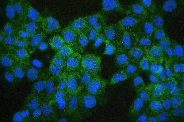
| IF/ICC Q&A: should the same pattern appear? | HPA ICC-IF places COMT mainly in vesicles and additionally in endoplasmic reticulum (HPA subcellular ICC-IF). Those observations come from cells imaged by IF; they do not prescribe a paraffin-section IHC pattern. For IHC, interpret the cytoplasmic and cell-level tissue observations reported by HPA tissue IHC. |
| Situation | Likely cause | Next action |
|---|---|---|
| Known-positive cell population has no chromogenic signal. | The region may lack the expected cells, or the IHC workflow may have failed (general IHC practice). HPA reports High staining in kidney collecting ducts and placental cytotrophoblasts (HPA tissue IHC). | Verify cell identity and section integrity, then review the catalog antibody's IHC-P instructions, retrieval, dilution and detection controls (general IHC practice). Do not infer a COMT-specific fixation effect from these sources. |
| Only nuclei are colored. | That compartment is discordant with cytoplasm/cell membrane localization (UniProt P21964 localization) and HPA's cytoplasmic tissue profile (HPA tissue IHC). | Compare with a no-primary control and inspect the counterstain. Recheck antibody specificity and detection background before assigning COMT positivity (general IHC practice). |
| Adipocytes or cardiomyocytes stain as strongly as the intended positive cells. | Both cell types are reported Not detected in HPA tissue IHC (HPA tissue IHC); cross-reactivity or endogenous detection activity is possible (general IHC practice). | Check a no-primary control and the detection-system controls. Score the unexpected cells separately from nearby positive structures rather than calling the whole section positive (general IHC practice). |
| Diffuse brown color masks cell borders. | Background from nonspecific binding or the detection system can prevent compartment assessment (general IHC practice). | Inspect the no-primary control, blocking and washes; adjust the detection workflow using its validated instructions (general IHC practice). Reassess whether cytoplasmic signal follows identifiable cells. |
| A low-staining population looks negative. | HPA lists low staining in hippocampal glia, thyroid glandular cells and liver cholangiocytes (HPA tissue IHC); weak signal may be hard to distinguish from background. | Compare it with a High-staining population and the negative control under the same scoring criteria (HPA tissue IHC; general IHC practice). Record weak or equivocal staining without upgrading it to High. |
| Punctate IF staining is used to judge the paraffin IHC slide. | HPA ICC-IF reports vesicles and additional endoplasmic reticulum localization (HPA subcellular ICC-IF), while HPA tissue IHC describes variable cytoplasmic staining (HPA tissue IHC). | Score the IHC slide against tissue cell types and compartments documented for IHC (HPA tissue IHC). Use the separate IF/ICC guide when interpreting cellular puncta. |


Comprehensive Human Protein Atlas IHC scoring per tissue (reliability: Enhanced — Medium consistency between antibody staining and RNA expression data. Paired antibodies with high similarity supports the protein expression profile.). Rows are taken directly from the HPA tissue chart — click any row's HPA link to view the source.
| Tissue | Cell type | Level | Evidence | Source |
|---|---|---|---|---|
| Cerebellum | Bergmann glia - cytoplasm/membrane | High | Protein (IHC) | HPA → |
| Epididymis | Glandular cells | High | Protein (IHC) | HPA → |
| Kidney | Collecting ducts | High | Protein (IHC) | HPA → |
| Placenta | Cytotrophoblasts | High | Protein (IHC) | HPA → |
| Skin | Melanocytes | Medium | Protein (IHC) | HPA → |
| Tissue | Cell type | Level | Evidence | Source |
|---|---|---|---|---|
| Adipose tissue | Adipocytes | Not detected | Protein (IHC) | HPA → |
| Heart muscle | Cardiomyocytes | Not detected | Protein (IHC) | HPA → |
| Skeletal muscle | Myocytes | Not detected | Protein (IHC) | HPA → |
| Vagina | Squamous epithelial cells | Not detected | Protein (IHC) | HPA → |
Use compartment, cell type, and control staining together when optimizing COMT chromogenic IHC in paraffin sections (UniProt P21964; HPA tissue IHC).
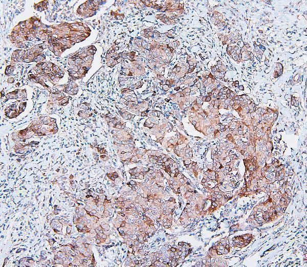
Anti-COMT IHC images cover human cancer and placenta sections and mouse and rat lung sections (catalog IHC captions); IF images cover A431 and MCF7 cells (M00464 IF captions).
M00464 will render with a human intestinal cancer paraffin-section IHC image; its catalog also documents human mammary cancer and placenta IHC and A431 and MCF7 IF (M00464 image captions). PB9539 will render with a mouse lung paraffin-section IHC image; its catalog also documents rat lung and human placenta IHC (PB9539 image captions).
Which to pick: For human tissue IHC, M00464 is a monoclonal option with human paraffin-section images (M00464 catalog and IHC captions). For IHC across species, PB9539 has human, mouse, and rat reactivity and paraffin-section images from each (PB9539 catalog and IHC captions); the captions do not report a fixative. For IF/ICC, choose M00464 because IF and ICC are listed applications with cell images, while PB9539 has no listed IF application (catalog applications; M00464 IF captions).