This website uses cookies to ensure you get the best experience on our website.


- Table of Contents
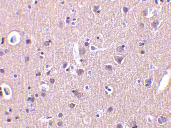
Plan chromogenic CRMP1 IHC on paraffin sections using brain neuronal staining as the reference pattern (HPA tissue IHC). The catalog antibody has a human brain IHC image at 2 μg/mL (datasheet: A05002).

Expected localisation, validated protocols, controls and antibodies — the at-a-glance facts below, then the full design guide.
| Expected localisation | Neuronal cytoplasm (HPA tissue IHC) | |
| Staining pattern | Cytoplasmic staining in neuronal cells (HPA tissue IHC) | |
| Antigen retrieval | Citrate pH 6.0 HIER, 95–98 °C, 20 min (rule: cytoplasmic / membrane antigen) | |
| Positive control | Caudate+3 more · see all | |
| Negative control | Adipose tissue+4 more · see all |
| Fixation | Keep fixation consistent across sections (standard IHC practice; not target-specific); optimize empirically. | |
| Caveat | Staining and RNA show medium consistency (HPA tissue IHC) | |
| Regulation | Brain-enriched expression (UniProt) | |
| Isoform / epitope | 2 isoforms; epitope coverage is unreported (UniProt) |
The catalog antibody protocol is followed by two published chromogenic IHC protocols for CRMP1 in prostate and brain tissue (PMC5290039; PMC5319751).
| Sample | Tissue sections; selected-image fixative not specified (standard IHC workflow) |
| Fixation | Image fixative and duration unreported (datasheet A05002); verify before use. |
| Sectioning | 4–5 µm sections on charged slides (standard) |
| Deparaffinisation | Xylene, graded ethanol series to water (standard) |
| Antigen retrieval | Heat-induced epitope retrieval in citrate buffer, pH 6.0, 20 min at 95–98 °C (standard rule: cytoplasmic / membrane antigen) |
| Peroxidase block | 3% H2O2, 10 min, room temperature (standard) |
| Blocking | 10% normal serum of the secondary host, 30 min, room temperature (standard) |
| Primary antibody | Rabbit anti-CRMP1, 2 μg/mL (datasheet A05002) |
| Primary incubation | Overnight at 4 °C (standard) |
| Detection | HRP-polymer secondary, DAB chromogen 5–10 min (standard) |
| Counterstain | Hematoxylin, blue, dehydrate and mount (standard) |
| Expected result | CRMP1-positive staining in neuronal cells of caudate (HPA tissue IHC: Medium). HPA tissue profile: Cytoplasmic expression in neuronal cells. No signal in the no-primary control. |
CRMP1 is a cytoplasmic protein with no transmembrane segment (UniProt Q14194: localisation and topology). In paraffin tissue sections, expect staining chiefly in neuronal cells, including cerebellar Purkinje cells (HPA tissue IHC). HPA rates the tissue staining Approved, with medium consistency between antibody staining and RNA expression; treat the pattern as a guide rather than proof of antibody specificity (HPA tissue IHC).
| Medium cytoplasmic staining in cortical, hippocampal or caudate neuronal cells, or cerebellar Purkinje cells (HPA tissue IHC). | This matches the reported positive cells and staining level (HPA tissue IHC). Compare cellular distribution with a negative control before scoring a section (standard IHC practice). |
| Predominantly nuclear staining, with little neuronal cytoplasmic signal. | A nuclear-dominant pattern conflicts with the reported cytoplasmic tissue pattern (HPA tissue IHC; UniProt Q14194: localisation). Check whether the same pattern appears in controls before interpreting it as CRMP1 (standard IHC practice). |
| Strong staining in adipocytes or in glandular cells of an HPA-negative tissue. | Those cells were reported as not detected in the listed adipose, adrenal or appendix samples (HPA tissue IHC). Consider cross-reactivity or endogenous detection activity, and compare antibody-omission and detection controls (standard IHC practice). |
| Widespread haze across cells and surrounding tissue, obscuring neuronal boundaries. | This is difficult to reconcile with the reported cell-selective cytoplasmic pattern (HPA tissue IHC). Assess background with a negative control; blocking, washes and detection chemistry can affect nonspecific signal (standard IHC practice). |
| No staining in neuronal cells of caudate, cortex, hippocampus or cerebellum. | These are HPA-positive reference tissues, although their reported level is Medium (HPA tissue IHC). First confirm tissue quality and detection controls, then review the catalog antibody’s IHC-P conditions (standard IHC practice). |
| Cell and tissue selection | HPA reports Medium staining in caudate, cerebral cortex and hippocampal neuronal cells and cerebellar Purkinje cells; these are useful positive references (HPA tissue IHC). Its sampled adipocytes and selected glandular cells were not detected, but a negative sample does not establish universal absence (HPA tissue IHC). |
| Subcellular interpretation | UniProt places CRMP1 in cytoplasm, cytoskeleton, centrosome, spindle, perikaryon and growth cone, and reports no transmembrane segment (UniProt Q14194: localisation and topology). HPA tissue IHC describes cytoplasmic neuronal staining; do not require a resolvable centrosomal dot in every paraffin section (HPA tissue IHC; standard IHC practice). |
| Antibody evidence | HPA lists IHC as Approved for HPA035640 and CAB037313, while its tissue summary reports medium agreement with RNA (HPA antibodies; HPA tissue IHC). Approval supports using the reported pattern as a reference, but a control remains necessary when staining departs from it (standard IHC practice). |
| Isoforms and epitope scope | UniProt lists 2 isoforms, isoform 1 and LCRMP-1 (UniProt Q14194: isoforms). The supplied evidence does not map either antibody’s epitope to an isoform; staining cannot identify which isoform is present without separate evidence (HPA antibodies; UniProt Q14194: isoforms). |
| Processing and modifications | UniProt reports a chain spanning residues 1–572, with no signal peptide or propeptide, and lists phosphorylation and other modified residues (UniProt Q14194: processing and modified residues). Epitope overlap and effects on antibody binding are unreported here; do not infer a staining change from these annotations alone. |
| IF/ICC Q: Where should CRMP1 appear? | A: Mainly at the centrosome, with additional cytosolic localisation in the HPA ICC-IF assessment; images are listed for REH and U2OS (HPA subcellular). This answers an IF localisation question; it supplies no IF protocol conditions. |
| Situation | Likely cause | Next action |
|---|---|---|
| Neuronal cytoplasm is absent in a positive reference section. | The observation conflicts with Medium staining in the listed neuronal populations (HPA tissue IHC); section quality or an IHC detection step may also account for lost signal (standard IHC practice). | Check tissue morphology, antibody-omission and detection controls, and the catalog antibody’s IHC-P instructions; review retrieval and detection settings without assuming CRMP1-specific fixation sensitivity (standard IHC practice). |
| Signal appears chiefly in nuclei. | This disagrees with HPA’s cytoplasmic neuronal tissue profile and UniProt’s cytoplasmic localisation (HPA tissue IHC; UniProt Q14194: localisation). | Compare the pattern with a positive neuronal reference and negative control, then reassess detection background before assigning a nuclear CRMP1 pattern (standard IHC practice). |
| Strong staining appears in HPA-negative cell types. | The selected adipocytes or glandular cells were not detected by HPA; cross-reactivity or endogenous detection activity is possible (HPA tissue IHC; standard IHC practice). | Compare a matched negative tissue and antibody-omission control; check whether the detection system itself produces staining in those cells (standard IHC practice). |
| Diffuse colour obscures which cells are positive. | Background can result from nonspecific antibody binding, incomplete blocking or inadequate washing (standard IHC practice); the expected tissue profile is cell-selective and cytoplasmic (HPA tissue IHC). | Use a negative control to locate the background source, then review blocking, washes and detection exposure under the established IHC-P workflow (standard IHC practice). |
| A few weakly stained cells are being called negative. | HPA reports Medium staining in specified neuronal cells but Low staining in some other sampled cell types (HPA tissue IHC); scoring depends on cell identity and the control baseline (standard IHC practice). | Score the named cell population and its cytoplasm against a matched control, recording intensity and proportion separately (HPA tissue IHC; standard IHC practice). |
| IHC staining is judged against an IF centrosome image. | HPA’s ICC-IF summary emphasizes centrosomes and cytosol, whereas its tissue IHC profile reports neuronal cytoplasmic staining (HPA subcellular; HPA tissue IHC). | Assess paraffin sections against the HPA tissue pattern and an IHC positive reference; use the ICC-IF summary only to interpret subcellular localisation in IF (HPA tissue IHC; HPA subcellular). |


Comprehensive Human Protein Atlas IHC scoring per tissue (reliability: Approved — Medium consistency between antibody staining and RNA expression data.). Rows are taken directly from the HPA tissue chart — click any row's HPA link to view the source.
| Tissue | Cell type | Level | Evidence | Source |
|---|---|---|---|---|
| Caudate | Neuronal cells | Medium | Protein (IHC) | HPA → |
| Cerebellum | Purkinje cells | Medium | Protein (IHC) | HPA → |
| Cerebral cortex | Neuronal cells | Medium | Protein (IHC) | HPA → |
| Hippocampus | Neuronal cells | Medium | Protein (IHC) | HPA → |
| Tissue | Cell type | Level | Evidence | Source |
|---|---|---|---|---|
| Adipose tissue | Adipocytes | Not detected | Protein (IHC) | HPA → |
| Adrenal gland | Glandular cells | Not detected | Protein (IHC) | HPA → |
| Appendix | Glandular cells | Not detected | Protein (IHC) | HPA → |
| Bone marrow | Hematopoietic cells | Not detected | Protein (IHC) | HPA → |
| Breast | Adipocytes | Not detected | Protein (IHC) | HPA → |
Troubleshoot CRMP1 staining in paraffin sections by checking retrieval, cellular distribution, controls and scoring before interpreting chromogenic signal.
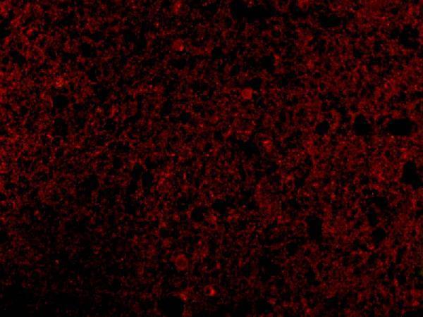
A05002 has IHC imagery from human brain tissue and IF imagery from human brain cells (catalog image captions). Both antibodies list human, mouse and rat reactivity (catalog reactivity).
A05002 is the only SKU with an IHC figure for its card; the caption shows human brain tissue stained at 2 μg/mL (A05002 IHC caption). Its IF caption shows human brain cells stained at 10 μg/mL (A05002 IF caption).
Which to pick: Choose A05002 for paraffin-section tissue IHC (catalog applications: IHC-P; A05002 IHC caption: human brain tissue); its image caption does not report the fixative (A05002 IHC caption). For IF/ICC, M05002-1 lists ICC/IF and is rabbit monoclonal clone 18C29 (catalog applications; catalog host/clone), while A05002 has a human brain-cell IF image (A05002 IF caption). Both list human, mouse and rat reactivity for cross-species planning (catalog reactivity).