CRTAP / Cartilage-associated protein · IHC design guide

Design Immunohistochemistry for CRTAP

Plan CRTAP chromogenic IHC on paraffin sections with the catalog antibody at 1:50–1:200 (datasheet). HPA reports widespread cytoplasmic tissue staining (HPA tissue IHC), while UniProt places CRTAP in the secreted extracellular matrix (UniProt).

Evidence assembled Oct 2026 · For research use; verify linked source records and product datasheet before use
Immunohistochemistry protocol sheet for CRTAP (IHC for CRTAP): expected localisation Cytoplasmic tissue staining (HPA tissue IHC); secreted ECM location (UniProt), antibody A05852, validated IHC image, and IHC protocol steps
Printable CRTAP IHC protocol sheet — expected localisation Cytoplasmic tissue staining (HPA tissue IHC); secreted ECM location (UniProt), antibody A05852, controls and protocol steps. Open the full CRTAP IHC guide →

CRTAP Immunohistochemistry Experimental Design Guide

Expected localisation, validated protocols, controls and antibodies — the at-a-glance facts below, then the full design guide.

Must know before staining
Expected localisation Cytoplasmic tissue staining (HPA tissue IHC); secreted ECM location (UniProt)
Staining pattern Widespread cytoplasmic staining; high in glandular cells (HPA tissue IHC)
Antigen retrieval Citrate pH 6.0 HIER, 95–98 °C, 20 min (rule: cytoplasmic / membrane antigen)
Positive control ⓘ Breast+4 more · see all
Negative control ⓘ None in HPA (detected in all 45 tissues); use no-primary + isotype controls
Important caveats
Reasons your staining may differ from the expected pattern.
Fixation Keep fixation consistent across sections. (standard IHC practice; not target-specific)
Caveat As a secreted protein, tissue RNA and protein locations may differ (HPA tissue IHC)
Regulation No specific expression regulator reported (UniProt)
Isoform / epitope No annotated isoforms; mature chain 27–401; no transmembrane epitope split (UniProt)
Section 1

Recommended CRTAP IHC & IF Protocols

The catalog antibody’s IHC-P protocol is accompanied by one published CRTAP paraffin-section method (PMC6221197).

Recommended immunohistochemistry (IHC-P) protocol parameters
SampleParaffin-embedded rat lung tissue; fixative not specified (datasheet A05852)
FixationImage fixative and duration unreported (datasheet A05852); verify before use.
Sectioning4–5 µm sections on charged slides (standard)
DeparaffinisationXylene, graded ethanol series to water (standard)
Antigen retrievalHeat-induced epitope retrieval in citrate buffer, pH 6.0, 20 min at 95–98 °C (standard rule: cytoplasmic / membrane antigen)
Peroxidase block3% H2O2, 10 min, room temperature (standard)
Blocking10% normal serum of the secondary host, 30 min, room temperature (standard)
Primary antibodyRabbit anti-CRTAP, 1:50-1:200 (datasheet A05852)
Primary incubationOvernight at 4 °C (standard)
DetectionHRP-polymer secondary, DAB chromogen 5–10 min (standard)
CounterstainHematoxylin, blue, dehydrate and mount (standard)
Expected resultCRTAP-positive staining in glandular cells of breast (HPA tissue IHC: High). HPA tissue profile: Ubiquitous cytoplasmic expression. No signal in the no-primary control.
💡Decision noteStart with citrate pH 6.0 retrieval at 95–98 °C for 20 min (page retrieval); the published method uses microwave heating in citrate (PMC6221197).
Section 2

What Is the Expected CRTAP Staining Pattern?

For IHC on paraffin sections, expect predominantly cytoplasmic CRTAP staining in glandular cells of breast, colon, duodenum, endometrium, fallopian tube, gallbladder and prostate, and in placental decidual cells (HPA tissue IHC: High; reliability Approved). CRTAP has no transmembrane segment and is also annotated as secreted into the extracellular space and matrix (UniProt O75718 topology and subcellular location).

What am I looking at on my slide?
Strong cytoplasmic staining in glandular cells, with tissue structure still readable.This matches the reported high glandular-cell signal in the listed tissues and the overall cytoplasmic IHC profile (HPA tissue IHC: High; ubiquitous cytoplasmic expression). Compare intensity within the same staining run; the HPA labels describe observed patterns, not a universal intensity cutoff (HPA tissue IHC: High; general IHC practice).
Staining appears extracellular as well as cytoplasmic.An extracellular component is biologically plausible because CRTAP is secreted and annotated in extracellular space and matrix (UniProt O75718 subcellular location). HPA reports a ubiquitous cytoplasmic tissue-IHC profile, so extracellular staining alone does not establish that the expected cellular pattern was reproduced (HPA tissue IHC: profile).
Signal is confined to nuclei or another compartment without the expected cytoplasmic pattern.Treat an isolated nuclear pattern as suspect: HPA reports cytoplasmic tissue staining, while UniProt annotates extracellular location and no transmembrane segment (HPA tissue IHC: profile; UniProt O75718 location and topology). Check morphology and controls before calling it CRTAP (general IHC practice).
Cells outside the expected stained population dominate the result.In a selected high-positive tissue, stronger staining outside the reported glandular or decidual population raises concern for cross-reactivity or endogenous detection activity; it is not proof of either cause (HPA tissue IHC: positive-cell listings; general IHC practice). Interpret weaker or mixed staining against tissue morphology and controls (general IHC practice).
The slide shows widespread haze, or no signal in a reported high-positive population.Haze obscures the cell-level pattern; absence of signal prevents confirmation of the reported high-positive pattern (HPA tissue IHC: High; general IHC practice). Either finding calls for a controlled repeat before interpreting CRTAP abundance (general IHC practice).
💡Expected CRTAP appearanceCall a section positive when glandular cells in a listed high-positive tissue, or placental decidual cells, show clear cytoplasmic staining above background; diffuse haze or isolated nuclear staining fails that pattern check (HPA tissue IHC: High and cytoplasmic profile; general IHC practice).
How each factor affects the staining
Tissue and cell selectionHPA lists High staining in glandular cells of breast, colon, duodenum, endometrium, fallopian tube, gallbladder and prostate, and decidual cells of placenta (HPA tissue IHC: positive listings). Its low-staining listings are not stated negatives (HPA tissue IHC: low listings; negative list empty).
Secreted protein and compartmentHPA warns that RNA and protein tissue locations may differ for a secreted protein; its tissue-IHC profile is cytoplasmic (HPA tissue IHC: reliability description and profile). UniProt also places CRTAP in extracellular space and matrix (UniProt O75718 subcellular location).
Processing and molecular featuresUniProt annotates a signal peptide at residues 1–26, a chain at 27–401, and glycosylation sites at 87 and 363 (UniProt O75718 processing and glycosylation). These annotations alone do not establish which compartment an antibody detects in a paraffin section (general IHC interpretation).
IHC antibody evidenceHPA lists HPA043598 and HPA044150 as IHC Approved (HPA antibodies). That supports using their recorded IHC patterns as comparisons, but supplies no antibody-specific dilution, retrieval condition or target-specific fixation effect for this section (HPA antibodies; supplied HPA tissue IHC).
IF/ICC Q&A: what pattern is reported?HPA reports cytosol, actin filaments and intermediate filaments in ICC-IF, each as an approved main location; HPA044150 is ICC Approved (HPA subcellular ICC-IF; HPA antibodies). This is an IF/ICC observation, not a paraffin-IHC protocol or a reason to require filament staining in tissue (HPA subcellular ICC-IF; general IHC interpretation).
Why is my staining missing, weak or wrong?
SituationLikely causeNext action
No staining in glandular cells of a selected high-positive tissue.The run may have insufficient detectable signal; the selected cell population is reported High, but that designation does not validate this run's assay conditions (HPA tissue IHC: High; general IHC practice).Confirm the expected cells are present, inspect the positive control, then review the catalog antibody's IHC-P dilution and retrieval instructions before repeating (HPA tissue IHC: positive-cell listings; general IHC practice).
Diffuse brown haze masks cell boundaries.Nonspecific background or excess detection signal can obscure a cytoplasmic pattern (general chromogenic IHC practice); HPA's reported profile requires discernible cytoplasmic staining (HPA tissue IHC: profile).Compare a no-primary control, inspect blocking and washes, and adjust the detection conditions according to the assay controls (general chromogenic IHC practice).
Signal appears in the no-primary control.The primary antibody cannot account for signal in that control; endogenous activity or nonspecific detection reagents are possible contributors (general chromogenic IHC practice).Check the detection system and its endogenous-activity blocking step, then repeat the control alongside the stained section (general chromogenic IHC practice).
Only nuclei are strongly stained.A nuclear-only result does not match HPA's cytoplasmic tissue profile or UniProt's extracellular annotation (HPA tissue IHC: profile; UniProt O75718 subcellular location). The cause remains undetermined without controls (general IHC practice).Verify cell morphology and compare the control sections; review the IHC-validated antibody's documented conditions before assigning the signal to CRTAP (general IHC practice).
A low-listed cell population appears as bright as the selected high-positive population.HPA reports Low staining in specific cerebellar, hippocampal, caudate, ovarian, splenic and lymph-node cell populations; it does not designate them negative (HPA tissue IHC: low listings; negative list empty).Compare corresponding cell types and control sections within the same run; investigate unexpected intensity without treating a low-listed tissue as a zero-signal control (HPA tissue IHC: low listings; general IHC practice).
Extracellular staining dominates while expected cytoplasmic cells are unclear.Extracellular CRTAP is plausible from the UniProt annotation, but HPA describes the tissue-IHC profile as cytoplasmic (UniProt O75718 subcellular location; HPA tissue IHC: profile). Dominant extracellular color alone is therefore inconclusive.Inspect tissue morphology and background controls, then repeat or compare with an independently validated IHC antibody if the cellular pattern remains unresolved (general IHC practice; HPA antibodies: two IHC Approved entries).

Sample controls for CRTAP IHC & IF

🧪Run breast first and score its glandular cells for CRTAP staining (HPA: High in breast glandular cells). HPA detects CRTAP in all 45 scored tissues, so there is no supported negative tissue; use no-primary and isotype controls for background, and treat unstained non-glandular areas on the breast slide only as internal background references, not validated negative cells (HPA: no negative tissue rows; High in breast glandular cells).
Positive control tissue: Breast (Glandular cells, HPA High)
Negative control tissue: None in HPA: CRTAP is detected in all 45 scored tissues. Use a no-primary (secondary-only) and an isotype control instead.
ICC-IF cell lines (HPA subcellular resource): HPA ICC-IF images show CRTAP in U-251MG, U2OS, NIH 3T3, with annotated localisation: Intermediate filaments (approved), Actin filaments (approved), Cytosol (approved) (HPA subcellular).
Technical controls: Include a no-primary, secondary-only slide, an isotype control matched to the primary antibody’s host species and clonality, and a CRTAP knockout or validated peptide-block control where available (standard IHC practice). For chromogenic HRP detection, block endogenous peroxidase; if using avidin–biotin detection, check endogenous biotin (standard IHC practice).
⚠️Feasibility: No supplied matched source reports a CRTAP-specific fixation window or fixation effect, and the A05852 rat-lung paraffin caption does not state its fixative (datasheet: A05852 rat-lung IHC caption). That caption uses microwave retrieval in 10 mM PBS, pH 7.2, at 1:100, but retrieval dependence in breast is unreported (datasheet: A05852 rat-lung IHC caption). HPA has ICC-IF images in U-251MG, U2OS and NIH 3T3, but the supplied evidence does not establish whether frozen sections or IF are easier than paraffin IHC; assess endogenous peroxidase background in breast when using HRP detection (HPA: ICC-IF image cell lines; standard IHC practice).

HPA tissue IHC evidence for CRTAP

Comprehensive Human Protein Atlas IHC scoring per tissue (reliability: Approved — Secreted protein, tissue location of RNA and protein is expected to differ.). Rows are taken directly from the HPA tissue chart — click any row's HPA link to view the source.

Positive expression · recommended positive controls

TissueCell typeLevelEvidenceSource
Breast Glandular cells High Protein (IHC) HPA →
Colon Glandular cells High Protein (IHC) HPA →
Duodenum Glandular cells High Protein (IHC) HPA →
Endometrium Glandular cells High Protein (IHC) HPA →
Fallopian tube Glandular cells High Protein (IHC) HPA →

Undetected expression · recommended negative controls

TissueCell typeLevelEvidenceSource
None in HPA: CRTAP is detected in all 45 scored tissues. Use a no-primary (secondary-only) and an isotype control instead.
Section 3

Advanced CRTAP IHC Tips

Troubleshoot CRTAP staining in paraffin sections by checking retrieval, compartment, controls, and scoring before interpreting chromogenic signal.

Which retrieval condition should I try first for CRTAP IHC?
Start with microwave antigen retrieval in 10 mM PBS, pH 7.2, as used for the catalog antibody on paraffin-embedded rat lung at 1:100 (caption A05852). The caption does not report a microwave temperature or duration, so record the settings used and compare sections processed together (caption A05852; standard IHC practice). If staining is weak, test citrate pH 6.0 at 95–98 °C for 20 min on a separate section as this page’s alternative retrieval condition (page retrieval specification). Compare signal and tissue preservation with a no-primary control before selecting a condition, since stronger chromogen alone does not establish CRTAP specificity (standard IHC practice).
How should I assess fixation-related loss of CRTAP staining?
CRTAP-specific sensitivity to fixation is unknown from the supplied evidence; the catalog image identifies paraffin-embedded rat lung but does not state its fixative (caption A05852). Record the fixative, fixation duration, section thickness, and processing history for each specimen before comparing staining intensity (standard IHC practice). If staining varies across specimens, stain adjacent sections in one run with the same retrieval and 1:100 catalog-antibody dilution to isolate processing differences (caption A05852; standard IHC practice). Check morphology and edge-to-center staining before attributing weak signal to fixation, and establish any fixation effect empirically with matched tissue (standard IHC practice).
Should CRTAP appear inside cells or in extracellular matrix?
Assess both cellular and extracellular signal: CRTAP is annotated as secreted to the extracellular space and matrix, without a transmembrane segment (UniProt O75718 localisation and topology). Tissue IHC is described as ubiquitously cytoplasmic, while cellular IF reports cytosol, actin filaments, and intermediate filaments (HPA tissue IHC profile; HPA subcellular). In paraffin sections, score cytoplasmic and matrix-associated chromogen separately rather than treating either pattern as automatically nonspecific (HPA tissue IHC profile; UniProt O75718 localisation; standard IHC practice). Use morphology and a no-primary control to distinguish cellular signal from diffuse precipitate, and avoid assigning a thin boundary stain to membrane CRTAP without independent support (standard IHC practice; UniProt O75718 topology).
How do processing and epitope uncertainty affect CRTAP IHC?
The supplied record lists no isoforms, a signal peptide at residues 1–26, and a mature chain spanning 27–401 (UniProt O75718 processing and isoforms). It also lists glycosylation sites at residues 87 and 363, but supplies no antibody epitope, so neither processing nor glycosylation establishes why a section stains weakly (UniProt O75718 glycosylation; supplied antibody evidence). Confirm the antibody’s epitope from its documentation before interpreting intracellular versus matrix signal as detection of a particular CRTAP form (standard IHC practice; UniProt O75718 localisation). If signal changes with retrieval, compare matched sections and controls rather than assigning the change to epitope masking or a specific modification without direct evidence (standard IHC practice).
How should I adapt CRTAP localisation checks for multiplex IF?
On the separate IF/ICC guide, pair CRTAP with a validated marker for the cell population under study, and assess overlap within cells rather than assuming nearby matrix signal belongs to them (UniProt O75718 localisation; standard IF practice). Choose fluorophores and filter sets after checking autofluorescence in an unstained section, reserving a spectrally distinct channel for the weaker signal (standard IF practice). For an intracellular CRTAP epitope, optimise mild permeabilisation against morphology and signal; for an extracellular epitope, first assess staining without detergent (HPA subcellular; UniProt O75718 localisation; standard IF practice). Include single-stain and no-primary controls to check bleed-through and background, and do not transfer the paraffin IHC caption’s retrieval condition directly to IF/ICC (caption A05852; standard IF practice).
What should I check when CRTAP chromogen is diffuse?
First compare a no-primary section with the stained section to identify chromogen deposition unrelated to primary-antibody binding (standard IHC practice). Apply an appropriate peroxidase block before chromogenic detection, then assess whether background follows tissue edges, damaged areas, or the counterstain rather than cell or matrix structures (standard IHC practice). Titrate the catalog antibody around the caption’s 1:100 condition and keep retrieval and detection settings constant during that comparison (caption A05852; standard IHC practice). CRTAP can be extracellular and HPA describes widespread cytoplasmic IHC staining, so diffuse staining alone cannot prove background or specificity; require reproducible morphology and clean controls (UniProt O75718 localisation; HPA tissue IHC profile; standard IHC practice).
How should I score CRTAP across paraffin sections? ⚠ ANSWER MARKED FOR VERIFICATION
Define the region and compartment before scoring: report cytoplasmic staining and matrix-associated staining separately because both are plausible CRTAP locations (HPA tissue IHC profile; UniProt O75718 localisation). For cellular staining, use an H-score or percent positive cells with fixed intensity thresholds; for extracellular staining, report stained area or optical density per mm² of evaluable tissue (standard IHC quantification practice). Normalise cellular results to the number of eligible cells and matrix results to intact tissue area, excluding folds, necrosis, and edges by a rule set before review (standard IHC quantification practice). Apply identical retrieval, detection, imaging, and threshold settings across comparison groups, and retain representative control images alongside numerical results (standard IHC practice).
When is CRTAP staining convincing rather than artefactual?
A convincing pattern repeats in intact tissue, follows interpretable cytoplasmic or extracellular structures, and exceeds the matched no-primary control (HPA tissue IHC profile; UniProt O75718 localisation; standard IHC practice). HPA reports high glandular-cell staining in breast, colon, and duodenum, but its tissue profile also describes widespread cytoplasmic staining, so a positive cell type alone does not establish antibody specificity (HPA tissue IHC). Treat isolated membrane outlines cautiously because CRTAP has no annotated transmembrane segment, and inspect edge staining, necrosis, and endogenous peroxidase signal before scoring (UniProt O75718 topology; standard IHC practice). Finally, secretion means tissue RNA and protein location can differ; interpret chromogenic distribution with morphology and controls rather than requiring cellular RNA and protein maps to coincide (HPA tissue IHC reliability description; standard IHC practice).
Boster reagents

Best CRTAP / Cartilage-associated protein IHC Antibodies

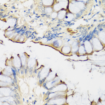
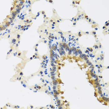
A05852 has IHC images of paraffin-embedded rat lung, human colon, and mouse lung (catalog image captions). No IF image is supplied (catalog: IF images).

Real IHC data Immunohistochemistry of paraffin-embedded rat lung using CRTAP antibody at dilution of 1:100 .Perform microwave antigen retrieval with 10 mM PBS buffer pH 7.2 before commencing with IHC staining protocol.
Anti-Cartilage-associated protein CRTAP Antibody
Cat # A05852

A05852 is listed for IHC and reacts with human, mouse, and rat (catalog: applications and reactivity). Its IHC images show paraffin-embedded rat lung, human colon, and mouse lung at 1:100 (catalog image captions).

Which to pick: For tissue IHC, choose A05852: it is a rabbit polyclonal antibody listed for IHC at 1:50–1:200, with paraffin-section images (catalog: host, dilution, applications; catalog image captions). For cross-species IHC, A05852 has an image in each listed species: human, mouse, and rat (catalog: reactivity; catalog image captions). No IF/ICC application or image is documented for A05852; its IHC captions specify paraffin embedding but do not report the fixative (catalog: applications, IF images, IHC image captions).

Each figure is that product's own IHC / IF validation image from its datasheet.

References

  1. UniProt Consortium. UniProt entry O75718 (CRTAP_HUMAN, Cartilage-associated protein).
  2. Human Protein Atlas. CRTAP tissue IHC expression (reliability: Approved).
  3. Human Protein Atlas. CRTAP subcellular location (ICC-IF): Localized to the cytosol, actin filaments and intermediate filaments..
  4. Human Protein Atlas. CRTAP antibody validation summary (2 antibodies).
  5. Dental and craniofacial defects in the Crtap(-/-) mouse model of osteogenesis imperfecta type VII. Developmental dynamics : an official publication of the American Association of Anatomists 2020 — PMC7727892.
  6. Tendon and motor phenotypes in the Crtap(-/-) mouse model of recessive osteogenesis imperfecta. eLife 2021 — PMC8186905.
  7. Expression characterization and functional implication of the collagen-modifying Leprecan proteins in mouse gonadal tissue and mature sperm. AIMS genetics 2018 — PMC6221197.
  8. Excessive transforming growth factor-β signaling is a common mechanism in osteogenesis imperfecta. Nature medicine 2014 — PMC4048326.
  9. PubMed PMID:10702664 — UniProt-cited evidence.
  10. PubMed PMID:14702039 — UniProt-cited evidence.
  11. PubMed PMID:15489334 — UniProt-cited evidence.