This website uses cookies to ensure you get the best experience on our website.


- Table of Contents
Plan paraffin CRY2 IHC with the catalog antibody at 0.5–1 μg/mL (datasheet PB9576). Assess granular cytoplasmic staining across tissues (HPA tissue IHC), and record collection time for intensity comparisons (UniProt).

Expected localisation, validated protocols, controls and antibodies — the at-a-glance facts below, then the full design guide.
| Expected localisation | Granular cytoplasm (HPA tissue IHC); nucleus possible (UniProt) | |
| Staining pattern | Granular cytoplasmic staining in most tissues (HPA tissue IHC) | |
| Antigen retrieval | EDTA pH 8.0 HIER, heat-mediated (datasheet PB9576) | |
| Positive control | Adrenal gland+4 more · see all | |
| Negative control | None in HPA (detected in all 45 tissues); use no-primary + isotype controls |
| Fixation | Matched tissue-IHC evidence does not establish the fixation claim. Validate the specimen-specific method before use. (selected-SKU IHC image PB9576) | |
| Caveat | Sampling time may affect intensity comparisons (UniProt) | |
| Regulation | Circadian feedback regulates CRY2 (UniProt) | |
| Isoform / epitope | 2 isoforms; epitope coverage is unspecified (UniProt) |
The catalog antibody’s IHC-P protocol (datasheet: PB9576) is accompanied by 3 published CRY2 IHC protocols (PMC5652816; PMC4458447; PMC7476803).
| Sample | Paraffin-embedded rat liver tissue; fixative not specified (datasheet PB9576) |
| Fixation | Image fixative and duration unreported (datasheet PB9576); verify before use. |
| Sectioning | 4–5 µm sections on charged slides (standard) |
| Deparaffinisation | Xylene, graded ethanol series to water (standard) |
| Antigen retrieval | Heat retrieval: EDTA pH 8.0 (datasheet PB9576); 20 min, 95–100 °C (standard) |
| Peroxidase block | 3% H2O2, 10 min, room temperature (standard) |
| Blocking | 10% goat serum (datasheet PB9576) |
| Primary antibody | Rabbit anti-CRY2, 0.5-1μg/ml (datasheet PB9576) |
| Primary incubation | Overnight at 4 °C (datasheet PB9576) |
| Detection | Streptavidin-biotin complex (SABC), DAB chromogen (datasheet PB9576) |
| Counterstain | Hematoxylin, blue, dehydrate and mount (standard) |
| Expected result | CRY2-positive staining in glandular cells of adrenal gland (HPA tissue IHC: High). HPA tissue profile: Granular cytoplasmic expression in a majority of tissues. No signal in the no-primary control. |
CRY2 is a soluble protein found in the cytoplasm and nucleus, with no transmembrane segment (UniProt Q49AN0: subcellular location and topology). In tissue IHC, expect granular cytoplasmic staining across many cell types rather than a tissue restricted pattern (HPA tissue IHC: Supported; granular cytoplasmic expression in a majority of tissues). HPA reports High staining in several specific cell populations (HPA tissue IHC).
| Granular cytoplasmic staining appears in cells from several tissues. | This matches the predominant tissue IHC pattern (HPA tissue IHC: granular cytoplasmic expression in a majority of tissues; Supported). Assess staining in identifiable cells against adjacent background. HPA reports High staining in adrenal glandular cells, bronchial respiratory epithelial cells and bone marrow hematopoietic cells, among others (HPA tissue IHC). |
| Staining is confined to an unexpected compartment, with no convincing cytoplasmic signal. | Recheck the result before assigning it to CRY2. Nuclear staining alone is not inherently wrong because CRY2 also localizes to the nucleus (UniProt Q49AN0: subcellular location). The predominant tissue IHC observation is granular cytoplasm (HPA tissue IHC); HPA nuclear speckles come from ICC-IF, a different preparation (HPA subcellular: nuclear speckles). |
| Strong staining appears only in a cell population outside the reported positive populations. | Consider cross-reactivity or endogenous detection activity, especially if the reported granular cytoplasmic pattern is absent (HPA tissue IHC: profile). The supplied HPA list gives positive examples, not an exhaustive cell map or a validated negative population (HPA tissue IHC: positive list; negative list empty). Check morphology and appropriate staining controls before interpreting the unexpected cells. |
| Diffuse colour covers cells and surrounding tissue without a clear granular pattern. | Treat the result as background until controls and microscopy support a cellular signal. Diffuse staining does not resemble the predominant granular cytoplasmic tissue pattern (HPA tissue IHC: profile). In chromogenic IHC, inadequate blocking, excessive antibody concentration or detection background are general possibilities; compare a primary antibody omission control and optimize the workflow. |
| A known HPA positive cell population has little or no signal. | A negative slide alone does not establish absent CRY2. Confirm that the expected cells are present, then check antibody and detection performance with a positive control. HPA reports High staining in breast glandular cells and cerebellar granular layer cells, for example (HPA tissue IHC). No CRY2 specific fixation sensitivity or retrieval condition is supplied. |
| Tissue and cell choice | Use an identifiable reported positive population to judge whether a slide worked: HPA lists High staining in adrenal glandular, bronchial respiratory epithelial and bone marrow hematopoietic cells (HPA tissue IHC). Its empty negative list does not identify a true negative tissue. UniProt describes expression across examined tissues, with highest levels in heart and skeletal muscle, but that statement is not an IHC intensity ranking (UniProt Q49AN0: tissue specificity). |
| Compartment and preparation | Tissue IHC is predominantly granular cytoplasmic (HPA tissue IHC: profile), while CRY2 can also occupy the nucleus (UniProt Q49AN0: subcellular location). Keep those observations distinct when reading paraffin sections: HPA's supported nuclear speckles and additional cytosol localization were observed by ICC-IF (HPA subcellular), so speckles are not a required tissue IHC feature. |
| IF/ICC Q&A: what pattern is reported? | HPA reports mainly nuclear speckles, with additional cytosol localization in ICC-IF (HPA subcellular: supported locations). Its images are listed for A-431, U-251MG and U2OS (HPA subcellular). That cell imaging result can inform interpretation, but it does not supply a paraffin IHC protocol or override the tissue IHC profile (HPA tissue IHC: granular cytoplasmic expression). |
| Isoforms and antibody coverage | UniProt lists 2 CRY2 isoforms (UniProt Q49AN0: isoforms). The payload gives no antibody epitope or isoform coverage, so do not assume that a negative result excludes both isoforms. It also gives no basis to predict an isoform specific compartment or cell pattern from this antibody. |
| Strength of validation | HPA marks the tissue IHC profile Supported, citing high consistency between antibody staining and RNA expression data (HPA tissue IHC: reliability). Antibody HPA037577 is listed as Supported for IHC and ICC (HPA antibodies). These labels support using the reported pattern as a reference; they do not make every unusual signal specific or establish a fixation effect. |
| Situation | Likely cause | Next action |
|---|---|---|
| Reported positive cells stain weakly or not at all. | Target-specific fixation effects are not established by the supplied assay evidence. Verify with a matched IHC source before attributing a result to fixation. | Locate the expected cells and compare a concurrently processed positive control. Check the antibody, detection reagents and counterstain, then optimize retrieval and antibody concentration as general paraffin IHC steps; do not infer a CRY2 fixation effect from this failure. |
| Colour appears across the section, including spaces between cells. | Non-specific reagent deposition or insufficient suppression of endogenous detection activity is possible in chromogenic IHC; this appearance differs from the granular cytoplasmic HPA profile (HPA tissue IHC). | Inspect a primary antibody omission control, review blocking and washes, and adjust detection conditions. Score cell associated granules only after background is controlled; HPA supplies no target specific dilution for this section. |
| Only nuclei stain in a tissue section. | CRY2 can enter the nucleus (UniProt Q49AN0: subcellular location), but nuclear only tissue staining differs from HPA's predominant granular cytoplasmic IHC profile (HPA tissue IHC). | Verify nuclear boundaries with the counterstain and examine a reported positive population on the same run. Reassess specificity if cytoplasmic staining is absent; do not require the nuclear speckles observed in ICC-IF (HPA subcellular). |
| A structure or cell population outside HPA's examples stains strongly. | Cross-reactivity or endogenous chromogen activity may contribute, but HPA's positive list is selective and its negative list is empty (HPA tissue IHC); novelty alone cannot prove an artefact. | Check tissue morphology, compartment and the primary antibody omission control. Compare with a reported positive population and confirm unexpected staining independently before presenting it as CRY2 localization. |
| Signal varies among tissue samples or fields. | Cell composition can change what is visible; HPA assigns levels to specified cell populations, not uniformly to every cell in an organ (HPA tissue IHC: positive list). CRY2 participates in a circadian clock (UniProt Q49AN0: function), but the supplied IHC data do not quantify time dependent staining. | Score the same identifiable cell type and compartment across sections, record collection context if relevant, and compare slides processed together. Avoid attributing variation to circadian timing without matched sampling evidence. |
| ICC-IF shows speckles while the tissue IHC slide looks granular. | The patterns come from different preparations: ICC-IF mainly shows nuclear speckles with additional cytosol (HPA subcellular), whereas tissue IHC reports granular cytoplasm in a majority of tissues (HPA tissue IHC). | Interpret each preparation against its own HPA reference and controls. Use the ICC-IF result as localization context; judge the paraffin chromogenic section by the tissue IHC profile and its identifiable positive cells. |


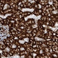


Comprehensive Human Protein Atlas IHC scoring per tissue (reliability: Supported — High consistency between antibody staining and RNA expression data.). Rows are taken directly from the HPA tissue chart — click any row's HPA link to view the source.
| Tissue | Cell type | Level | Evidence | Source |
|---|---|---|---|---|
| Adrenal gland | Glandular cells | High | Protein (IHC) | HPA → |
| Appendix | Glandular cells | High | Protein (IHC) | HPA → |
| Bone marrow | Hematopoietic cells | High | Protein (IHC) | HPA → |
| Breast | Glandular cells | High | Protein (IHC) | HPA → |
| Bronchus | Respiratory epithelial cells | High | Protein (IHC) | HPA → |
| Tissue | Cell type | Level | Evidence | Source |
|---|---|---|---|---|
| None in HPA: CRY2 is detected in all 45 scored tissues. Use a no-primary (secondary-only) and an isotype control instead. | ||||
Use the PB9576 paraffin-section workflow as the starting point, then assess CRY2 staining by cell type, compartment and collection time (datasheet PB9576; HPA; UniProt Q49AN0).
PB9576 has real CRY2 IHC images from paraffin sections of rat liver, mouse kidney and human heart (catalog IHC image captions).
PB9576 is listed for IHC in human, mouse and rat samples (catalog: applications and reactivity). Its IHC captions show staining in rat liver, mouse kidney and human heart paraffin sections (PB9576 IHC image captions).
Which to pick: For tissue IHC, choose PB9576 for paraffin sections; its rat liver and mouse kidney captions use 1 μg/ml, while its human heart caption uses 2 μg/ml (PB9576 IHC image captions). PB9576 is also the cross-species choice for human, mouse and rat IHC (catalog: applications and reactivity). No IF/ICC option is supported by this payload (catalog: applications and IF images); the IHC captions do not report the fixative (PB9576 IHC image captions).