DHPS / Deoxyhypusine synthase · IHC design guide

Design Immunohistochemistry for DHPS

Plan DHPS staining in paraffin sections with the IHC-validated antibody at 2–5 μg/mL (datasheet A02106-2). Assess cytoplasmic and membranous staining while accounting for presumed off-target binding (HPA tissue IHC).

Evidence assembled Oct 2026 · For research use; verify linked source records and product datasheet before use
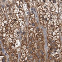
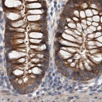
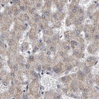
Immunohistochemistry protocol sheet for DHPS (IHC for DHPS): expected localisation Cytoplasmic and membranous staining (HPA tissue IHC), antibody A02106-2, validated IHC image, and IHC protocol steps
Printable DHPS IHC protocol sheet — expected localisation Cytoplasmic and membranous staining (HPA tissue IHC), antibody A02106-2, controls and protocol steps. Open the full DHPS IHC guide →

DHPS Immunohistochemistry Experimental Design Guide

Expected localisation, validated protocols, controls and antibodies — the at-a-glance facts below, then the full design guide.

Must know before staining
Expected localisation Cytoplasmic and membranous staining (HPA tissue IHC)
Staining pattern Cytoplasmic and membranous staining in several tissues (HPA tissue IHC)
Antigen retrieval EDTA pH 8.0 HIER, heat-mediated (datasheet A02106-2)
Positive control ⓘ Adrenal gland+4 more · see all
Negative control ⓘ Caudate+4 more · see all
Important caveats
Reasons your staining may differ from the expected pattern.
Fixation Keep paraffin-section fixation consistent (standard IHC practice; not target-specific)
Caveat Presumed off-target binding may confound staining (HPA tissue IHC)
Regulation No expression regulator specified (UniProt)
Isoform / epitope 3 isoforms; verify epitope coverage (UniProt)
Section 1

Recommended DHPS IHC & IF Protocols

The catalog antibody’s IHC-P protocol (datasheet A02106-2) is accompanied by one published chromogenic DHPS protocol for FFPE ovarian serous carcinoma (PMC11704301 methods).

Recommended immunohistochemistry (IHC-P) protocol parameters
SampleParaffin-embedded human lung cancer tissue; fixative not specified (datasheet A02106-2)
FixationImage fixative and duration unreported (datasheet A02106-2); verify before use.
Sectioning4–5 µm sections on charged slides (standard)
DeparaffinisationXylene, graded ethanol series to water (standard)
Antigen retrievalHeat retrieval: EDTA pH 8.0 (datasheet A02106-2); 20 min, 95–100 °C (standard)
Peroxidase block3% H2O2, 10 min, room temperature (standard)
Blocking10% goat serum (datasheet A02106-2)
Primary antibodyRabbit anti-DHPS, 2-5μg/ml (datasheet A02106-2)
Primary incubationOvernight at 4 °C (datasheet A02106-2)
DetectionStreptavidin-biotin complex (SABC), DAB chromogen (datasheet A02106-2)
CounterstainHematoxylin, blue, dehydrate and mount (standard)
Expected resultDHPS-positive staining in glandular cells of adrenal gland (HPA tissue IHC: High). HPA tissue profile: Cytoplasmic and membranous expression in several tissues. No signal in the no-primary control.
💡Decision noteStart with heat-mediated EDTA pH 8.0 retrieval for the catalog antibody (datasheet A02106-2). The published ovarian protocol does not specify retrieval (PMC11704301 methods).
Section 2

What Is the Expected DHPS Staining Pattern?

In paraffin-section IHC, expect predominantly cytoplasmic, sometimes membranous, DHPS staining in selected glandular and ciliated cells and cells in seminiferous ducts (HPA tissue IHC). DHPS has no annotated transmembrane segment or signal peptide (UniProt P49366 topology/processing). HPA rates its tissue IHC evidence Approved, with medium agreement between staining and RNA data and presumed off-target staining disregarded (HPA tissue IHC reliability).

What am I looking at on my slide?
Clear cytoplasmic staining in adrenal or colon glandular cells, bronchial ciliated cell bodies, or cells in seminiferous ducts.These are high-staining cell populations in HPA tissue IHC. Judge the intended cells against neighboring cells and the counterstain; a recognizable cellular pattern is more useful than overall section darkness (HPA tissue IHC; general IHC practice).
Staining appears exclusively in extracellular material, luminal contents, or sharp cell-surface outlines, with little cell-body signal.That distribution conflicts with the cytoplasmic and membranous tissue-IHC profile (HPA tissue IHC). DHPS has no annotated signal peptide or transmembrane segment (UniProt P49366 topology/processing). Check section morphology and controls before interpreting it as DHPS; compartment mismatch alone does not identify the artifact (general IHC practice).
Strong staining favors cells reported as undetected, such as caudate glia or prostate glandular cells.This is discordant with those HPA observations, so consider cross-reactivity or endogenous chromogenic activity (HPA tissue IHC; general IHC practice). HPA's Approved rating includes a warning that presumed off-target staining was observed and disregarded; an HPA 'Not detected' entry is a reference observation, not proof that every specimen must be negative (HPA tissue IHC reliability).
Brown color spreads evenly across tissue, stroma, or blank areas without outlining identifiable positive cells.A diffuse pattern cannot establish the reported cell-specific DHPS distribution (HPA tissue IHC). Consider nonspecific antibody binding, insufficient washing, or detection-system background; inspect a matched negative reagent control and the tissue morphology (general IHC practice).
No staining appears in a section containing an expected high-staining cell population.First confirm that the relevant cells are present and preserved: HPA reports high staining in adrenal glandular cells, colon glandular cells, and bronchial ciliated cell bodies (HPA tissue IHC). If the run control also fails, investigate the IHC workflow; if it succeeds, investigate the specimen and antibody conditions (general IHC practice).
💡Expected DHPS appearanceCall a positive IHC result when identifiable adrenal or colon glandular cells, bronchial ciliated cell bodies, or seminiferous-duct cells show clear cytoplasmic, sometimes membranous, staining consistent with HPA's high-scoring examples; diffuse extracellular color or equally strong staining of HPA-undetected cell populations warrants control-based review (HPA tissue IHC; general IHC practice).
How each factor affects the staining
Cell-specific reference controlsUse the named cell population, not the organ alone: HPA scores colon glandular cells High, appendix glandular cells Medium, and prostate glandular cells Not detected (HPA tissue IHC). These are comparative reference patterns, not universal intensity thresholds for every section (general IHC practice).
Localization and processingHPA describes cytoplasmic and membranous staining across several tissues (HPA tissue IHC). UniProt lists a single 1–369 chain, with no annotated signal peptide, propeptide, or transmembrane segment (UniProt P49366 processing/topology); it does not establish a membrane-spanning or shed DHPS pool.
Isoform coverageUniProt lists Long, Short, and 3 isoforms (UniProt P49366 isoforms). The supplied evidence gives no antibody epitope or isoform-specific IHC performance, so the observed pattern cannot be assigned to one isoform.
Strength of the tissue referenceThe HPA tissue-IHC rating is Approved, with medium staining–RNA consistency and presumed off-target binding disregarded (HPA tissue IHC reliability). Treat its positive and undetected cells as useful comparisons while requiring slide controls for unexpected staining (general IHC practice).
Antigen retrievalTarget-specific fixation effects are not established by the supplied assay evidence. Verify with a matched IHC source before attributing a result to fixation.
Why is my staining missing, weak or wrong?
SituationLikely causeNext action
A high-reference cell population is present but unstained.The IHC run may have failed, or the specimen or antibody conditions may differ; HPA reports high staining in colon glandular cells (HPA tissue IHC; general IHC practice).Check a concurrently processed positive control, section morphology, primary-antibody conditions, and the catalog IHC-P instructions before scoring the specimen negative (general IHC practice).
An HPA-undetected population stains strongly.The pattern may reflect off-target binding or endogenous chromogenic activity; HPA notes presumed off-target staining in its validation summary (HPA tissue IHC reliability; general IHC practice).Compare a negative reagent control and an HPA high-reference cell population on the same run; investigate the detection system if color persists without primary antibody (general IHC practice).
Color is diffuse across cells and surrounding tissue.Nonspecific binding or detection background can obscure the reported cytoplasmic and membranous pattern (HPA tissue IHC; general IHC practice).Inspect a matched negative reagent control, verify blocking and washes, and score only staining that can be assigned to intact cells (general IHC practice).
Only luminal or extracellular deposits stain.That location differs from HPA's tissue-IHC profile; UniProt annotates no signal peptide or transmembrane segment (HPA tissue IHC; UniProt P49366 topology/processing).Review morphology, negative controls, and detection deposits before assigning the signal to DHPS (general IHC practice).
Adjacent sections vary greatly in staining intensity.Differences in the proportion of relevant cells can alter an apparent whole-section result; HPA reports staining by cell population (HPA tissue IHC).Compare the same identifiable cell type and confirm its presence in each section before changing the IHC conditions (general IHC practice).
Q: IF/ICC shows nuclear signal; is that incompatible with DHPS?A: HPA approves nucleoplasm and cytosol localization in ICC-IF, while its tissue-IHC summary describes cytoplasmic and membranous staining (HPA subcellular; HPA tissue IHC).Interpret each application against its own HPA reference and controls; nuclear IF signal alone does not establish a paraffin-IHC staining pattern (HPA subcellular; general IHC practice).

Sample controls for DHPS IHC & IF

🧪Run colon first and score its glandular cells for DHPS staining (HPA: High in colon glandular cells). Use liver cholangiocytes as the negative comparator (HPA: Not detected in liver cholangiocytes); on the colon slide, unstained non-glandular cells are provisional background references because no internal negative cell type is identified in the supplied colon row (HPA: colon glandular cells only).
Positive control tissue: Adrenal gland (Glandular cells, HPA High)
Negative control tissue: Caudate (HPA Not detected)
ICC-IF cell lines (HPA subcellular resource): HPA ICC-IF images show DHPS in A-431, SH-SY5Y, U2OS, with annotated localisation: Nucleoplasm (approved), Cytosol (approved) (HPA subcellular).
Technical controls: Include no-primary (secondary-only) and concentration-matched rabbit IgG isotype controls, plus independently verified DHPS-knockout material or peptide competition if the antigen peptide is available (caption: rabbit primary; standard IHC specificity controls). For the reported SABC/DAB method, quench endogenous peroxidase and check for endogenous biotin background (caption: SABC/DAB; standard IHC practice).
⚠️Feasibility: A target-specific fixation window or fixation effect is unreported, and the selected paraffin-section caption does not state the fixative (caption: fixative not stated). Heat retrieval with EDTA at pH 8.0 was used, but whether retrieval is required has not been established (caption: EDTA retrieval). No supplied comparison shows frozen sections or IF/ICC are easier; inspect colonic mucus and luminal debris for nonspecific DAB deposit (standard IHC practice).

HPA tissue IHC evidence for DHPS

Comprehensive Human Protein Atlas IHC scoring per tissue (reliability: Approved — Medium consistency between antibody staining and RNA expression data. Presumed off target binding observed and disregarded.). Rows are taken directly from the HPA tissue chart — click any row's HPA link to view the source.

Positive expression · recommended positive controls

TissueCell typeLevelEvidenceSource
Adrenal gland Glandular cells High Protein (IHC) HPA →
Bronchus Ciliated cells (cell body) High Protein (IHC) HPA →
Colon Glandular cells High Protein (IHC) HPA →
Gallbladder Glandular cells High Protein (IHC) HPA →
Nasopharynx Ciliated cells (cell body) High Protein (IHC) HPA →

Undetected expression · recommended negative controls

TissueCell typeLevelEvidenceSource
Caudate Glial cells Not detected Protein (IHC) HPA →
Cerebral cortex Glial cells Not detected Protein (IHC) HPA →
Hippocampus Glial cells Not detected Protein (IHC) HPA →
Liver Cholangiocytes Not detected Protein (IHC) HPA →
Oral mucosa Squamous epithelial cells Not detected Protein (IHC) HPA →
Section 3

Advanced DHPS IHC Tips

Troubleshoot DHPS staining in paraffin sections by keeping retrieval and detection conditions consistent, then checking cell identity, compartment, and controls.

What retrieval should I use when DHPS staining is weak in paraffin sections?
Use heat-mediated antigen retrieval in EDTA at pH 8.0 for paraffin sections (datasheet A02106-2). In the illustrated section, the catalog antibody was applied at 2 μg/ml overnight at 4°C, after blocking with 10% goat serum (datasheet A02106-2). If staining is weak, check heating uniformity, cooling, and section adhesion, then compare retrieval times on adjacent sections while keeping antibody and detection conditions fixed (standard IHC practice). If signal remains poor, assess another buffer or pH as a documented fallback, using the EDTA condition as the reference and reviewing morphology for retrieval damage (standard IHC practice).
How should I investigate weak DHPS staining when fixation conditions are unclear?
The illustrated paraffin-section caption does not report a fixative, so target-specific DHPS fixation sensitivity is unknown (datasheet A02106-2). Record the fixative, fixation duration, tissue thickness, processing date, and any decalcification for each block before attributing weak signal to fixation (standard IHC practice). Compare adjacent sections from differently processed blocks using the same EDTA pH 8.0 retrieval and 2 μg/ml antibody concentration, alongside a consistently processed positive control (datasheet A02106-2; standard IHC practice). Review tissue morphology and internal controls with the staining: uneven fixation or processing can affect paraffin-section signal, but these observations alone cannot establish a DHPS-specific fixation effect (standard IHC practice).
Should I accept membranous or nuclear DHPS staining in chromogenic IHC?
HPA describes cytoplasmic and membranous DHPS staining across several tissues, while its subcellular IF data place DHPS in the nucleoplasm and cytosol (HPA tissue IHC; HPA subcellular). UniProt provides no subcellular annotation and reports no transmembrane segment, so assess sharply outlined membrane staining against cell boundaries before assigning a membrane compartment (UniProt P49366 topology; standard IHC practice). Evaluate nuclear DAB against the counterstain and neighboring cells, and compare serial sections or a second validated antibody where available (standard IHC practice). Record cytoplasmic, nuclear, and apparent membranous signal separately, since agreement across cell types and controls is more informative than compartment appearance alone (standard IHC practice).
Can this IHC stain distinguish DHPS isoforms or a modified epitope?
DHPS has 3 listed isoforms—Long, Short, and 3—and a recorded phosphoserine at residue 78 (UniProt P49366). The supplied antibody caption gives no mapped epitope or isoform selectivity, so do not assign an IHC staining pattern to a particular isoform or phosphorylation state (datasheet A02106-2). Check whether any separately obtained epitope information covers a shared or variant region before making isoform claims, and document that information with the scoring method (standard IHC practice). For ambiguous staining, compare an independently validated antibody or appropriate loss-of-target control on similarly processed sections, while retaining the same retrieval and detection settings (standard IHC practice).
How can multiplex IF help assess a disputed DHPS IHC pattern?
Use IF as a complementary localisation check: HPA reports approved nucleoplasmic and cytosolic DHPS localisation in cell imaging (HPA subcellular). Pair DHPS with a validated marker of the cell population under review, such as a ciliated-cell marker when examining bronchial ciliated cells, which HPA scores high (HPA tissue IHC; standard IF practice). Select a spectrally separated, preferably far-red fluorophore after measuring tissue autofluorescence, and include single-color and no-primary controls for bleed-through and background (standard IF practice). Optimize permeabilisation for access to cytosolic or nucleoplasmic epitopes; the supplied caption does not map this antibody’s epitope or provide an IF condition (datasheet A02106-2).
What should I check when DHPS DAB staining is diffuse or widespread?
The illustrated workflow uses 10% goat serum blocking, a biotinylated secondary antibody, an avidin–biotin complex, and DAB (datasheet A02106-2). Check a no-primary section and an appropriate secondary-only control to separate antibody-associated background from tissue or detection-system signal (standard IHC practice). Include an endogenous peroxidase block for DAB detection and assess endogenous biotin contribution when using avidin–biotin amplification; document these as general detection controls (standard IHC practice). If background remains high, titrate the primary around the documented 2 μg/ml condition, inspect wash stringency, and compare signal in expected cells with matched low-staining cells (datasheet A02106-2; HPA tissue IHC; standard IHC practice).
How should I score DHPS IHC across samples with different cell mixtures? ⚠ ANSWER MARKED FOR VERIFICATION
Define the cell population and compartment before scoring, since HPA reports differing DHPS intensity among tissue cell types (HPA tissue IHC). For viable target cells, record the percentage at each intensity from 0 to 3 and calculate an H-score from 0–300, or report percent positive cells with a fixed threshold (standard IHC practice). Normalize counts to the number of evaluable target cells; for spatial analyses, report positive-cell density per mm² of viable annotated tissue (standard IHC practice). Keep retrieval, staining batch, counterstain, imaging, and threshold settings consistent, and exclude necrotic or damaged regions from the denominator using prespecified rules (standard IHC practice).
How can I distinguish convincing DHPS positivity from IHC artefact?
Compare signal with cell identity: HPA scores DHPS high in bronchial ciliated cells and colonic glandular cells, while reporting no detection in cerebral-cortex glial cells (HPA tissue IHC). HPA calls its tissue staining approved with medium RNA agreement and notes presumed off-target binding, so an unexpected positive population needs independent checking (HPA tissue IHC). Inspect strong nuclear-only or sharply membranous staining against the reported cytosolic and nucleoplasmic IF localisation and the absence of a transmembrane segment (HPA subcellular; UniProt P49366 topology). Discount staining confined to section edges, necrosis, or endogenous peroxidase activity using morphology, no-primary controls, and a matched positive section (standard IHC practice).
Boster reagents

Best DHPS / Deoxyhypusine synthase IHC Antibodies

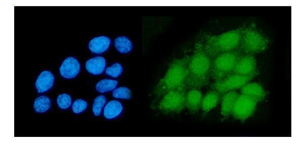
The IHC-validated antibody A02106-2 has images from human paraffin tissue sections and IF data from MCF-7 cells (catalog image captions); its listed reactivity covers human, mouse and rat (catalog reactivity).

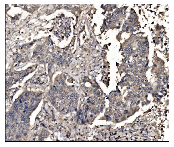
Real IHC data IHC analysis of DHPS/DHS using anti-DHPS/DHS antibody (A02106-2). DHPS/DHS was detected in a paraffin-embedded section of human lung cancer tissue. Heat mediated antigen retrieval was performed in EDTA buffer (pH 8.0, epitope retrieval solution). The tissue section was blocked with 10% goat serum. The tissue section was then incubated with 2 μg/ml rabbit anti-DHPS/DHS Antibody (A02106-2) overnight at 4°C. Biotinylated goat anti-rabbit IgG was used as secondary antibody and incubated for 30 minutes at 37°C. The tissue section was developed using Strepavidin-Biotin-Complex (SABC) (Catalog # SA1022) with DAB as the chromogen.
Anti-DHPS/DHS Antibody ®
Cat # A02106-2

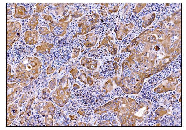
A02106-2 has IHC images from human lung cancer and gallbladder adenocarcinoma paraffin sections (catalog IHC image captions). A02106-2 also has an IF image from MCF-7 cells and lists ICC and IF applications (catalog IF image caption; catalog applications).

Which to pick: Choose A02106-2 for paraffin-section IHC: its own captions document staining in human lung cancer and gallbladder adenocarcinoma sections, but do not report the fixative (catalog IHC image captions). Choose A02106-2 for IF/ICC based on its MCF-7 IF image and listed ICC/IF applications; its clonality is unreported (catalog IF image caption; catalog applications; catalog clone field). For cross-species work, A02106-2 lists mouse and rat reactivity, while its supplied IHC images show human tissue only (catalog reactivity; catalog IHC image captions).

Each figure is that product's own IHC / IF validation image from its datasheet.

References

  1. UniProt Consortium. UniProt entry P49366 (DHYS_HUMAN, Deoxyhypusine synthase).
  2. Human Protein Atlas. DHPS tissue IHC expression (reliability: Approved).
  3. Human Protein Atlas. DHPS subcellular location (ICC-IF): Localized to the nucleoplasm and cytosol..
  4. Human Protein Atlas. DHPS antibody validation summary (2 antibodies).
  5. Knockout or inhibition of DHPS suppresses ovarian tumor growth and metastasis by attenuating the TGFβ pathway. Scientific reports 2025 — PMC11704301.
  6. Epithelial Hypusination Regulates Helicobacter pylori-induced Gastric Inflammation. Research square 2025 — PMC12637816.
  7. Epithelial hypusination regulates Helicobacter pylori-induced gastric inflammation. Amino acids 2026 — PMC13194257.
  8. The transition from monocyte to tissue-resident macrophage requires DHPS. Nature 2026 — PMC12999486.
  9. PubMed PMID:7673224 — UniProt-cited evidence.
  10. PubMed PMID:8549832 — UniProt-cited evidence.
  11. PubMed PMID:8615810 — UniProt-cited evidence.