DLG1 / Disks large homolog 1 · IHC design guide

Design Immunohistochemistry for DLG1

Plan DLG1 IHC-P around membranous and cytoplasmic tissue staining (HPA tissue IHC). The catalog antibody has a paraffin-section dilution of 0.5–1 μg/mL (datasheet PB9552); HPA reports high staining in glandular cells and no detected staining in adipocytes (HPA tissue IHC).

Evidence assembled Oct 2026 · For research use; verify linked source records and product datasheet before use
Immunohistochemistry protocol sheet for DLG1 (IHC for DLG1): expected localisation Membranous and cytoplasmic tissue staining (HPA tissue IHC), antibody PB9552, validated IHC image, and IHC protocol steps
Printable DLG1 IHC protocol sheet — expected localisation Membranous and cytoplasmic tissue staining (HPA tissue IHC), antibody PB9552, controls and protocol steps. Open the full DLG1 IHC guide →

DLG1 Immunohistochemistry Experimental Design Guide

Expected localisation, validated protocols, controls and antibodies — the at-a-glance facts below, then the full design guide.

Must know before staining
Expected localisation Membranous and cytoplasmic tissue staining (HPA tissue IHC)
Staining pattern Glandular cells show membranous and cytoplasmic staining (HPA tissue IHC)
Antigen retrieval EDTA pH 8.0 HIER, heat-mediated (datasheet PB9552)
Positive control ⓘ Appendix+4 more · see all
Negative control ⓘ Adipose tissue+1 more · see all
Important caveats
Reasons your staining may differ from the expected pattern.
Fixation Matched tissue-IHC evidence does not establish the fixation claim. Validate the specimen-specific method before use. (selected-SKU IHC image PB9552)
Caveat Adipocytes may lack signal despite the broad tissue profile (HPA tissue IHC)
Regulation MARCHF2 promotes DLG1 degradation (UniProt)
Isoform / epitope 9 isoforms; check epitope coverage across variants (UniProt)
Section 1

Recommended DLG1 IHC & IF Protocols

The catalog antibody’s IHC-P protocol is accompanied by published DLG1 staining protocols for endometrial cancer and OPSCC tissue (PMC4984904; PMC11201649).

Recommended immunohistochemistry (IHC-P) protocol parameters
SampleParaffin-embedded human prostatic cancer tissue; fixative not specified (datasheet PB9552)
FixationImage fixative and duration unreported (datasheet PB9552); verify before use.
Sectioning4–5 µm sections on charged slides (standard)
DeparaffinisationXylene, graded ethanol series to water (standard)
Antigen retrievalHeat retrieval: EDTA pH 8.0 (datasheet PB9552); 20 min, 95–100 °C (standard)
Peroxidase block3% H2O2, 10 min, room temperature (standard)
Blocking10% goat serum (datasheet PB9552)
Primary antibodyRabbit anti-DLG1, 0.5-1μg/ml (datasheet PB9552)
Primary incubationOvernight at 4 °C (datasheet PB9552)
DetectionStreptavidin-biotin complex (SABC), DAB chromogen (datasheet PB9552)
CounterstainHematoxylin, blue, dehydrate and mount (standard)
Expected resultDLG1-positive staining in glandular cells of appendix (HPA tissue IHC: High). HPA tissue profile: Ubiquitous membranous and cytoplasmic expression. No signal in the no-primary control.
💡Decision noteStart with heat-mediated EDTA retrieval at pH 8.0 (datasheet: PB9552); citric acid retrieval is a published alternative for endometrial cancer sections (PMC4984904).
Section 2

What Is the Expected DLG1 Staining Pattern?

DLG1 is expected at cell membranes, especially epithelial cell contacts and basolateral surfaces, with cytoplasmic staining also observed (UniProt Q12959 subcellular location; HPA tissue IHC: ubiquitous membranous and cytoplasmic expression). Glandular and respiratory epithelial cells are useful positive populations (HPA tissue IHC: High). HPA rates the tissue staining profile Supported because antibody staining is consistent with RNA expression (HPA tissue IHC: Supported). DLG1 has no transmembrane segment (UniProt Q12959 topology).

What am I looking at on my slide?
Membranous staining outlines glandular or respiratory epithelial cells, with some cytoplasmic signal.This fits the reported tissue pattern; enrichment at epithelial contacts or basolateral surfaces also fits DLG1 localization (HPA tissue IHC: ubiquitous membranous and cytoplasmic expression; UniProt Q12959 subcellular location).
Strong nuclear-only staining replaces the expected membranous and cytoplasmic pattern.Treat this as a localization mismatch and investigate artefact before scoring cells as DLG1 positive; nuclear localization is absent from the supplied location summaries (UniProt Q12959 subcellular location; HPA tissue IHC profile).
Adipocytes or caudate glial cells stain strongly while expected positive cells do not.This conflicts with their reported Not detected status and may reflect cross-reactivity or endogenous detection activity; it does not establish either cause on its own (HPA tissue IHC: Not detected; general IHC practice).
Color is spread evenly across cells, stroma, and the section edge.A diffuse deposit lacks the expected cellular distribution. Assess background with a primary-omission control and inspect blocking, washing, and detection conditions (HPA tissue IHC profile; general IHC practice).
Glandular cells in a colon or appendix control show no detectable staining.Both are reported High, so a negative control section raises concern about the run or reagent performance; a single negative result does not establish absent DLG1 expression (HPA tissue IHC: High in colon and appendix glandular cells; general IHC practice).
💡Expected DLG1 appearanceCall a result positive when glandular or respiratory epithelial cells show clear membranous staining, often with cytoplasmic signal, against lower background; strong nuclear-only or indiscriminate staining is suspect (HPA tissue IHC: High in these cells; HPA tissue IHC profile; UniProt Q12959 subcellular location).
How each factor affects the staining
Cell population and tissueCompare like cells: glandular cells in colon, appendix, breast, cervix, duodenum, and endometrium are High, whereas adipocytes are Not detected; a tissue-wide average can obscure that contrast (HPA tissue IHC).
Membrane polarityDLG1 can concentrate at epithelial basolateral membranes and intercellular contacts. Uneven staining around a polarized cell can therefore be compatible with its location (UniProt Q12959 subcellular location).
Cytoplasmic signalDo not reject a result solely for cytoplasmic staining: HPA describes a membranous and cytoplasmic tissue profile, and UniProt also lists cytoplasm (HPA tissue IHC profile; UniProt Q12959 subcellular location).
Isoforms and epitopeNine isoforms have differing expression profiles, but the supplied record gives no catalog antibody epitope. Isoform coverage and any effect of retrieval on that epitope cannot be predicted here (UniProt Q12959 isoforms; supplied antibody evidence).
Antibody validationTissue IHC and ICC-IF validation are application specific: CAB016307 is IHC Supported, while HPA069593 is ICC Approved. Those labels do not make their staining patterns interchangeable (HPA antibodies).
Fixation sensitivityTarget-specific fixation sensitivity is unreported in the supplied evidence. Choose and document retrieval conditions as a general IHC optimization step, without attributing an outcome to DLG1 (supplied UniProt and HPA evidence; general IHC practice).
Why is my staining missing, weak or wrong?
SituationLikely causeNext action
No staining in a High glandular-cell control.An unsuccessful staining run or reagent problem is possible; absence alone does not identify the failed step (HPA tissue IHC: High; general IHC practice).Check control-section integrity, antibody and detection steps, then compare documented retrieval conditions in a repeat run (general IHC practice).
Weak staining in cardiomyocytes or hepatocytes.Both cell populations are listed Low, so weak signal can agree with the reference profile (HPA tissue IHC: Low).Judge sensitivity with a High epithelial control before changing the protocol; compare equivalent cell populations (HPA tissue IHC: High in colon glandular cells; general IHC practice).
Strong color appears in adipocytes or caudate glial cells.These populations are Not detected in HPA; cross-reactivity or endogenous detection activity is possible (HPA tissue IHC: Not detected; general IHC practice).Compare primary-omission and detection controls, then reassess localization and the IHC validation status (general IHC practice; HPA antibodies: CAB016307 IHC Supported).
Membranous borders are obscured by diffuse color.High background can make the reported membrane pattern hard to interpret (HPA tissue IHC profile; general IHC practice).Inspect primary-omission controls and review blocking, washes, antibody concentration, and detection time as general IHC variables (general IHC practice).
Only nuclear staining remains after background controls.The compartment does not match the supplied DLG1 tissue or UniProt location summaries; its identity is unresolved (HPA tissue IHC profile; UniProt Q12959 subcellular location).Do not score it as the expected pattern; repeat alongside a High epithelial control and review antibody specificity (HPA tissue IHC: High; general IHC practice).
Does IF/ICC have to reproduce the tissue IHC pattern?No direct equivalence is established: HPA ICC-IF reports mainly vesicles, with plasma membrane also supported, whereas tissue IHC reports membranous and cytoplasmic expression (HPA subcellular ICC-IF; HPA tissue IHC).Interpret IF/ICC with its own validated antibody and controls; keep this IHC-P assessment anchored to tissue IHC evidence (HPA antibodies; HPA tissue IHC; general IF practice).

Sample controls for DLG1 IHC & IF

🧪Run appendix first: its glandular cells should stain strongly (HPA: High in appendix glandular cells). Use adipose tissue as the negative tissue, focusing on adipocytes (HPA: Not detected in adipocytes); on the appendix slide, assess non-glandular areas as provisional internal background rather than assuming those cells lack DLG1 (HPA: appendix row identifies glandular cells only).
Positive control tissue: Appendix (Glandular cells, HPA High)
Negative control tissue: Adipose tissue (HPA Not detected)
ICC-IF cell lines (HPA subcellular resource): HPA ICC-IF images show DLG1 in HaCaT, SiHa, U2OS, ASC52telo, RPTEC/TERT1, hTERT-RPE1 (serum starved), with annotated localisation: Vesicles (approved) (HPA subcellular).
Technical controls: Include a no-primary, secondary-only control and a rabbit IgG isotype control matched to the primary’s clonality where applicable (caption: rabbit primary; standard IHC practice). Confirm specificity with DLG1 knockout material or peptide competition if the immunizing peptide is available, and block endogenous peroxidase and check endogenous biotin when using the caption’s biotin–SABC/DAB detection on appendix (caption: biotin–SABC/DAB; standard IHC practice).
⚠️Feasibility: A target-specific fixation window or fixation effect is unreported, and the selected PB9552 paraffin-section caption does not state the fixative (caption: fixative unreported). Heat retrieval with EDTA at pH 8.0 is documented for that IHC example, but retrieval dependence is unreported (caption: heat-mediated EDTA retrieval). Frozen sections or IF cannot be judged easier from the supplied evidence; for appendix, check gland-associated background against the no-primary slide when scoring chromogenic staining (HPA: High in appendix glandular cells; standard IHC practice).

HPA tissue IHC evidence for DLG1

Comprehensive Human Protein Atlas IHC scoring per tissue (reliability: Supported — High consistency between antibody staining and RNA expression data.). Rows are taken directly from the HPA tissue chart — click any row's HPA link to view the source.

Positive expression · recommended positive controls

TissueCell typeLevelEvidenceSource
Appendix Glandular cells High Protein (IHC) HPA →
Breast Glandular cells High Protein (IHC) HPA →
Bronchus Respiratory epithelial cells High Protein (IHC) HPA →
Cerebellum Synaptic glomeruli - core High Protein (IHC) HPA →
Cervix Glandular cells High Protein (IHC) HPA →

Undetected expression · recommended negative controls

TissueCell typeLevelEvidenceSource
Adipose tissue Adipocytes Not detected Protein (IHC) HPA →
Caudate Glial cells Not detected Protein (IHC) HPA →
Section 3

Advanced DLG1 IHC Tips

Troubleshoot DLG1 staining in paraffin sections by checking retrieval, compartment-specific signal, controls and consistent scoring before interpreting biological differences.

Which retrieval condition should I try first for DLG1 in paraffin sections?
Start with heat-mediated retrieval in EDTA at pH 8.0 for the IHC-validated antibody (datasheet PB9552). The selected paraffin-section example used this condition before primary antibody at 1 μg/ml overnight at 4°C (datasheet PB9552). If staining is weak, compare modest changes in heating and cooling while keeping section thickness, detection chemistry and exposure to DAB constant (standard IHC practice). Include a known positive epithelial section in each comparison, because absent staining in both test and control sections points to a processing or detection problem (HPA: high staining in colon glandular cells; standard IHC practice).
Could fixation explain weak or diffuse DLG1 staining in my paraffin sections?
Target-specific fixation sensitivity is unknown: the selected paraffin-section caption does not state its fixative (datasheet PB9552). Record the fixative, fixation duration and processing history for each block before comparing staining across specimens (standard IHC practice). Keep retrieval at EDTA pH 8.0 and primary antibody at 1 μg/ml while testing sections with different documented processing histories (datasheet PB9552; standard IHC practice). If a positive control also weakens, investigate tissue preservation, retrieval and detection together; junctional and cytoplasmic patterns alone cannot identify fixation as the cause (HPA: ubiquitous membranous and cytoplasmic expression; standard IHC practice).
Where should convincing DLG1 staining appear in epithelial tissue?
Look for membranous staining at cell contacts, often along basolateral borders, with possible cytoplasmic staining (UniProt Q12959: basolateral epithelial localisation; HPA: ubiquitous membranous and cytoplasmic expression). DLG1 also associates with junctions and endoplasmic reticulum membranes, so a single compartment need not dominate every specimen (UniProt Q12959: subcellular localisation). Compare glandular cells with adjacent structures on the same section and assess whether borders remain crisp where tissue architecture is intact (HPA: high staining in colon glandular cells; standard IHC practice). Uniform nuclear-only staining warrants a specificity and detection review before being scored as DLG1 (UniProt Q12959: subcellular localisation; standard IHC practice).
How can epitope location and DLG1 isoforms affect IHC interpretation?
DLG1 has 9 reported isoforms, and their expression profiles can differ (UniProt Q12959: isoforms and tissue specificity). Confirm the catalog antibody's immunogen or mapped epitope, then check whether that sequence is retained in the isoforms relevant to the specimen (standard IHC practice). The protein has PDZ, SH3 and guanylate kinase-like regions, but the supplied caption does not identify which region this antibody recognizes (UniProt Q12959: domains; datasheet PB9552). If two validated antibodies produce different patterns, compare their epitopes and control tissues before attributing the difference to isoform expression (UniProt Q12959: isoforms; standard IHC practice).
How should I extend this DLG1 IHC assessment to multiplex IF?
Treat IF as a separate assay requiring its own antibody and fixation validation; the supplied product example establishes staining in paraffin sections with chromogenic detection (datasheet PB9552; standard IF practice). Multiplex DLG1 with a marker of the expected epithelial cell population and assess junctional signal within marker-positive cells (UniProt Q12959: basolateral epithelial localisation; standard IF practice). Choose fluorophores in channels with low tissue autofluorescence, and include single-stain controls to assess bleed-through (standard IF practice). Because DLG1 has no transmembrane segment, select and validate permeabilisation for the intracellular side bearing the antibody's epitope, whose position is unspecified here (UniProt Q12959: topology; standard IF practice).
What should I adjust when DLG1 DAB staining is widespread?
Check a no-primary control alongside the test section to distinguish detection-system background from antibody-dependent staining (standard IHC practice). The selected method used 10% goat serum blocking, 1 μg/ml primary antibody overnight at 4°C, and biotinylated secondary detection with DAB (datasheet PB9552). For diffuse precipitate, review wash quality, secondary-antibody controls and DAB development, and apply a peroxidase block as a general chromogenic IHC step (standard IHC practice). Retain genuine membranous or cytoplasmic patterns in the assessment rather than treating all widespread staining as background, since DLG1 is broadly expressed (HPA: ubiquitous membranous and cytoplasmic expression).
How should I score DLG1 when membrane and cytoplasm both stain? ⚠ ANSWER MARKED FOR VERIFICATION
Define membrane and cytoplasmic compartments separately before scoring, since both are reported for tissue DLG1 (HPA: ubiquitous membranous and cytoplasmic expression). For each compartment, record the percentage of positive cells and an intensity category, then calculate an H-score using the same thresholds across slides (standard IHC practice). Normalize to the number of evaluable cells in the chosen cell population, excluding folds, necrosis and poorly preserved edges (standard IHC practice). If measuring stained area instead, report positive area per mm² of viable, annotated tissue and keep the image-analysis threshold fixed across comparison groups (standard IHC practice).
How can I distinguish true DLG1 signal from tissue artefact?
Favor reproducible cell-border and cytoplasmic signal in intact expected cells over staining confined to cut edges or damaged areas (UniProt Q12959: cell-junction and cytoplasmic localisation; standard IHC practice). Strong staining in glandular epithelium is plausible, while an isolated claim of strong adipocyte staining deserves extra validation (HPA: high in colon glandular cells; not detected in adipocytes). Compare serial sections and controls to identify necrotic deposits, endogenous peroxidase activity and nonspecific DAB accumulation (standard IHC practice). An exclusively nuclear pattern should prompt antibody-specific controls before interpretation, because the supplied localisation evidence emphasizes membranes, junctions and cytoplasm (UniProt Q12959: subcellular localisation; HPA: tissue IHC profile).
Boster reagents

Best DLG1 / Disks large homolog 1 IHC Antibodies

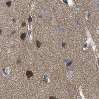
PB9552 has a real DLG1 IHC image from a human paraffin section; its listed reactivity includes Human, Mouse and Rat (PB9552 IHC image caption; catalog: reactivity).

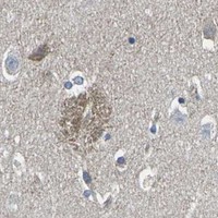
Real IHC data IHC analysis of SAP97 using anti-SAP97 antibody (PB9552). SAP97 was detected in a paraffin-embedded section of human prostatic cancer tissue. Heat mediated antigen retrieval was performed in EDTA buffer (pH 8.0, epitope retrieval solution). The tissue section was blocked with 10% goat serum. The tissue section was then incubated with 1 μg/ml rabbit anti-SAP97 Antibody (PB9552) overnight at 4°C. Biotinylated goat anti-rabbit IgG was used as secondary antibody and incubated for 30 minutes at 37°C. The tissue section was developed using Strepavidin-Biotin-Complex (SABC) (Catalog # SA1022) with DAB as the chromogen.
Anti-SAP97/DLG1 Antibody ®
Cat # PB9552

PB9552 is listed for IHC and WB and has an IHC image from human prostatic cancer tissue in a paraffin section (catalog: applications; PB9552 IHC image caption). Its listed reactivity is Human, Mouse and Rat, but the supplied IHC image documents Human tissue only (catalog: reactivity; PB9552 IHC image caption).

Which to pick: Choose PB9552 for human paraffin-section IHC, the preparation shown in its own image; the fixative is unreported (PB9552 IHC image caption). No IF/ICC antibody is supported by this payload: PB9552 lists IHC and WB, has no IF figure or dilution, and has no reported clone (catalog: applications, IF fields and clone). For Mouse or Rat samples, PB9552 lists reactivity, but its supplied tissue IHC evidence is Human (catalog: reactivity; PB9552 IHC image caption).

Each figure is that product's own IHC / IF validation image from its datasheet.

References

  1. UniProt Consortium. UniProt entry Q12959 (DLG1_HUMAN, Disks large homolog 1).
  2. Human Protein Atlas. DLG1 tissue IHC expression (reliability: Supported).
  3. Human Protein Atlas. DLG1 subcellular location (ICC-IF): Mainly localized to vesicles. In addition localized to the plasma membrane and basal body..
  4. Human Protein Atlas. DLG1 antibody validation summary (2 antibodies).
  5. Requirement of DLG1 for cardiovascular development and tissue elongation during cochlear, enteric, and skeletal development: possible role in convergent extension. PloS one 2015 — PMC4393223.
  6. Loss of the cell polarity determinant human Discs-large is a novel molecular marker of nodal involvement and poor prognosis in endometrial cancer. British journal of cancer 2016 — PMC4984904.
  7. The SF3B1(R625H) mutation promotes prolactinoma tumor progression through aberrant splicing of DLG1. Journal of experimental & clinical cancer research : CR 2022 — PMC8762886.
  8. Moderate SCRIB Expression Levels Correlate with Worse Prognosis in OPSCC Patients Regardless of HPV Status. Cells 2024 — PMC11201649.
  9. PubMed PMID:7937897 — UniProt-cited evidence.
  10. PubMed PMID:14702039 — UniProt-cited evidence.
  11. PubMed PMID:17974005 — UniProt-cited evidence.