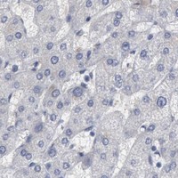
DMC1 / Meiotic recombination protein DMC1 homolog · IHC design guide

Design Immunohistochemistry for DMC1

Plan DMC1 IHC in paraffin sections using testis as a positive tissue, where pachytene spermatocytes show high nuclear staining (HPA tissue IHC). Compare nuclear signal across cell populations and keep fixation conditions consistent.

Evidence assembled Oct 2026 · For research use; verify linked source records and product datasheet before use
Immunohistochemistry protocol sheet for DMC1 (IHC for DMC1): expected localisation Nuclear staining in pachytene spermatocytes (HPA tissue IHC), antibody A02978-3, validated IHC image, and IHC protocol steps
Printable DMC1 IHC protocol sheet — expected localisation Nuclear staining in pachytene spermatocytes (HPA tissue IHC), antibody A02978-3, controls and protocol steps. Open the full DMC1 IHC guide →

DMC1 Immunohistochemistry Experimental Design Guide

Expected localisation, validated protocols, controls and antibodies — the at-a-glance facts below, then the full design guide.

Must know before staining
Expected localisation Nuclear staining in pachytene spermatocytes (HPA tissue IHC)
Staining pattern High nuclear signal in pachytene spermatocytes (HPA tissue IHC)
Antigen retrieval EDTA pH 8.0 HIER, heat-mediated (datasheet A02978-3)
Positive control ⓘ Testis
Negative control ⓘ Adipose tissue+4 more · see all
Important caveats
Reasons your staining may differ from the expected pattern.
Fixation Keep fixation conditions consistent across sections. (standard IHC practice; not target-specific)
Caveat Signal depends on the proportion of pachytene spermatocytes (HPA tissue IHC)
Regulation High in pachytene spermatocytes (HPA tissue IHC)
Isoform / epitope 2 isoforms; epitope coverage is unspecified (UniProt)
Section 1

Recommended DMC1 IHC & IF Protocols

The catalog antibody uses heat-mediated EDTA retrieval at pH 8.0 (datasheet A02978-3). Published DMC1 IHC examples cover cervical cancer, skin, and mouse testis (PMC2657811; PMC6554469; PMC4125102).

Recommended immunohistochemistry (IHC-P) protocol parameters
SampleParaffin-embedded mouse testis tissue; fixative not specified (datasheet A02978-3)
FixationImage fixative and duration unreported (datasheet A02978-3); verify before use.
Sectioning4–5 µm sections on charged slides (standard)
DeparaffinisationXylene, graded ethanol series to water (standard)
Antigen retrievalHeat retrieval: EDTA pH 8.0 (datasheet A02978-3); 20 min, 95–100 °C (standard)
Peroxidase block3% H2O2, 10 min, room temperature (standard)
Blocking10% goat serum (datasheet A02978-3)
Primary antibodyRabbit anti-DMC1, 2-5 μg/ml (datasheet A02978-3)
Primary incubationOvernight at 4 °C (datasheet A02978-3)
DetectionHRP-conjugated secondary, DAB chromogen (datasheet A02978-3)
CounterstainHematoxylin, blue, dehydrate and mount (standard)
Expected resultDMC1-positive staining in pachytene spermatocytes of testis (HPA tissue IHC: High). HPA tissue profile: High nuclear expression in spermatocytes. No signal in the no-primary control.
💡Decision noteStart with EDTA at pH 8.0 for the catalog antibody (datasheet A02978-3); optimize retrieval against a positive control if staining is weak (standard IHC practice).
Section 2

What Is the Expected DMC1 Staining Pattern?

DMC1 is a nuclear, chromosome-associated meiotic recombinase that binds single-stranded DNA near break sites (UniProt Q14565). In paraffin-section IHC, expect strong nuclear staining in testicular pachytene spermatocytes (HPA tissue IHC: High; reliability: Enhanced). DMC1 has no transmembrane segment, so a dominant membrane pattern would conflict with its annotated location (UniProt Q14565 topology).

What am I looking at on my slide?
Strong nuclear chromogen in pachytene spermatocytes, with adjacent cells distinguishable.This matches the reported positive cell and compartment (HPA tissue IHC: High nuclear expression in spermatocytes; pachytene spermatocytes: High). Read staining against section morphology and the counterstain before scoring.
Predominantly membrane or cytoplasmic staining, with little convincing nuclear signal.This conflicts with the annotated nuclear and chromosome location and the lack of a transmembrane segment (UniProt Q14565). Consider nonspecific staining or a detection artefact; location alone cannot identify its cause.
Strong staining in cell types reported as undetected, such as adipocytes or adrenal glandular cells.That distribution conflicts with those HPA tissue observations (HPA tissue IHC: Not detected in adipocytes and adrenal glandular cells). Check morphology, cross-reactivity and endogenous detection activity before interpreting it as DMC1.
Diffuse chromogen covers several compartments or obscures nuclear boundaries.The slide cannot support a compartment-specific DMC1 call (UniProt Q14565: nucleus; HPA tissue IHC: high nuclear expression). Excess detection or inadequate blocking or washing can produce background (general IHC practice); compare negative controls.
No nuclear signal in a morphologically adequate testis section containing pachytene spermatocytes.This is a failed expected-positive result under the reported tissue pattern (HPA tissue IHC: pachytene spermatocytes High). Check the IHC-validated antibody and detection controls before treating the sample as DMC1-negative (general IHC practice).
💡Expected DMC1 appearanceCall the result positive when high nuclear staining identifies pachytene spermatocytes in testis (HPA tissue IHC: High); dominant membrane staining or strong signal in reported undetected cells warrants investigation (UniProt Q14565 topology; HPA tissue IHC).
How each factor affects the staining
Tissue and cell selectionTestis containing pachytene spermatocytes offers a reported positive reference (HPA tissue IHC: High). Adipocytes and adrenal glandular cells are reported as undetected (HPA tissue IHC); neither establishes a universal negative result for every specimen.
Compartment and molecular formDMC1 is nuclear and chromosome-associated, binds single-stranded DNA, and forms a nucleoprotein filament (UniProt Q14565). These annotations support nuclear interpretation but do not specify a required chromogen texture or puncta count.
Isoforms and processingTwo isoforms are listed, with no signal peptide, propeptide or annotated glycosylation sites (UniProt Q14565). The supplied record does not map the antibody epitope, so it cannot establish isoform coverage or an epitope-specific retrieval setting.
Antibody evidenceHPA lists enhanced IHC validation for HPA001232 and CAB015397 (HPA antibodies). This supports the reported tissue pattern; it does not determine how an unspecified catalog antibody will perform in the user's section.
What should IF/ICC show?Nucleoplasmic localisation is approved in HPA ICC-IF images from A-431 and U2OS (HPA subcellular). That is an IF/ICC localisation reference; it does not supply an IF protocol or replace the testis IHC pattern.
Why is my staining missing, weak or wrong?
SituationLikely causeNext action
Expected testis nuclei are blank.The staining run may have failed, or the section may lack identifiable pachytene spermatocytes (HPA tissue IHC: pachytene spermatocytes High).Confirm cell morphology and run controls; verify primary antibody use, antigen retrieval and detection steps against the IHC protocol (general IHC practice).
Signal is strongest at membranes or in cytoplasm.That location conflicts with nuclear and chromosome annotation and absent transmembrane topology (UniProt Q14565).Compare with a no-primary control, inspect nuclear counterstaining, and review detection background before assigning DMC1 positivity (general IHC practice).
Many cells stain strongly outside the expected testis population.A broad pattern conflicts with high nuclear expression in spermatocytes (HPA tissue IHC); cross-reactivity or endogenous activity is possible.Identify stained cell types by morphology, compare reported undetected tissues, and inspect no-primary and detection controls (HPA tissue IHC; general IHC practice).
Diffuse brown haze obscures nuclei.Nonspecific detection, inadequate blocking or insufficient washing can obscure localisation (general IHC practice).Check a no-primary control, then review blocking, primary dilution, detection and washes within the validated IHC workflow (general IHC practice).
Only faint staining appears in heart muscle.Low staining of cardiomyocytes is reported (HPA tissue IHC: Low); it is a poor substitute for the high testis reference.Assess the run with testis pachytene spermatocytes as the positive reference and avoid scoring faint heart signal in isolation (HPA tissue IHC: High in pachytene spermatocytes; Low in cardiomyocytes).
The IF/ICC image looks nucleoplasmic but the IHC section is negative.HPA's approved nucleoplasmic ICC-IF location and its testis tissue IHC pattern are observations from different applications (HPA subcellular; HPA tissue IHC).Evaluate the paraffin-section IHC with its own testis positive and detection controls; use the separate IF/ICC guide for fluorescence workflow decisions (general IHC practice).

Sample controls for DMC1 IHC & IF

🧪Run testis first and confirm staining in pachytene spermatocytes (HPA: High in pachytene spermatocytes). Use adipose tissue adipocytes as a negative tissue control (HPA: Not detected in adipocytes); compare other cells on the testis slide with the pachytene signal, without assuming they are DMC1-negative (HPA: positive call is specific to pachytene spermatocytes).
Positive control tissue: Testis (Pachytene spermatocytes, HPA High)
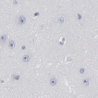
Negative control tissue: Adipose tissue (HPA Not detected)
ICC-IF cell lines (HPA subcellular resource): HPA ICC-IF images show DMC1 in A-431, U2OS, with annotated localisation: Nucleoplasm (approved) (HPA subcellular).
Technical controls: Include no-primary (secondary-only) and rabbit IgG isotype controls matched to the primary’s clonality once verified, plus DMC1-knockout tissue as a biological negative when available (hero caption: rabbit primary; standard IHC practice). Quench endogenous peroxidase and check background before interpreting DAB staining in testis (hero caption: peroxidase detection with DAB; standard IHC practice).
⚠️Feasibility: A target-specific fixation window or fixation effect is unreported, and the selected A02978-3 tissue-IHC caption does not state the fixative (hero caption: fixative not stated). The paraffin-section example used heat retrieval in EDTA at pH 8.0, but whether DMC1 staining depends on that retrieval condition is unreported (hero caption: EDTA retrieval). No matched evidence establishes whether frozen sections or IF are easier; assess nuclear staining against background in testis (UniProt Q14565: nucleus and chromosome; HPA: High in pachytene spermatocytes).

HPA tissue IHC evidence for DMC1

Comprehensive Human Protein Atlas IHC scoring per tissue (reliability: Enhanced — High consistency between antibody staining and RNA expression data.). Rows are taken directly from the HPA tissue chart — click any row's HPA link to view the source.

Positive expression · recommended positive controls

TissueCell typeLevelEvidenceSource
Testis Pachytene spermatocytes High Protein (IHC) HPA →

Undetected expression · recommended negative controls

TissueCell typeLevelEvidenceSource
Adipose tissue Adipocytes Not detected Protein (IHC) HPA →
Adrenal gland Glandular cells Not detected Protein (IHC) HPA →
Appendix Glandular cells Not detected Protein (IHC) HPA →
Bone marrow Hematopoietic cells Not detected Protein (IHC) HPA →
Breast Adipocytes Not detected Protein (IHC) HPA →
Section 3

Advanced DMC1 IHC Tips

Troubleshoot DMC1 staining in paraffin sections by checking retrieval, nuclear localisation, spermatocyte specificity and matched controls before scoring.

What retrieval conditions should I try first for DMC1 staining in paraffin sections?
Start with heat-mediated retrieval in EDTA at pH 8.0 for paraffin sections (datasheet A02978-3). The selected mouse testis image used that condition before incubation with 2 μg/ml primary antibody overnight at 4°C, so keep those conditions together when reproducing the example (datasheet A02978-3). If nuclear staining is weak, check section heating and compare a modest adjustment in retrieval time on adjacent sections while keeping EDTA at pH 8.0 (standard IHC practice). Evaluate staining against a testis positive control and a no-primary control, because excessive retrieval can increase nonspecific signal or damage tissue morphology (HPA tissue IHC: high nuclear expression in spermatocytes; standard IHC practice).
Could fixation explain weak DMC1 staining in my paraffin sections?
Target-specific fixation sensitivity is unknown: the selected paraffin-section caption does not report a fixative (datasheet A02978-3: fixative not stated). Record the fixative, fixation duration and processing schedule for each specimen, then compare sections processed under the same conditions before changing antibody concentration (standard IHC practice). Use the documented EDTA pH 8.0 retrieval as the starting condition, and include a testis section processed alongside the sample to separate a run problem from a specimen problem (datasheet A02978-3; HPA tissue IHC: testis expression). Do not attribute weak staining to a DMC1-specific fixation effect without a controlled comparison (standard IHC practice).
Where should convincing DMC1 staining appear in a testis section?
Prioritise nuclear staining in spermatocytes: the tissue profile reports high nuclear expression, including high staining in pachytene spermatocytes (HPA tissue IHC). DMC1 is associated with chromosomes and binds single-stranded DNA near a break site, while the subcellular profile lists nucleoplasm as its approved location (UniProt Q14565: localisation; HPA subcellular). Compare nuclear signal with a counterstain and inspect the same cell classes in a matched no-primary section before accepting faint chromogen deposits (standard IHC practice). Diffuse staining across unrelated cell compartments deserves investigation, particularly when the expected spermatocyte nuclear pattern is absent (HPA tissue IHC; standard IHC interpretation).
Could isoforms or epitope accessibility alter my DMC1 IHC result?
DMC1 has 2 annotated isoforms, but the supplied antibody caption does not identify its epitope or establish isoform-specific staining (UniProt Q14565: isoforms; datasheet A02978-3). Check the antibody’s documented immunogen or epitope before interpreting a changed staining pattern as an isoform difference (standard IHC practice). DMC1 has no annotated transmembrane segment, glycosylation sites or modified residues in the supplied record; those annotations do not establish how this antibody performs after tissue processing (UniProt Q14565: topology and annotations). Compare adjacent sections with the documented EDTA pH 8.0 retrieval and matched controls before assigning an epitope-based explanation (datasheet A02978-3; standard IHC practice).
How can I check DMC1 localisation by multiplex IF?
For a separate IF experiment, pair DMC1 with an independently validated pachytene spermatocyte marker and a nuclear counterstain, because that cell class shows high nuclear expression in tissue IHC (HPA tissue IHC). Choose spectrally separated fluorophores and place the weaker signal in a channel with low tissue autofluorescence; inspect unstained tissue and single-colour controls before interpreting overlap (standard IF practice). DMC1 is nuclear and has no transmembrane segment, so use permeabilisation suitable for access to an intracellular nuclear epitope (UniProt Q14565: localisation and topology; standard IF practice). Establish IF fixation, retrieval and antibody concentration experimentally; the selected paraffin-section IHC caption does not validate an IF protocol (datasheet A02978-3).
How do I reduce diffuse or granular DMC1 chromogenic background?
First compare the stained section with no-primary and secondary-only controls to locate signal caused by detection reagents or endogenous peroxidase activity (standard IHC practice). The selected mouse testis procedure used 10% goat serum, 2 μg/ml primary antibody overnight at 4°C, a 30-minute secondary incubation at 37°C, and DAB development (datasheet A02978-3). If background persists, check blocking, wash stringency, peroxidase blocking and DAB development time one variable at a time, preserving a comparable testis section in each run (standard chromogenic IHC practice). Treat broad cytoplasmic colour cautiously when it lacks the expected spermatocyte nuclear pattern (HPA tissue IHC: high nuclear expression in spermatocytes).
How should I quantify DMC1-positive cells across testis sections? ⚠ ANSWER MARKED FOR VERIFICATION
Define the spermatocyte population and score its nuclei, because the tissue profile places strong DMC1 expression in pachytene spermatocytes (HPA tissue IHC). Report the percentage of positive spermatocyte nuclei or calculate an H-score from intensity categories 0–3, giving a possible score of 0–300 (standard IHC scoring practice). Normalise counts to the number of evaluable spermatocyte nuclei, or report positive-cell density per mm² of a consistently defined tissue region (standard histology practice). Keep section thickness, retrieval, imaging and positivity thresholds consistent, and exclude folds or damaged regions before comparing specimens (standard IHC quantification practice).
How can I distinguish true DMC1 staining from artefact?
A credible result aligns with nuclear staining in spermatocytes, particularly pachytene spermatocytes, and with DMC1’s chromosome and nuclear localisation (HPA tissue IHC; UniProt Q14565: localisation). Check whether similarly strong colour appears in unrelated cells, cytoplasm or the no-primary control before calling a specimen positive (HPA tissue IHC; standard IHC practice). Discount staining confined to section edges, folds or necrotic areas, and investigate brown deposits that persist without primary antibody as possible detection or endogenous enzyme artefacts (standard chromogenic IHC practice). Interpret a weak testis result alongside a processed positive control; the selected product image documents staining in a paraffin-embedded mouse testis section (datasheet A02978-3).
Boster reagents

Best DMC1 / Meiotic recombination protein DMC1 homolog IHC Antibodies

Two anti-DMC1 antibodies have paraffin-section IHC figures for mouse and rat testis or human heart (catalog IHC captions). A02978 lists IF/ICC applications, but supplies no IF figure (catalog applications/images).

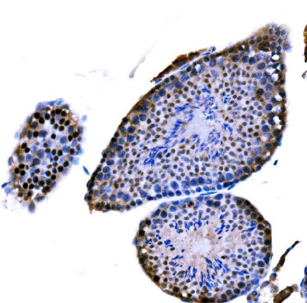
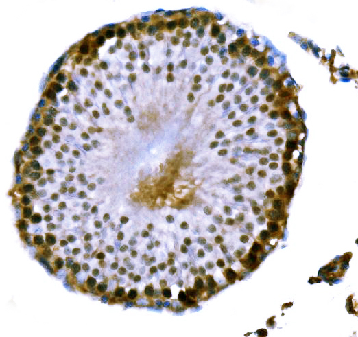
Real IHC data IHC analysis of DMC1 using anti-DMC1 antibody (A02978-3). DMC1 was detected in a paraffin-embedded section of mouse testis tissue. Heat mediated antigen retrieval was performed in EDTA buffer (pH 8.0, epitope retrieval solution). The tissue section was blocked with 10% goat serum. The tissue section was then incubated with 2 μg/ml rabbit anti-DMC1 Antibody (A02978-3) overnight at 4°C. Peroxidase Conjugated Goat Anti-rabbit IgG was used as secondary antibody and incubated for 30 minutes at 37°C. The tissue section was developed using HRP Conjugated Rabbit IgG Super Vision Assay Kit (Catalog # SV0002) with DAB as the chromogen.
Anti-DMC1 Antibody ®
Cat # A02978-3
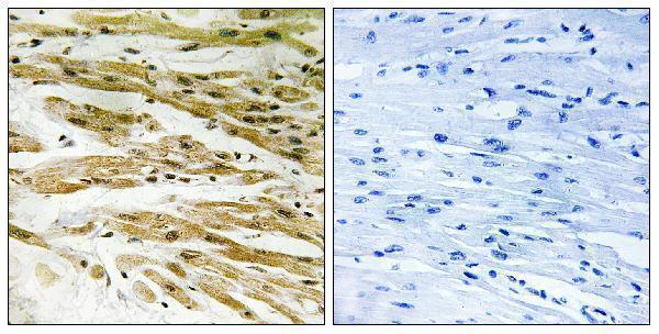
Real IHC data Immunohistochemistry analysis of paraffin-embedded human heart tissue, using DMC1 Antibody. The picture on the right is blocked with the synthesized peptide.
Anti-DMC1 Antibody
Cat # A02978

A02978-3 has IHC figures from paraffin-embedded mouse and rat testis; its listed reactivity is human, mouse and rat (A02978-3 IHC captions; catalog reactivity). A02978 has a peptide-blocked IHC figure from paraffin-embedded human heart and lists IF/ICC for human and mouse (A02978 IHC caption; catalog applications/reactivity).

Which to pick: For testis paraffin-section IHC, choose A02978-3: its mouse and rat figures use EDTA retrieval at pH 8.0, and the catalog recommends 2–5 μg/ml; the fixative is unreported (A02978-3 IHC captions; catalog dilution). For human heart paraffin-section IHC, A02978 has a peptide-blocked figure and a 1:100–1:300 IHC range; its fixative is unreported (A02978 IHC caption; catalog dilution). For IF/ICC, choose polyclonal A02978 based on its listed applications, though no IF figure is supplied; for the widest listed species reactivity, choose A02978-3 for human, mouse and rat, while its IHC figures document mouse and rat only (A02978 catalog applications/images and dilution details; A02978-3 catalog reactivity and IHC captions).

Each figure is that product's own IHC / IF validation image from its datasheet.

References

  1. UniProt Consortium. UniProt entry Q14565 (DMC1_HUMAN, Meiotic recombination protein DMC1 homolog).
  2. Human Protein Atlas. DMC1 tissue IHC expression (reliability: Enhanced).
  3. Human Protein Atlas. DMC1 subcellular location (ICC-IF): Localized to the nucleoplasm..
  4. Human Protein Atlas. DMC1 antibody validation summary (2 antibodies).
  5. Characterization of meiotic recombination intermediates through gene knockouts in founder hybrid mice. Genome research 2023 — PMC10760447.
  6. Activation of meiosis-specific genes is associated with depolyploidization of human tumor cells following radiation-induced mitotic catastrophe. Cancer research 2009 — PMC2657811.
  7. The Ectopic Expression of Meiosis Regulatory Genes in Cutaneous T-Cell Lymphomas (CTCL). Frontiers in oncology 2019 — PMC6554469.
  8. Retinoic acid activates two pathways required for meiosis in mice. PLoS genetics 2014 — PMC4125102.
  9. PubMed PMID:8590282 — UniProt-cited evidence.
  10. PubMed PMID:8602360 — UniProt-cited evidence.
  11. PubMed PMID:15461802 — UniProt-cited evidence.