DNM1 / Dynamin-1 · IHC design guide

Design Immunohistochemistry for DNM1

Plan DNM1 chromogenic IHC around neuronal cytoplasmic and neuropil staining (HPA tissue IHC). Start with 0.5–1 μg/mL of the IHC-validated antibody for paraffin sections (datasheet A02536-2), and score neuronal processes separately from cell bodies (HPA tissue IHC).

Evidence assembled Oct 2026 · For research use; verify linked source records and product datasheet before use
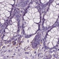
Immunohistochemistry protocol sheet for DNM1 (IHC for DNM1): expected localisation Neuronal cytoplasm and neuropil (HPA tissue IHC), antibody A02536-2, validated IHC image, and IHC protocol steps
Printable DNM1 IHC protocol sheet — expected localisation Neuronal cytoplasm and neuropil (HPA tissue IHC), antibody A02536-2, controls and protocol steps. Open the full DNM1 IHC guide →

DNM1 Immunohistochemistry Experimental Design Guide

Expected localisation, validated protocols, controls and antibodies — the at-a-glance facts below, then the full design guide.

Must know before staining
Expected localisation Neuronal cytoplasm and neuropil (HPA tissue IHC)
Staining pattern Selective staining of neuronal cytoplasm and neuropil (HPA tissue IHC)
Antigen retrieval Citrate pH 6 HIER, heat-mediated (datasheet A02536-2)
Positive control ⓘ Cerebellum+4 more · see all
Negative control ⓘ Adipose tissue+4 more · see all
Important caveats
Reasons your staining may differ from the expected pattern.
Fixation Keep fixation consistent across sections (standard IHC practice; not target-specific)
Caveat Dense neuropil can complicate cell-level scoring (HPA tissue IHC)
Regulation Staining-intensity regulation unknown (UniProt)
Isoform / epitope 4 isoforms; no transmembrane segment; map the epitope (UniProt)
Section 1

Recommended DNM1 IHC & IF Protocols

Compare the catalog antibody’s IHC-P protocol (datasheet: A02536-2) with one published DNM1 tissue protocol (PMC12903489).

Recommended immunohistochemistry (IHC-P) protocol parameters
SampleParaffin-embedded rat small intestine tissue; fixative not specified (datasheet A02536-2)
FixationImage fixative and duration unreported (datasheet A02536-2); verify before use.
Sectioning4–5 µm sections on charged slides (standard)
DeparaffinisationXylene, graded ethanol series to water (standard)
Antigen retrievalHeat retrieval: Citrate pH 6, 20 min (datasheet A02536-2)
Peroxidase block3% H2O2, 10 min, room temperature (standard)
Blocking10% goat serum (datasheet A02536-2)
Primary antibodyRabbit anti-DNM1, 0.5-1μg/ml (datasheet A02536-2)
Primary incubationOvernight at 4 °C (datasheet A02536-2)
DetectionStreptavidin-biotin complex (SABC), DAB chromogen (datasheet A02536-2)
CounterstainHematoxylin, blue, dehydrate and mount (standard)
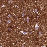
Expected resultDNM1-positive staining in processes in molecular layer of cerebellum (HPA tissue IHC: High). HPA tissue profile: Selective cytoplasmic expression in neuronal cells, neuropil and peripheral nerves. No signal in the no-primary control.
💡Decision noteStart with heat-mediated citrate retrieval at pH 6 (datasheet: A02536-2); the published protocol also uses sodium citrate buffer (PMC12903489).
Section 2

What Is the Expected DNM1 Staining Pattern?

DNM1 should stain mainly the cytoplasm and processes of neuronal cells, including neuropil and peripheral nerves (HPA: selective cytoplasmic expression; IHC reliability Enhanced). Membrane-associated puncta are plausible because dynamin-1 visits clathrin-coated pits and cytoplasmic vesicles (UniProt Q05193: subcellular location). It has no transmembrane segment, so a continuous membrane outline is not its defining pattern (UniProt Q05193: topology).

What am I looking at on my slide?
Strong staining in cerebral cortex neuropil or cerebellar molecular-layer processes, with weaker staining in some hippocampal neurons.This fits the reported distribution: cortex neuropil and cerebellar processes are High, while hippocampal neuronal cells are Medium (HPA: tissue IHC). Judge the relevant cells and processes, rather than assigning one intensity to an entire section (HPA: tissue IHC).
Predominantly nuclear staining, or a uniform sharp outline around every cell, with little neuronal cytoplasmic or process staining.Treat this as a compartment mismatch requiring validation: HPA describes selective cytoplasmic staining, while UniProt places DNM1 at pits, vesicles and presynapses (HPA: tissue IHC; UniProt Q05193: subcellular location). A membrane association does not establish uniform surface staining (UniProt Q05193: transient recruitment).
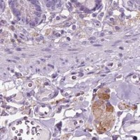
Strong signal in adipocytes, respiratory epithelial cells or glial cells, especially without the expected neuronal pattern.Consider antibody cross-reactivity or endogenous chromogenic activity; those cell groups are reported Not detected in the specified HPA tissues (HPA: tissue IHC; general IHC practice). A single discordant section cannot establish the cause, so compare controls and detection components.
Diffuse colour covers tissue and blank areas, obscuring cell boundaries and the expected neuronal processes.Interpret this as background until controls resolve it, rather than scoring it as DNM1. The expected HPA pattern is selective within neuronal cells, neuropil and peripheral nerves (HPA: tissue IHC). Check the no-primary control and whether chromogen developed outside tissue (general IHC practice).
No signal in cerebral cortex neuropil or cerebellar molecular-layer processes despite intact tissue morphology.These are reported High-staining structures, so a negative result raises a workflow or reagent concern before it supports biological absence (HPA: tissue IHC). Compare an IHC-validated antibody and its documented paraffin-section conditions, plus a positive control processed in the same run (HPA: IHC Enhanced; general IHC practice).
💡Expected DNM1 appearanceCall positive when neuronal cytoplasm and processes show selective staining, strongest in cortex neuropil or cerebellar molecular-layer processes; widespread nuclear or unrelated-cell colour is suspect (HPA: tissue IHC; UniProt Q05193: subcellular location).
How each factor affects the staining
Where staining should be judgedHPA reports High staining in cortex neuropil and cerebellar molecular-layer processes, Medium in hippocampal neuronal cells, and selective staining in peripheral nerves (HPA: tissue IHC). These named structures provide clearer comparisons than whole-tissue averages.
Membrane and vesicle associationDNM1 is transiently recruited to clathrin-coated pits and associates with vesicle membranes; it lacks a transmembrane segment (UniProt Q05193: subcellular location and topology). Local puncta can fit this biology, but their exact visibility in paraffin IHC is not established by these sources.
Antibody validation and isoformsTwo listed antibodies, HPA049910 and CAB005920, have Enhanced IHC status, and UniProt lists four DNM1 isoforms (HPA: antibodies; UniProt Q05193: isoforms). Without a supplied epitope map, do not infer that either antibody detects every isoform.
Antigen retrieval and target-specific sensitivityThe supplied sources give no DNM1-specific retrieval condition or fixation-sensitivity result (HPA: tissue IHC; UniProt Q05193). Select retrieval and dilution from the IHC-validated antibody's actual paraffin-section instructions; optimise against positive tissue and controls (general IHC practice).
IF/ICC: what localisation is established?HPA supplies no ICC-IF images or main subcellular location, so it cannot validate an IF staining pattern here (HPA: subcellular record). UniProt reports pits, vesicles and presynapses as biological locations; assess IF/ICC performance on its separate guide page (UniProt Q05193: subcellular location).
Why is my staining missing, weak or wrong?
SituationLikely causeNext action
Expected brain structures remain blank.Possible staining-run failure, antibody mismatch or unsuitable retrieval; the supplied sources do not identify a DNM1-specific fixation effect (HPA: tissue IHC; general IHC practice).Run cortex or cerebellum as a same-run positive control and follow the IHC-validated antibody's paraffin-section instructions for retrieval, dilution and detection (HPA: High structures; general IHC practice).
Colour appears in the no-primary control.Detection reagents or endogenous activity can create chromogenic signal without primary antibody (general IHC practice).Check endogenous enzyme blocking, secondary-reagent specificity and chromogen development before interpreting tissue staining (general IHC practice).
Staining is diffuse across most cells and tissue regions.Nonspecific binding or excess detection signal may overwhelm the selective neuronal pattern reported by HPA (HPA: tissue IHC; general IHC practice).Compare the no-primary control; review blocking, washes, antibody dilution and development time using the antibody's instructions (general IHC practice).
Nuclei dominate while neuronal processes are faint.This conflicts with HPA's cytoplasmic pattern and UniProt's pit, vesicle and presynaptic locations; the staining alone does not identify its cause (HPA: tissue IHC; UniProt Q05193: subcellular location).Recheck counterstain versus chromogen, then compare an independent IHC-validated antibody and a same-run positive tissue (HPA: antibodies; general IHC practice).
Adipocytes or respiratory epithelium stain strongly.Cross-reactivity or endogenous detection activity is plausible because these specified cells are Not detected in HPA tissue IHC (HPA: tissue IHC; general IHC practice).Use a no-primary control and compare the neuronal positive pattern; do not score the unexpected cells as DNM1 solely from colour (HPA: tissue IHC; general IHC practice).
A supposedly negative tissue shows a small stained cell population.HPA levels apply to named cells, not every cell in a tissue: colon enterocyte microvilli and kidney Bowman's capsule are Medium examples (HPA: tissue IHC).Identify the stained cell type and compartment before calling a false positive; compare with the corresponding HPA cell-level observation (HPA: tissue IHC).

Sample controls for DNM1 IHC & IF

🧪Run cerebellum first and require staining in processes of the molecular layer (HPA: High); use adipose tissue adipocytes as the negative tissue (HPA: Not detected). Within the cerebellum section, cells and compartments outside the stained processes should remain at background, judged against the no-primary control (HPA: High in processes of the molecular layer).
Positive control tissue: Cerebellum (Processes in molecular layer, HPA High)
Negative control tissue: Adipose tissue (HPA Not detected)
ICC-IF cell lines (HPA subcellular resource): HPA carries no ICC-IF cell line for DNM1; derive a cell-line control from the positive tissue's cell type (Processes in molecular layer) and confirm it by RNA or western blot first.
Technical controls: Include a no-primary (secondary-only) control, a matched rabbit IgG isotype control (selected-SKU IHC caption: rabbit primary antibody), and DNM1-knockout tissue as a biological negative if available. For chromogenic cerebellum IHC, quench endogenous peroxidase and check for endogenous biotin when using the caption’s streptavidin–biotin detection system (selected-SKU IHC caption: SABC with DAB).
⚠️Feasibility: A target-specific fixation window or fixation effect is unreported, and the fixative for the selected-SKU paraffin-section image is unreported (selected-SKU IHC caption: paraffin section; fixative not stated). The reported IHC procedure uses heat retrieval in citrate buffer at pH 6 for 20 minutes, but the caption does not establish whether retrieval is essential (selected-SKU IHC caption). Frozen-section or IF/ICC ease cannot be ranked from the supplied evidence; the cerebellum’s stained processes require careful separation from background when scoring (HPA: High in processes of the molecular layer).

HPA tissue IHC evidence for DNM1

Comprehensive Human Protein Atlas IHC scoring per tissue (reliability: Enhanced — High consistency between antibody staining and RNA expression data.). Rows are taken directly from the HPA tissue chart — click any row's HPA link to view the source.

Positive expression · recommended positive controls

TissueCell typeLevelEvidenceSource
Cerebellum Processes in molecular layer High Protein (IHC) HPA →
Cerebral cortex Neuropil High Protein (IHC) HPA →
Colon Enterocytes - Microvilli Medium Protein (IHC) HPA →
Hippocampus Neuronal cells Medium Protein (IHC) HPA →
Kidney Bowman's capsule Medium Protein (IHC) HPA →

Undetected expression · recommended negative controls

TissueCell typeLevelEvidenceSource
Adipose tissue Adipocytes Not detected Protein (IHC) HPA →
Adrenal gland Glandular cells Not detected Protein (IHC) HPA →
Bone marrow Hematopoietic cells Not detected Protein (IHC) HPA →
Breast Adipocytes Not detected Protein (IHC) HPA →
Bronchus Respiratory epithelial cells Not detected Protein (IHC) HPA →
Section 3

Advanced DNM1 IHC Tips

Troubleshoot DNM1 staining in paraffin sections by checking retrieval, compartment, controls and scoring before interpreting chromogenic signal.

Which retrieval condition should I start with for DNM1 paraffin IHC?
Start with heat mediated retrieval in citrate buffer at pH 6 for 20 minutes (datasheet A02536-2). The documented paraffin section used 10% goat serum blocking, 1 μg/ml primary antibody overnight at 4°C, and biotin based detection with DAB (datasheet A02536-2). If staining is weak, first check heating consistency and section adhesion, then compare a more intensive retrieval condition on adjacent sections while keeping detection constant (standard IHC practice). Judge any change against the expected neuronal and neuropil pattern, since stronger overall brown staining alone does not establish better DNM1 detection (HPA tissue IHC; standard IHC practice).
Could fixation explain weak or uneven DNM1 staining?
The selected antibody caption identifies a paraffin section but does not report its fixative, so DNM1 sensitivity to fixation remains unknown for this assay (datasheet A02536-2). Record the actual fixative, duration, tissue thickness and processing history before comparing staining across blocks (standard IHC practice). If a batch stains weakly, compare sections processed under documented conditions using the same pH 6 citrate retrieval and matched detection settings (datasheet A02536-2; standard IHC practice). Do not attribute a fixation effect to DNM1 phosphorylation or membrane association without a controlled comparison, even though both features are recorded for the protein (UniProt Q05193; standard IHC practice).
Where should convincing DNM1 staining appear in tissue sections?
Expect cytoplasmic staining in neuronal cells, neuropil and peripheral nerves, with particularly strong signal reported in cerebellar molecular layer processes and cerebral cortical neuropil (HPA tissue IHC). DNM1 also associates with cytoplasmic vesicles, presynapses and clathrin coated pits, so fine puncta near membranes can fit its biology (UniProt Q05193). It has no transmembrane segment; a continuous cell border without compatible cytoplasmic or process staining warrants scrutiny (UniProt Q05193 topology; standard IHC practice). Compare candidate signal with adjacent morphology and a hematoxylin counterstain, then confirm that the pattern survives matched controls and reasonable changes in antibody concentration (standard IHC practice).
How do isoforms and epitope location affect DNM1 IHC interpretation?
DNM1 has 4 recorded isoforms, but the supplied antibody caption does not identify its epitope or establish which isoforms it detects (UniProt Q05193; datasheet A02536-2). Its GTPase, PH and GED domains span residues 28–294, 519–625 and 659–750, respectively, providing landmarks if epitope information becomes available (UniProt Q05193). Reported modified residues include phosphotyrosine and phosphoserine sites, yet these annotations alone do not show that modification alters this antibody’s tissue staining (UniProt Q05193; standard IHC practice). When comparing samples, keep retrieval and detection matched and describe the result as antibody staining unless isoform or modification specificity has been independently established (standard IHC practice).
How should I investigate DNM1 localisation by multiplex IF?
Use IF as a separate optimization from the documented chromogenic paraffin assay, whose caption provides no IF conditions (datasheet A02536-2). Pair DNM1 with a marker for the expected neuronal cell or process population and inspect whether puncta align with cytoplasm, neuropil or presynaptic regions (HPA tissue IHC; UniProt Q05193; standard IF practice). Select fluorophores after checking tissue autofluorescence in unstained sections, favoring channels with clear separation from that background (standard IF practice). Because DNM1 has no transmembrane segment and associates with the cytoplasmic face of membranes and vesicles, optimize permeabilisation for intracellular epitope access while checking that morphology and puncta are retained (UniProt Q05193 topology and localisation; standard IF practice).
What should I check when DNM1 DAB staining looks diffuse?
First inspect a no primary control and tissue edges; widespread brown signal there suggests detection background or an edge effect rather than a resolved cellular pattern (standard IHC practice). The documented method uses a biotinylated secondary, streptavidin biotin complex and DAB, so assess endogenous biotin and peroxidase contributions with appropriate controls and blocking steps (datasheet A02536-2; standard IHC practice). Recheck serum blocking, primary concentration and wash consistency against the reported 10% goat serum and 1 μg/ml primary conditions (datasheet A02536-2). Compare the final pattern with neuronal processes and neuropil, and treat homogeneous staining across unrelated compartments cautiously (HPA tissue IHC; standard IHC practice).
How can I quantify DNM1 IHC across samples? ⚠ ANSWER MARKED FOR VERIFICATION
Define the anatomical region and cell compartment before scoring, because DNM1 staining can occupy neuronal cytoplasm, processes and neuropil (HPA tissue IHC). For discrete cells, record percent positive and an H-score from 0–300; for process rich regions, measure DAB positive area or signal density per mm² under fixed imaging thresholds (standard IHC practice). Normalize cell based scores to the number of eligible cells and process measurements to sampled tissue area, excluding folds, necrosis and section edges by the same rules across groups (standard IHC practice). Keep retrieval, antibody incubation, DAB development and counterstaining consistent, and report the chosen compartment separately from background measurements (datasheet A02536-2; standard IHC practice).
How do I distinguish true DNM1 signal from artefact?
A credible positive should follow the expected cytoplasmic, neuronal process or neuropil distribution and remain interpretable against local tissue morphology (HPA tissue IHC; standard IHC practice). Vesicular or pit associated puncta are plausible, whereas predominantly nuclear staining or uniform labeling of unrelated cells needs independent validation (UniProt Q05193 localisation; standard IHC practice). Check whether signal concentrates at section edges or necrotic areas, or persists in a no primary control; those findings support an artefact assessment (standard IHC practice). Because the documented assay uses biotin based DAB detection, inspect endogenous biotin and peroxidase controls before calling diffuse brown deposits DNM1 positive (datasheet A02536-2; standard IHC practice).
Boster reagents

Best DNM1 / Dynamin-1 IHC Antibodies

The catalog antibody has paraffin-section IHC images from human, mouse and rat samples (A02536-2 image captions). IF/ICC is listed for M02536-1, but no IF image is supplied (M02536-1 applications and image alts).

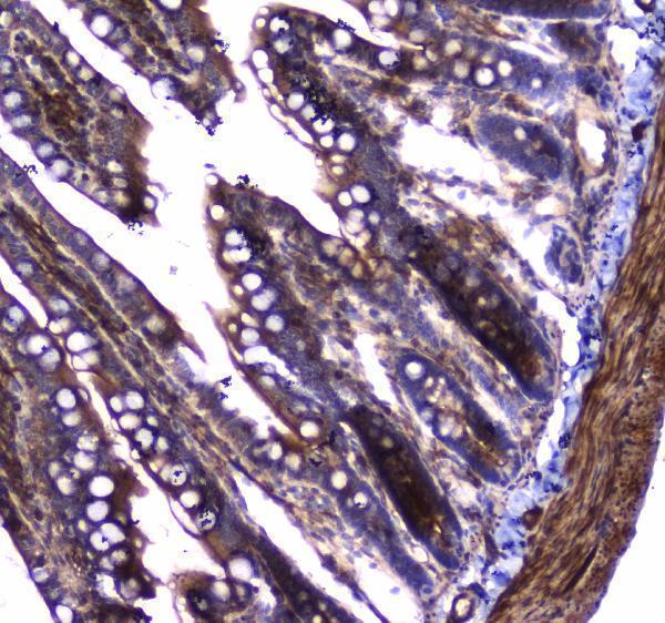
Real IHC data IHC analysis of Dynamin 1 using anti-Dynamin 1 antibody (A02536-2). Dynamin 1 was detected in paraffin-embedded section of rat small intestine tissue. Heat mediated antigen retrieval was performed in citrate buffer (pH6, epitope retrieval solution) for 20 mins. The tissue section was blocked with 10% goat serum. The tissue section was then incubated with 1μg/ml rabbit anti-Dynamin 1 Antibody (A02536-2) overnight at 4°C. Biotinylated goat anti-rabbit IgG was used as secondary antibody and incubated for 30 minutes at 37°C. The tissue section was developed using Strepavidin-Biotin-Complex (SABC)(Catalog # SA1022) with DAB as the chromogen.
Anti-Dynamin 1/DNM1 Antibody ®
Cat # A02536-2

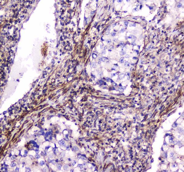
A02536-2 will render with its rat small-intestine paraffin-section IHC figure; its additional captions show human lung cancer, human mammary cancer and mouse small intestine (A02536-2 image captions). M02536-1 will render as a rabbit monoclonal listed for IHC and IF/ICC in human, mouse and rat, without a supplied IHC or IF image (M02536-1 catalog entry).

Which to pick: Choose A02536-2 for paraffin-section tissue IHC because its own captions document that preparation and detection; the fixative is unreported (A02536-2 image captions). Choose M02536-1 when an antibody explicitly listed for IF/ICC or a rabbit monoclonal is preferred, while treating its imaging performance as undocumented here (M02536-1 catalog entry). Both list human, mouse and rat reactivity for cross-species planning; A02536-2 supplies IHC images from all three species (catalog reactivity; A02536-2 image captions).

Each figure is that product's own IHC / IF validation image from its datasheet.

References

  1. UniProt Consortium. UniProt entry Q05193 (DYN1_HUMAN, Dynamin-1).
  2. Human Protein Atlas. DNM1 tissue IHC expression (reliability: Enhanced).
  3. Human Protein Atlas. DNM1 subcellular location (ICC-IF): Highest expression in A-549: 84.1 nTPM.
  4. Human Protein Atlas. DNM1 antibody validation summary (2 antibodies).
  5. Quantitative Fluorescent in situ Hybridization Reveals Differential Transcription Profile Sharpening of Endocytic Proteins in Cochlear Hair Cells Upon Maturation. Frontiers in cellular neuroscience 2021 — PMC7952526.
  6. Dynamin 1 promotes colorectal cancer progression by activating the PI3K/Akt signaling pathway. Journal of translational medicine 2025 — PMC12903489.
  7. RNAi-Based Gene Therapy Rescues Developmental and Epileptic Encephalopathy in a Genetic Mouse Model. Molecular therapy : the journal of the American Society of Gene Therapy 2020 — PMC7335739.
  8. Independent Neuronal Origin of Seizures and Behavioral Comorbidities in an Animal Model of a Severe Childhood Genetic Epileptic Encephalopathy. PLoS genetics 2015 — PMC4488318.
  9. PubMed PMID:8101525 — UniProt-cited evidence.
  10. PubMed PMID:15164053 — UniProt-cited evidence.
  11. PubMed PMID:15489334 — UniProt-cited evidence.