This website uses cookies to ensure you get the best experience on our website.


- Table of Contents
Plan DVL1 paraffin section IHC around the generally cytoplasmic tissue staining reported by HPA (HPA tissue IHC). Use the reported high staining in adrenal glandular cells and absent staining in smooth muscle cells to guide control selection (HPA tissue IHC).

Expected localisation, validated protocols, controls and antibodies — the at-a-glance facts below, then the full design guide.
| Expected localisation | General cytoplasmic tissue staining (HPA tissue IHC) | |
| Staining pattern | Generally cytoplasmic staining in sampled tissues (HPA tissue IHC) | |
| Antigen retrieval | Citrate pH 6.0 HIER, 95–98 °C, 20 min (rule: cytoplasmic / membrane antigen) | |
| Positive control | Adrenal gland+4 more · see all | |
| Negative control | Smooth muscle+1 more · see all |
| Fixation | Keep fixation conditions consistent across sections. (standard IHC practice; not target-specific) | |
| Caveat | Staining has low consistency with RNA expression (HPA tissue IHC) | |
| Regulation | Expression regulation not annotated (UniProt) | |
| Isoform / epitope | Two isoforms; epitope differences unspecified (UniProt) |
The catalog antibody’s IHC-P protocol is accompanied by published DVL1 IHC methods for colorectal cancer and lung tissue (PMC3821627; PMC9891876).
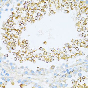
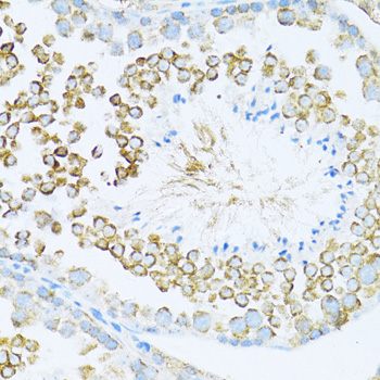
| Sample | Paraffin-embedded rat testis tissue; fixative not specified (datasheet A03533) |
| Fixation | Image fixative and duration unreported (datasheet A03533); verify before use. |
| Sectioning | 4–5 µm sections on charged slides (standard) |
| Deparaffinisation | Xylene, graded ethanol series to water (standard) |
| Antigen retrieval | Heat-induced epitope retrieval in citrate buffer, pH 6.0, 20 min at 95–98 °C (standard rule: cytoplasmic / membrane antigen) |
| Peroxidase block | 3% H2O2, 10 min, room temperature (standard) |
| Blocking | 10% normal serum of the secondary host, 30 min, room temperature (standard) |
| Primary antibody | Rabbit anti-DVL1, 1:50-1:200 (datasheet A03533) |
| Primary incubation | Overnight at 4 °C (standard) |
| Detection | HRP-polymer secondary, DAB chromogen 5–10 min (standard) |
| Counterstain | Hematoxylin, blue, dehydrate and mount (standard) |
| Expected result | DVL1-positive staining in glandular cells of adrenal gland (HPA tissue IHC: High). HPA tissue profile: General cytoplasmic expression. No signal in the no-primary control. |
DVL1 is a cytosolic and vesicle-associated protein that can localize to the cell membrane; it has no transmembrane segment (UniProt O14640). In paraffin-section IHC, expect chiefly cytoplasmic staining in the cell populations HPA rates High, including colon glandular cells and cerebral cortex neurons (HPA tissue IHC). HPA describes a general cytoplasmic pattern but rates its tissue IHC evidence Approved with low consistency between antibody staining and RNA expression (HPA tissue IHC).
| Clear cytoplasmic staining in colon glandular cells, with distinguishable cell borders and limited background. | This fits HPA's High call for colon glandular cells and its general cytoplasmic IHC profile (HPA tissue IHC). Membrane-associated signal can also fit DVL1 localization, especially where it interacts with frizzled family members (UniProt O14640). Judge intensity against neighboring cells on the same section; HPA's High category does not specify a universal chromogen intensity or staining threshold (HPA tissue IHC). |
| Predominantly nuclear staining in a paraffin section, with little cytoplasmic signal. | Treat this as a compartment discrepancy for the proposed IHC readout: HPA describes general cytoplasmic tissue staining, while UniProt lists cytosol, vesicles and cell membrane (HPA tissue IHC; UniProt O14640). Nuclear signal alone does not establish a false positive, because HPA reports approved nucleoplasmic localization in ICC-IF (HPA ICC-IF). Compare antibody controls and tissue morphology before interpreting it. |
| Strong staining in smooth muscle cells or soft-tissue fibroblasts. | HPA lists these cell populations as Not detected in its tissue IHC profile (HPA tissue IHC). Unexpected staining may reflect cross-reactivity, endogenous detection activity or a preparation-dependent result; the observation alone cannot identify which cause applies (general IHC practice). Recheck cell identity and compare controls before assigning DVL1 expression, especially given HPA's reported staining–RNA inconsistency (HPA tissue IHC). |
| Diffuse chromogen over cells, stroma and empty spaces, without a readable intracellular pattern. | This cannot be scored as the reported DVL1 cytoplasmic distribution (HPA tissue IHC). Broad background can arise from detection reagents or nonspecific primary-antibody binding (general IHC practice). A no-primary control and a suitable detection control help separate those possibilities; interpret any remaining cellular signal against morphology rather than total section darkness (general IHC practice). |
| No detectable staining in colon glandular cells or another HPA High population. | That conflicts with the reported positive tissue pattern but does not by itself prove DVL1 is absent (HPA tissue IHC). Check whether the IHC-validated antibody and detection system work on a known-positive section, then review retrieval, reagent activity and section quality as general IHC checks (general IHC practice). HPA's low staining–RNA consistency limits how strongly one negative specimen can be interpreted (HPA tissue IHC). |
| Compartment and topology | UniProt places DVL1 in cytosol, cytoplasmic vesicles and cell membrane, and reports no transmembrane segment (UniProt O14640). Membrane-associated staining need not form a continuous transmembrane-protein outline; compare it with the dominant cytoplasmic IHC pattern reported by HPA (HPA tissue IHC). |
| Tissue evidence and RNA agreement | HPA reports High IHC staining in specified glandular, neuronal, respiratory epithelial and hematopoietic populations, but rates staining–RNA consistency low (HPA tissue IHC). Its skeletal-muscle-enhanced RNA classification is an RNA finding, so it does not supply an IHC-positive skeletal muscle cell population (HPA tissue IHC). |
| Antibody validation | Both listed antibodies, HPA073607 and CAB011538, have Approved IHC status; the supplied record does not report Enhanced IHC validation (HPA antibodies). Approved status supports use of the reported pattern while leaving unexpected compartments or cell types to be checked with appropriate controls (HPA antibodies; general IHC practice). |
| Isoforms and modification | UniProt lists two DVL1 isoforms and a phosphoserine at residue 194 (UniProt O14640). These facts do not identify the antibodies' epitopes or establish different IHC staining between isoforms or phosphorylation states; avoid using them to explain a weak or absent signal without further evidence (UniProt O14640; HPA antibodies). |
| Situation | Likely cause | Next action |
|---|---|---|
| A High-reference population is blank. | A technical failure is possible; HPA reports High staining in colon glandular cells and cerebral cortex neurons, but low staining–RNA consistency (HPA tissue IHC). | Check a known-positive section, IHC-validated antibody, detection reagents and the run's retrieval settings before interpreting the specimen as negative (general IHC practice). |
| Chromogen covers the section without clear cell boundaries. | Nonspecific binding or endogenous detection activity can obscure cellular localization (general IHC practice). | Compare a no-primary control and the detection controls; review blocking, washing and detection conditions, then score only morphology-resolved signal (general IHC practice). |
| Smooth muscle cells or fibroblasts stain strongly. | These populations are Not detected in the HPA tissue IHC profile; cross-reactivity or endogenous activity is possible (HPA tissue IHC; general IHC practice). | Verify the cell population on the section and compare control slides before reporting DVL1 positivity (general IHC practice). |
| Signal appears chiefly nuclear in IHC. | This differs from HPA's general cytoplasmic tissue profile, although approved ICC-IF images also place DVL1 in nucleoplasm (HPA tissue IHC; HPA ICC-IF). | Review controls and the accompanying cytoplasmic pattern; report the compartment explicitly rather than equating nuclear IHC signal with confirmed specificity (general IHC practice). |
| Two IHC-positive tissues show different intensities. | HPA's High calls describe named cell populations, while its staining–RNA consistency is low; they do not define equal intensity across tissues (HPA tissue IHC). | Compare the same cell types and staining compartments under matched run conditions, and record intensity separately for each specimen (general IHC practice). |
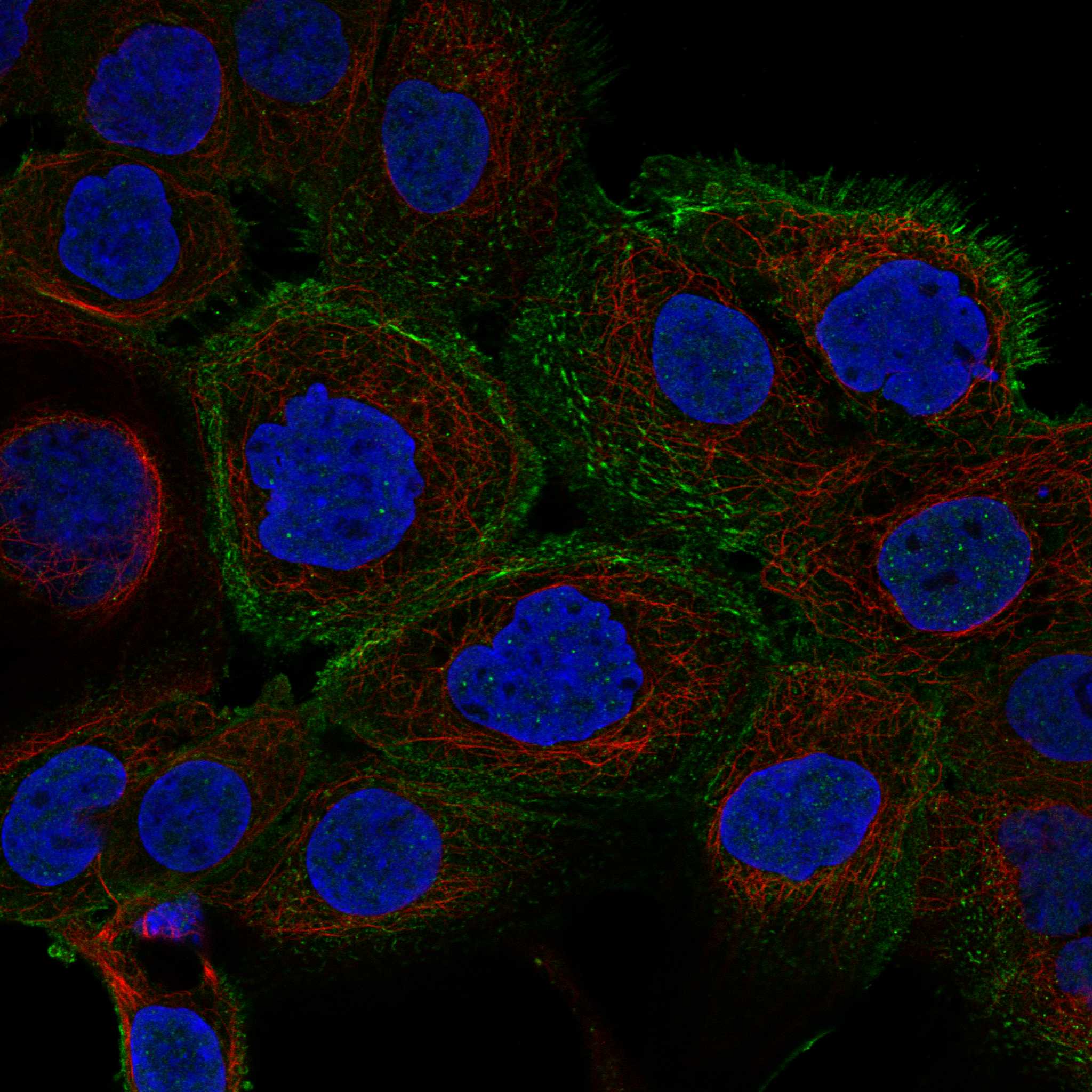
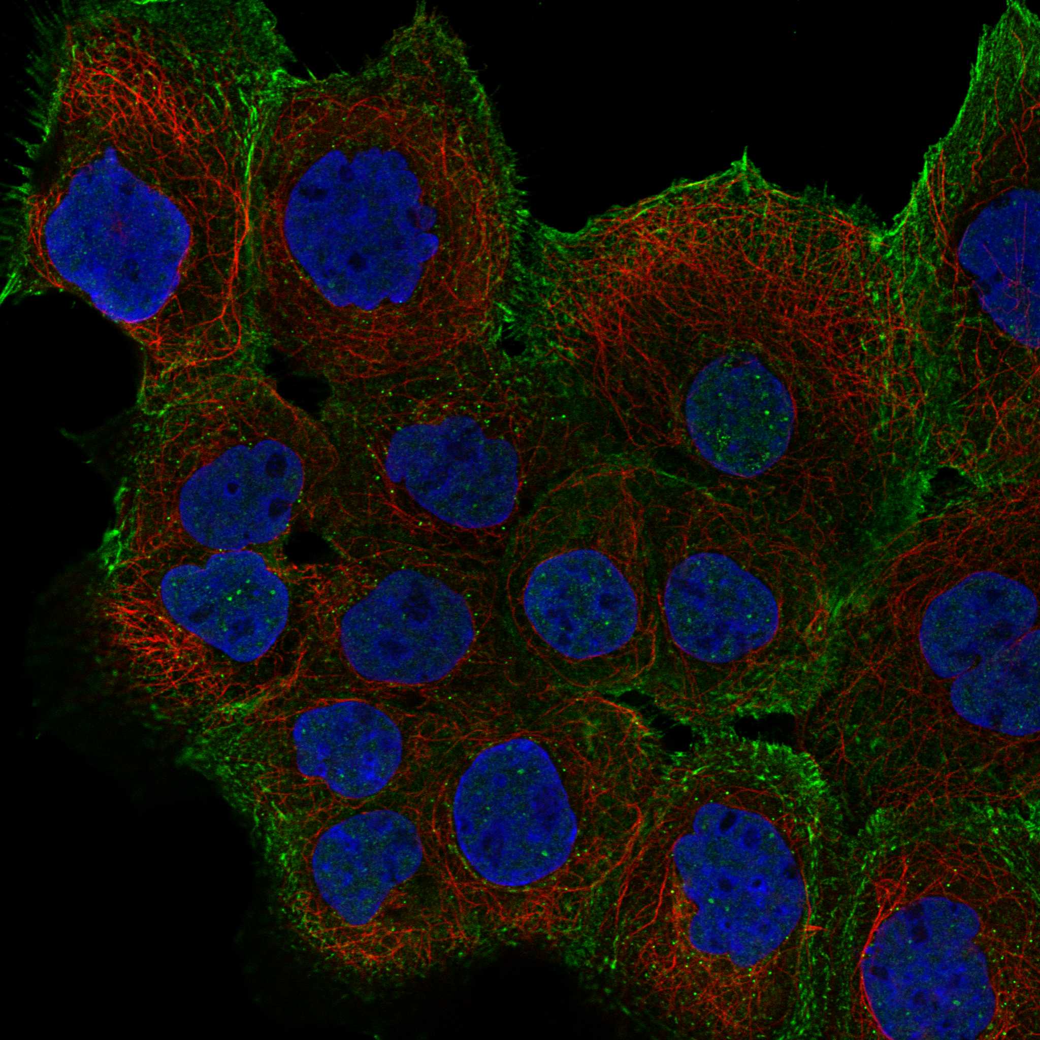
| Can ICC-IF show DVL1 in the nucleus? | HPA reports approved nucleoplasmic and plasma-membrane localization, with additional vesicle and focal-adhesion localization in ICC-IF (HPA ICC-IF). | Yes; interpret those ICC-IF compartments using that evidence and the dedicated IF/ICC guide, while evaluating paraffin-section IHC against its reported general cytoplasmic profile (HPA ICC-IF; HPA tissue IHC). |


Comprehensive Human Protein Atlas IHC scoring per tissue (reliability: Approved — Low consistency between antibody staining and RNA expression data.). Rows are taken directly from the HPA tissue chart — click any row's HPA link to view the source.
| Tissue | Cell type | Level | Evidence | Source |
|---|---|---|---|---|
| Adrenal gland | Glandular cells | High | Protein (IHC) | HPA → |
| Appendix | Glandular cells | High | Protein (IHC) | HPA → |
| Bone marrow | Hematopoietic cells | High | Protein (IHC) | HPA → |
| Bronchus | Respiratory epithelial cells | High | Protein (IHC) | HPA → |
| Cerebral cortex | Neuronal cells | High | Protein (IHC) | HPA → |
Troubleshoot DVL1 staining in paraffin sections by checking retrieval, compartment, controls, and scoring before interpreting a chromogenic signal.
A03533 has paraffin-section IHC images from rat and mouse testis (IHC image captions); IF/ICC is listed for human, mouse, and rat, but no IF image is supplied (catalog applications; IF image alts).
A03533 is the sole product card and shows paraffin-section IHC in rat and mouse testis at 1:100, with microwave retrieval in 10 mM PBS, pH 7.2 (IHC image captions). Human, mouse, and rat reactivity and IF/ICC use are listed, but no IF validation image is supplied (catalog reactivity; applications; IF image alts).
Which to pick: Choose A03533 for paraffin-section tissue IHC because its own images document rat and mouse testis staining (IHC image captions); the fixative is unreported (IHC image captions). For IF/ICC or samples from human, mouse, or rat, A03533 is the listed option, with an IF dilution range of 1:50–1:200; assess performance in your sample because no IF image is supplied (catalog applications; reactivity; IF dilution; IF image alts).