This website uses cookies to ensure you get the best experience on our website.


- Table of Contents
Plan EDEM2 chromogenic IHC in paraffin sections using the catalog antibody and high staining glandular tissues as reference points (HPA tissue IHC; datasheet A08174). Interpret the observed cytoplasmic pattern alongside EDEM2’s annotated ER lumen location, while accounting for medium consistency between antibody staining and RNA data (HPA tissue IHC; UniProt).

Expected localisation, validated protocols, controls and antibodies — the at-a-glance facts below, then the full design guide.
| Expected localisation | Cytoplasmic tissue staining; ER lumen is the molecular location (HPA tissue IHC; UniProt) | |
| Staining pattern | Cytoplasmic staining in glandular cells; texture unspecified (HPA tissue IHC) | |
| Antigen retrieval | Tris-EDTA pH 9.0 HIER, heat-mediated (datasheet A08174) | |
| Positive control | Appendix+4 more · see all | |
| Negative control | Soft tissue+1 more · see all |
| Fixation | Keep fixation consistent across sections. (standard IHC practice; not target-specific) | |
| Caveat | Antibody staining and RNA show medium consistency (HPA tissue IHC) | |
| Regulation | Broad expression; modest tissue differences (UniProt) | |
| Isoform / epitope | 2 isoforms; signal peptide 1–21 is removed; epitope coverage unknown (UniProt) |
Compare the catalog antibody’s IHC-P protocol with one published EDEM2 IHC protocol for glioma tissue (PMC9885217).
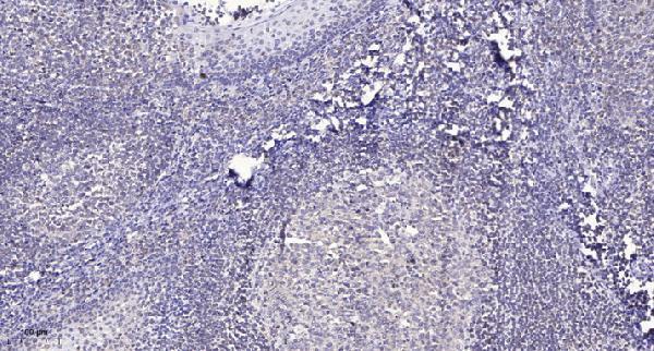
| Sample | Paraffin-embedded human tonsil tissue; fixative not specified (datasheet A08174) |
| Fixation | Image fixative and duration unreported (datasheet A08174); verify before use. |
| Sectioning | 4–5 µm sections on charged slides (standard) |
| Deparaffinisation | Xylene, graded ethanol series to water (standard) |
| Antigen retrieval | Heat retrieval: Tris-EDTA pH 9.0 (datasheet A08174); 20 min, 95–100 °C (standard) |
| Peroxidase block | 3% H2O2, 10 min, room temperature (standard) |
| Blocking | 10% normal serum of the secondary host, 30 min, room temperature (standard) |
| Primary antibody | Rabbit anti-EDEM2, 1:100-1:300 (datasheet A08174) |
| Primary incubation | Overnight at 4 °C (standard) |
| Detection | HRP-polymer secondary, DAB chromogen 5–10 min (standard) |
| Counterstain | Hematoxylin, blue, dehydrate and mount (standard) |
| Expected result | EDEM2-positive staining in glandular cells of appendix (HPA tissue IHC: High). HPA tissue profile: General cytoplasmic expression. No signal in the no-primary control. |
EDEM2 is an endoplasmic reticulum (ER) lumen protein with no transmembrane segment (UniProt Q9BV94 topology). In paraffin IHC, expect cytoplasmic staining, especially in glandular cells of the appendix, cervix, colon, and duodenum (HPA tissue IHC: High; general cytoplasmic expression). HPA rates the tissue staining Approved, with medium consistency against RNA data and external verification pending (HPA tissue IHC: reliability).
| Cytoplasmic staining in appendix or colon glandular cells, with little staining in adjacent unstained areas. | This fits the reported high glandular cell signal (HPA tissue IHC: High) and ER lumen localisation (UniProt Q9BV94). Chromogenic IHC alone cannot establish that individual stained structures are ER (general IHC practice). |
| Predominantly nuclear or sharply cell-surface staining in an otherwise positive glandular cell population. | That compartment conflicts with ER lumen localisation (UniProt Q9BV94). Treat it as a suspect pattern; review morphology and controls before calling it EDEM2 staining (general IHC practice). |
| Strong signal appears chiefly in a cell type reported as unstained, such as soft tissue chondrocytes. | HPA reports EDEM2 as Not detected in chondrocytes (HPA tissue IHC). Check cross-reactivity and endogenous detection activity; the HPA result alone does not prove that a new signal is false (general IHC practice; HPA tissue IHC: medium consistency). |
| Brown deposit spreads across tissue, empty spaces, or many unrelated cell types without clear cell boundaries. | A widespread deposit is difficult to assign to the reported cytoplasmic pattern (HPA tissue IHC). Check background in a control without primary antibody and review blocking, washes, and detection chemistry (general IHC practice). |
| No glandular cell signal appears in a well-preserved appendix or colon section. | These are reported high-signal cell populations (HPA tissue IHC: High). First check the assay and tissue controls; one negative section cannot establish absent EDEM2 expression, particularly given HPA's medium consistency rating (general IHC practice; HPA tissue IHC: reliability). |
| Subcellular resolution | EDEM2 is assigned to the ER lumen (UniProt Q9BV94), while HPA describes a general cytoplasmic IHC pattern (HPA tissue IHC). Interpret chromogenic signal at the cytoplasmic level; ER identity needs a more resolving assay (general IHC practice). |
| Choice of reference tissue and cell | Appendix, cervix, colon, and duodenum glandular cells are reported High; parathyroid glandular cells are Low (HPA tissue IHC). Compare the named cell population, rather than scoring a whole section as uniformly positive (general IHC practice). |
| Reported low or absent signal | HPA lists chondrocytes and spleen red pulp cells as Not detected, and several other cell populations as Low (HPA tissue IHC). These observations are comparison points, not universal negative controls: HPA reports medium consistency and pending external verification (HPA tissue IHC: reliability). |
| Protein forms | UniProt lists two isoforms, a signal peptide at residues 1–21, and four glycosylation sites (UniProt Q9BV94). The supplied sources do not map the antibody epitopes, so they do not establish which forms either IHC antibody detects (UniProt Q9BV94; HPA antibody record). |
| Antibody evidence | HPA lists HPA048353 and CAB037238 as IHC Approved (HPA antibody record). HPA's tissue profile has medium consistency with RNA data and awaits external verification; interpret an unexpected pattern with controls rather than assuming target specificity (HPA tissue IHC: reliability; general IHC practice). |
| Situation | Likely cause | Next action |
|---|---|---|
| Expected glandular cells show no chromogenic signal. | The run may have failed, or the selected cells may differ from the reported high-signal populations (HPA tissue IHC: High; general IHC practice). | Check tissue preservation and the run's positive and detection controls; compare appendix or colon glandular cells using the catalog antibody's IHC-P instructions (HPA tissue IHC: High; general IHC practice). |
| Every cell appears similarly dark. | Uniform deposit may obscure the reported cytoplasmic, cell-dependent pattern (HPA tissue IHC; general IHC practice). | Inspect a no-primary control, review blocking and washes, and assess whether detection intensity prevents cell-level scoring (general IHC practice). |
| Signal is mainly nuclear or outlines cell membranes. | Those distributions disagree with the ER lumen assignment (UniProt Q9BV94 topology). | Recheck cell morphology and controls; report the pattern as unconfirmed unless independent evidence supports the compartment assignment (general IHC practice). |
| Chondrocytes or spleen red pulp cells stain strongly. | HPA reports these populations as Not detected, although its tissue profile has medium consistency (HPA tissue IHC: reliability). | Review the no-primary control and antibody specificity evidence before assigning the signal to EDEM2 (general IHC practice). |
| A weak sample is being scored as negative. | HPA reports Low staining in parathyroid glandular cells, smooth muscle cells, and adipocytes (HPA tissue IHC). | Use a reported High glandular cell population as the positive comparison and score the relevant cell type separately (HPA tissue IHC: High; general IHC practice). |
| Can the IHC result specify an IF/ICC pattern? | HPA supplies no ICC-IF location or cell-line images for EDEM2 (HPA subcellular record). | Use ER lumen localisation only as a starting expectation (UniProt Q9BV94); assess IF/ICC localisation with its own controls and guide (general IF practice). |


Comprehensive Human Protein Atlas IHC scoring per tissue (reliability: Approved — Medium consistency between antibody staining and RNA expression data. Pending external verification.). Rows are taken directly from the HPA tissue chart — click any row's HPA link to view the source.
| Tissue | Cell type | Level | Evidence | Source |
|---|---|---|---|---|
| Appendix | Glandular cells | High | Protein (IHC) | HPA → |
| Cervix | Glandular cells | High | Protein (IHC) | HPA → |
| Colon | Glandular cells | High | Protein (IHC) | HPA → |
| Duodenum | Glandular cells | High | Protein (IHC) | HPA → |
| Endometrium | Glandular cells | High | Protein (IHC) | HPA → |
Troubleshoot EDEM2 chromogenic IHC in paraffin sections using the selected antibody conditions, cell-specific tissue patterns, and appropriate controls.
A08174 has real IHC data on paraffin-embedded human tonsil (A08174 IHC image caption). Human and mouse reactivity and IF are listed, but no IF image is supplied (catalog: reactivity; applications; IF image alts).
A08174 will render with its IHC figure from paraffin-embedded human tonsil (A08174 IHC image caption). It lists IHC and IF applications and human and mouse reactivity (catalog: applications; reactivity).
Which to pick: For tissue IHC, choose A08174: its own image documents human tonsil paraffin sections stained at 1:200 after Tris-EDTA retrieval at pH 9.0; the fixative is unreported (A08174 IHC image caption). For IF, A08174 lists a 1:50 dilution, but ICC validation and an IF image are unreported (catalog: IF dilution; applications; IF image alts). For cross-species planning, A08174 lists human and mouse reactivity and a rabbit host (catalog: reactivity; host); its tissue image documents human tonsil only (A08174 IHC image caption).