This website uses cookies to ensure you get the best experience on our website.


- Table of Contents
Plan chromogenic EIF4B IHC in paraffin sections with the IHC-validated antibody at 2–5 μg/ml (datasheet A00846-1). Expect general cytoplasmic staining, with high signal reported in cervical squamous epithelial cells and duodenal glandular cells (HPA tissue IHC).

Expected localisation, validated protocols, controls and antibodies — the at-a-glance facts below, then the full design guide.
| Expected localisation | General cytoplasmic staining (HPA tissue IHC) | |
| Staining pattern | Cytoplasmic staining in squamous and glandular cells (HPA tissue IHC) | |
| Antigen retrieval | EDTA pH 8.0 HIER, heat-mediated (datasheet A00846-1) | |
| Positive control | Cervix+4 more · see all | |
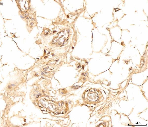
| Negative control | Adipose tissue+3 more · see all |
| Fixation | Keep fixation consistent across sections (standard IHC practice; not target-specific) | |
| Caveat | Staining and RNA show medium consistency (HPA tissue IHC) | |
| Regulation | Low tissue specificity (HPA tissue RNA) | |
| Isoform / epitope | 2 isoforms; verify epitope coverage (UniProt) |
The catalog antibody protocol (datasheet A00846-1) is accompanied by published IHC methods for human brain sections (PMC8339060) and tissue microarrays (PMC5827760).
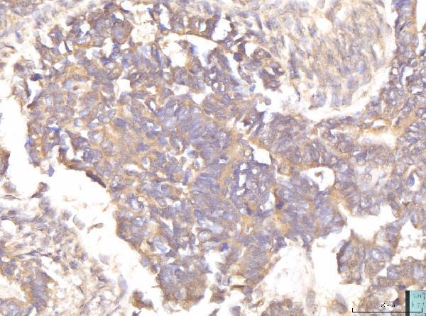
| Sample | Paraffin-embedded human cervical cancer tissue; fixative not specified (datasheet A00846-1) |
| Fixation | Image fixative and duration unreported (datasheet A00846-1); verify before use. |
| Sectioning | 4–5 µm sections on charged slides (standard) |
| Deparaffinisation | Xylene, graded ethanol series to water (standard) |
| Antigen retrieval | Heat retrieval: EDTA pH 8.0 (datasheet A00846-1); 20 min, 95–100 °C (standard) |
| Peroxidase block | 3% H2O2, 10 min, room temperature (standard) |
| Blocking | 10% goat serum (datasheet A00846-1) |
| Primary antibody | Rabbit anti-EIF4B, 2-5 μg/ml (datasheet A00846-1) |
| Primary incubation | Overnight at 4 °C (datasheet A00846-1) |
| Detection | Streptavidin-biotin complex (SABC), DAB chromogen (datasheet A00846-1) |
| Counterstain | Hematoxylin, blue, dehydrate and mount (standard) |
| Expected result | EIF4B-positive staining in squamous epithelial cells of cervix (HPA tissue IHC: High). HPA tissue profile: General cytoplasmic expression. No signal in the no-primary control. |
EIF4B should show predominantly cytoplasmic staining in paraffin tissue sections, especially in squamous epithelial cells of cervix, esophagus, oral mucosa and vagina, and glandular cells of duodenum, rectum and small intestine (HPA tissue IHC). HPA rates its tissue staining Approved, with medium consistency against RNA data and external verification pending (HPA tissue IHC). EIF4B has no transmembrane segment; UniProt does not annotate a subcellular location (UniProt P23588 topology; UniProt P23588 subcellular).
| Strong cytoplasmic staining in squamous epithelial cells of cervix, esophagus, oral mucosa or vagina (HPA tissue IHC). | This matches the cell type, compartment and High staining reported in those tissues (HPA tissue IHC). Assess the epithelial cells themselves; staining elsewhere on the section does not substitute for the expected pattern (HPA tissue IHC; general IHC practice). |
| Predominantly nuclear staining in cells expected to be positive (HPA tissue IHC). | A nuclear-dominant pattern conflicts with HPA's general cytoplasmic tissue profile and should prompt an artefact check (HPA tissue IHC; general IHC practice). UniProt provides no subcellular annotation that would independently support a nuclear interpretation (UniProt P23588 subcellular). |
| Strong staining confined to adipocytes, cholangiocytes, smooth muscle cells or chondrocytes (HPA tissue IHC). | HPA reports these specified cells as Not detected in adipose tissue, liver, smooth muscle and soft tissue, respectively (HPA tissue IHC). Check for cross-reactivity or endogenous detection activity before calling their signal EIF4B; these observations do not make every cell in those tissues a negative control (HPA tissue IHC; general IHC practice). |
| Diffuse color across cells and surrounding tissue, without a clear cytoplasmic boundary (general IHC practice). | This is difficult to score as specific staining against HPA's general cytoplasmic profile (HPA tissue IHC). Background may arise from nonspecific antibody binding or the detection system; inspect a matched control and the counterstained morphology (general IHC practice). |
| No convincing signal in squamous epithelium of esophagus or glandular cells of duodenum (HPA tissue IHC). | HPA reports High staining in those cells, so the result warrants a technical review (HPA tissue IHC; general IHC practice). It does not by itself prove EIF4B absence: review tissue identity, controls and the IHC workflow before interpreting a negative result (general IHC practice). |
| Tissue and cell choice | HPA reports High staining in selected squamous epithelia and intestinal glandular cells, but Low staining in several neuronal and glandular cell groups (HPA tissue IHC). Choose the exact cell population when judging a control; a weak result in a Low group is less informative than one in a High group (HPA tissue IHC; general IHC practice). |
| Evidence and antibody validation | The HPA tissue profile is Approved, with medium consistency against RNA expression and external verification pending (HPA tissue IHC). HPA046164 is IHC Approved; its ICC Enhanced status applies to ICC, while HPA039072 has ICC Approved and no listed IHC status (HPA antibodies). Do not transfer ICC validation to paraffin IHC (HPA antibodies). |
| Isoforms and epitope coverage | UniProt lists two EIF4B isoforms and an RRM at residues 96–173, but the supplied record gives no antibody epitope (UniProt P23588 isoforms; UniProt P23588 domains). Whether an antibody recognizes both isoforms cannot be concluded from these facts; use the actual antibody documentation when interpreting discordant staining (UniProt P23588 isoforms; general IHC practice). |
| Phosphorylation and processing | UniProt lists multiple modified residues, including phosphoserines at 93, 192, 207, 219 and 283; it lists one 1–611 chain and no signal peptide or propeptide (UniProt P23588 modified residues; UniProt P23588 processing). These annotations do not establish a phospho-dependent staining pattern or target-specific fixation sensitivity (UniProt P23588; general IHC practice). |
| IF/ICC Q: What localisation is expected on its separate guide page? | A: HPA places EIF4B mainly in endoplasmic reticulum and cytosol, with additional plasma membrane localisation; those locations are Approved in ICC-IF (HPA subcellular). This cell-imaging evidence can inform interpretation, but it does not turn a paraffin IHC result into an IF protocol or establish identical visual detail in tissue sections (HPA subcellular; general IHC practice). |
| Situation | Likely cause | Next action |
|---|---|---|
| Expected epithelium or intestinal glands show no signal (HPA tissue IHC). | The High HPA pattern was not reproduced; tissue selection or an IHC workflow step may need review (HPA tissue IHC; general IHC practice). | Confirm the expected cells on the counterstain, review a known-positive section and check the antibody and detection workflow (HPA tissue IHC; general IHC practice). |
| Nuclei dominate while cytoplasm is faint (HPA tissue IHC). | This differs from the general cytoplasmic tissue pattern; UniProt supplies no independent nuclear localisation annotation (HPA tissue IHC; UniProt P23588 subcellular). | Compare the signal with morphology and controls, then review staining and scoring before assigning EIF4B localisation (general IHC practice). |
| Color appears mainly in HPA-listed Not detected cell groups (HPA tissue IHC). | Cross-reactivity or endogenous detection activity is possible; the HPA negatives refer to specified cells, not whole tissues (HPA tissue IHC; general IHC practice). | Check the cell identity and compare appropriate negative and detection controls with an expected positive cell population (HPA tissue IHC; general IHC practice). |
| Diffuse haze obscures cell boundaries (general IHC practice). | Nonspecific binding or detection background can prevent reliable cytoplasmic scoring (general IHC practice). | Inspect matched controls and review blocking, antibody dilution, washes and detection conditions as general IHC workflow checks; no EIF4B-specific settings are supplied (general IHC practice). |
| Only weak staining appears in a chosen tissue (HPA tissue IHC). | HPA reports Low staining in some cell groups, including colon glandular cells, so the chosen cells may offer limited contrast (HPA tissue IHC). | Compare the exact cell type with its HPA level and assess a High-staining reference such as duodenal glandular cells before judging assay performance (HPA tissue IHC; general IHC practice). |
| A change in antigen retrieval seems to change staining (general IHC practice). | Target-specific fixation effects are not established by the supplied assay evidence. Verify with a matched IHC source before attributing a result to fixation. | Compare conditions with the same control tissue and score cell type, compartment and background together; avoid attributing a change to a specific EIF4B epitope without antibody evidence (HPA tissue IHC; general IHC practice). |


Comprehensive Human Protein Atlas IHC scoring per tissue (reliability: Approved — Medium consistency between antibody staining and RNA expression data. Pending external verification.). Rows are taken directly from the HPA tissue chart — click any row's HPA link to view the source.
| Tissue | Cell type | Level | Evidence | Source |
|---|---|---|---|---|
| Cervix | Squamous epithelial cells | High | Protein (IHC) | HPA → |
| Duodenum | Glandular cells | High | Protein (IHC) | HPA → |
| Esophagus | Squamous epithelial cells | High | Protein (IHC) | HPA → |
| Oral mucosa | Squamous epithelial cells | High | Protein (IHC) | HPA → |
| Rectum | Glandular cells | High | Protein (IHC) | HPA → |
| Tissue | Cell type | Level | Evidence | Source |
|---|---|---|---|---|
| Adipose tissue | Adipocytes | Not detected | Protein (IHC) | HPA → |
| Liver | Cholangiocytes | Not detected | Protein (IHC) | HPA → |
| Smooth muscle | Smooth muscle cells | Not detected | Protein (IHC) | HPA → |
| Soft tissue | Chondrocytes | Not detected | Protein (IHC) | HPA → |
Troubleshoot EIF4B staining in paraffin-section chromogenic IHC using the catalog antibody’s documented workflow and the reported tissue patterns (caption A00846-1; HPA tissue IHC).
The catalog includes antibodies reactive with human, mouse and rat (catalog: reactivity). IHC images show EIF4B in human paraffin sections; no IF images are supplied (catalog: image captions).
A00846-1 has IHC images from human cervical cancer, colorectal cancer, thyroid papillary carcinoma and tonsil paraffin sections (A00846-1: IHC image captions). P00846 is listed for IHC and recognizes phospho-eIF4B S406, but has no IHC image supplied (P00846: applications; title; image captions).
Which to pick: For tissue IHC, choose A00846-1 when an illustrated paraffin-section protocol matters: its caption specifies 2 μg/ml and EDTA retrieval at pH 8.0; the fixative is unreported (A00846-1: IHC image caption). Choose P00846 when the question is specifically S406 phosphorylation; it is a rabbit monoclonal with a listed IHC dilution of 1:50–1:200, but no IHC image is supplied (P00846: title; host; clone; dilution; image captions). Neither SKU lists IF/ICC validation; both list human, mouse and rat reactivity, while A00846-1’s pictured IHC samples are human only (catalog: applications; reactivity; A00846-1 IHC image captions).