This website uses cookies to ensure you get the best experience on our website.


- Table of Contents
This guide covers paraffin-section ESRRG staining, controls and interpretation, with stomach glandular and kidney tubule cells as reported positives (HPA tissue IHC). It flags the mainly cytoplasmic tissue pattern and uncertain agreement between staining and RNA data (HPA tissue IHC).

Expected localisation, validated protocols, controls and antibodies — the at-a-glance facts below, then the full design guide.
| Expected localisation | Mainly cytoplasmic in tissue IHC; annotated nuclear (HPA tissue IHC; UniProt) | |
| Staining pattern | Mainly cytoplasmic; stomach glands and kidney tubules stain (HPA tissue IHC) | |
| Antigen retrieval | Citrate pH 6 HIER, heat-mediated (datasheet A01470-1) | |
| Positive control | Stomach+1 more · see all | |
| Negative control | Adipose tissue+4 more · see all |
| Fixation | Matched tissue-IHC evidence does not establish the fixation claim. Validate the specimen-specific method before use. (selected-SKU IHC image A01470-1) | |
| Caveat | Staining–RNA discordance makes specificity uncertain (HPA tissue IHC) | |
| Regulation | Expression regulation not specified (UniProt) | |
| Isoform / epitope | 5 isoforms; verify epitope coverage (UniProt) |
The catalog antibody uses citrate pH 6 retrieval (datasheet A01470-1). The published IHC protocols below cover mouse embryos, gallbladder tissues, and patient spinal motor neuron sections (PMC3033182; PMC12066295; PMC7885352).
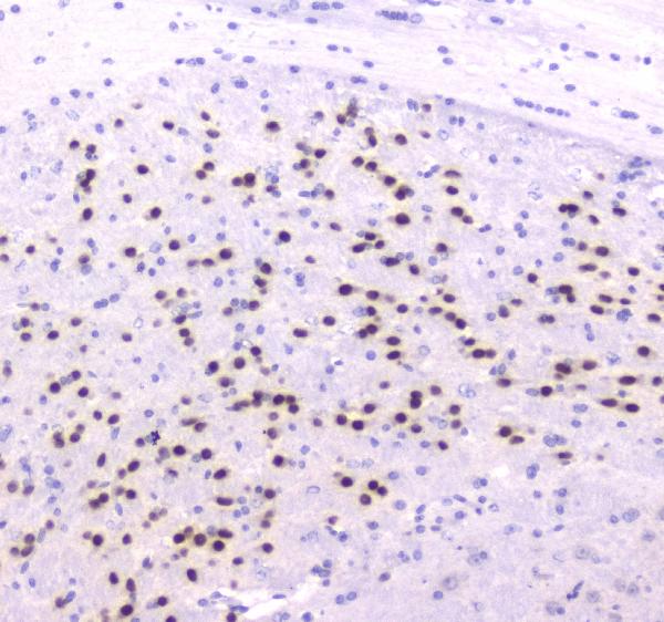
| Sample | Paraffin-embedded mouse brain tissue; fixative not specified (datasheet A01470-1) |
| Fixation | Image fixative and duration unreported (datasheet A01470-1); verify before use. |
| Sectioning | 4–5 µm sections on charged slides (standard) |
| Deparaffinisation | Xylene, graded ethanol series to water (standard) |
| Antigen retrieval | Heat retrieval: Citrate pH 6, 20 min (datasheet A01470-1) |
| Peroxidase block | 3% H2O2, 10 min, room temperature (standard) |
| Blocking | 10% goat serum (datasheet A01470-1) |
| Primary antibody | Rabbit anti-ESRRG, 0.5-1μg/ml (datasheet A01470-1) |
| Primary incubation | Overnight at 4 °C (datasheet A01470-1) |
| Detection | Streptavidin-biotin complex (SABC), DAB chromogen (datasheet A01470-1) |
| Counterstain | Hematoxylin, blue, dehydrate and mount (standard) |
| Expected result | ESRRG-positive staining in glandular cells of stomach (HPA tissue IHC: Medium). HPA tissue profile: Mainly cytoplasmic expression in few tissues. No signal in the no-primary control. |
ESRRG is a nuclear receptor with no transmembrane segment (UniProt P62508: nucleus, topology). In paraffin-section IHC, HPA reports medium staining in stomach glandular cells and kidney tubule cells, but describes its overall tissue pattern as mainly cytoplasmic and its IHC reliability as Uncertain because staining and RNA data have low consistency (HPA tissue IHC). Interpret compartment and cell identity together; the observed cytoplasmic pattern does not by itself establish specific ESRRG staining.
| Stomach glandular cells or kidney tubule cells show medium chromogenic staining, with identifiable nuclear signal. | These cell populations match HPA's reported IHC positives, and nuclear signal fits UniProt localization (HPA tissue IHC; UniProt P62508: nucleus). Treat the result as a candidate positive: HPA rates tissue IHC Uncertain, so morphology and control sections still matter. |
| Signal is exclusively cytoplasmic, with nuclei remaining clear in the cells being assessed. | HPA describes tissue staining as mainly cytoplasmic, while UniProt places ESRRG in the nucleus and HPA ICC-IF places it in the nucleoplasm (HPA tissue IHC; UniProt P62508; HPA subcellular). Record the mismatch; cytoplasmic staining alone cannot settle target specificity. |
| Strong staining appears in an unexpected cell population, including cells HPA reports as Not detected. | Compare the stained cells with section morphology before assigning identity. HPA reports adipocytes and adrenal glandular cells as Not detected in tissue IHC (HPA tissue IHC). Cross-reactivity or endogenous chromogen-producing activity are possible explanations to test (general IHC practice). |
| Brown signal spreads across nuclei, cytoplasm, and surrounding tissue without a clear cell-level pattern. | This distribution is hard to score as ESRRG because the expected biological compartment is nuclear and the HPA tissue result is uncertain (UniProt P62508: nucleus; HPA tissue IHC: Uncertain). Examine a primary-antibody omission control for detection-system background (general IHC practice). |
| No convincing signal appears in stomach glandular cells or kidney tubule cells. | Both are HPA-reported medium IHC populations, so a blank section prompts a check of tissue identity and assay performance (HPA tissue IHC). It does not prove ESRRG absence: HPA calls tissue IHC Uncertain, and its reported positives are observations, not guaranteed controls. |
| Compartment evidence | UniProt lists the nucleus, and HPA ICC-IF approves nucleoplasmic localization, but HPA tissue IHC reports mainly cytoplasmic staining (UniProt P62508; HPA subcellular; HPA tissue IHC). This disagreement makes a cytoplasmic-only paraffin-section result an interpretation question, not a confirmed localization. |
| Tissue and antibody evidence | HPA reports medium staining in stomach glandular and kidney tubule cells, yet assigns overall tissue IHC Uncertain reliability; antibody HPA044678 is also IHC Uncertain (HPA tissue IHC; HPA antibodies). Keep the reliability qualification beside any proposed positive tissue. |
| Isoforms and epitope coverage | UniProt lists 5 ESRRG isoforms (UniProt P62508: isoforms 1–5). The supplied record gives no antibody epitope, so it cannot establish which isoforms an IHC stain detects; avoid explaining a tissue difference by isoform recognition without separate evidence. |
| Antigen retrieval | Retrieval conditions are a general paraffin-section IHC variable (general IHC practice). No ESRRG-specific retrieval response or fixation sensitivity is supplied; optimize against appropriate control sections without attributing a stronger or weaker ESRRG signal to a documented target-specific effect. |
| IF/ICC Q&A: where should ESRRG appear? | Nucleoplasm is the approved HPA ICC-IF location, with images listed for A-431, SH-SY5Y, and U2OS; HPA044678 is ICC Approved (HPA subcellular; HPA antibodies). These ICC-IF findings inform compartment interpretation but do not establish the paraffin-section IHC pattern. |
| Situation | Likely cause | Next action |
|---|---|---|
| The putative positive stomach or kidney section is blank. | A section, retrieval, antibody, or detection problem is possible (general IHC practice); HPA's medium observations are not guaranteed positive controls because tissue IHC is Uncertain (HPA tissue IHC). | Confirm the expected glandular or tubular cells are present, then review reagent controls and the documented IHC conditions before interpreting the blank result (general IHC practice). |
| Only cytoplasm stains in the proposed positive cells. | HPA reports mainly cytoplasmic tissue staining, which conflicts with nuclear UniProt and approved nucleoplasmic ICC-IF localization (HPA tissue IHC; UniProt P62508; HPA subcellular). | Score nuclear and cytoplasmic compartments separately, retain the discordance in the report, and seek independent support before calling cytoplasmic signal specific (general IHC practice). |
| Cells outside the expected population stain strongly. | Cross-reactivity or endogenous detection activity may produce signal in an unexpected population (general IHC practice); HPA reports some populations as Not detected (HPA tissue IHC). | Recheck cell identity on the counterstained section and compare primary-antibody omission and detection controls; evaluate endogenous activity according to the detection chemistry used (general IHC practice). |
| Diffuse chromogen obscures cell boundaries. | Nonspecific antibody binding or detection background can prevent compartment-level scoring (general IHC practice). | Compare the primary-antibody omission control, review blocking and wash conditions, and adjust antibody or detection exposure using the assay documentation (general IHC practice); do not score diffuse color as ESRRG. |
| Staining differs between adjacent areas of the same section. | Section handling or uneven reagent coverage can create a spatial pattern unrelated to cell identity (general IHC practice). | Inspect whether the pattern follows tissue architecture or a section edge, then repeat with uniform reagent coverage if needed (general IHC practice); compare the same cell type across the section. |
| Nuclear staining is difficult to distinguish from the counterstain. | Overlapping chromogen and counterstain can obscure subcellular assignment (general IHC practice), which matters because ESRRG is nuclear in UniProt (UniProt P62508: nucleus). | Review the slide at higher magnification and adjust counterstain intensity in a repeat section if needed (general IHC practice); report an unresolved compartment as indeterminate. |


Comprehensive Human Protein Atlas IHC scoring per tissue (reliability: Uncertain — Low consistency between antibody staining and RNA expression data.). Rows are taken directly from the HPA tissue chart — click any row's HPA link to view the source.
| Tissue | Cell type | Level | Evidence | Source |
|---|---|---|---|---|
| Stomach | Glandular cells | Medium | Protein (IHC) | HPA → |
| Kidney | Cells in tubules | Medium | Protein (IHC) | HPA → |
| Tissue | Cell type | Level | Evidence | Source |
|---|---|---|---|---|
| Adipose tissue | Adipocytes | Not detected | Protein (IHC) | HPA → |
| Adrenal gland | Glandular cells | Not detected | Protein (IHC) | HPA → |
| Appendix | Glandular cells | Not detected | Protein (IHC) | HPA → |
| Bone marrow | Hematopoietic cells | Not detected | Protein (IHC) | HPA → |
| Caudate | Glial cells | Not detected | Protein (IHC) | HPA → |
Troubleshoot ESRRG staining in paraffin sections by checking retrieval, compartment, antibody specificity, and whether the measured signal fits the tissue evidence.
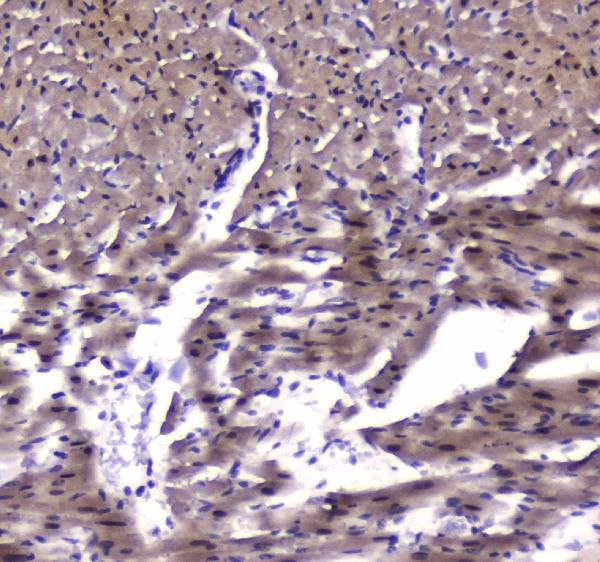
A01470-1 has IHC images from paraffin sections of mouse brain, rat cardiac muscle and human stomach, plus an IF/ICC image from A549 cells (catalog image captions).
A01470-1 will render with its mouse brain paraffin-section IHC figure (catalog IHC image caption). Its other captions show IHC in rat cardiac muscle and human stomach paraffin sections and IF/ICC in A549 cells (catalog image captions).
Which to pick: Choose A01470-1 for paraffin-section IHC, supported by its own IHC captions; the fixative is unreported (catalog IHC image captions). Choose the same SKU for IF/ICC based on its application list and A549 image, or for human, mouse and rat work based on its stated reactivity (catalog applications; catalog IF image caption; catalog reactivity). Clonality is unreported (catalog clone field).