FAM3D / Protein FAM3D · IHC design guide

Design Immunohistochemistry for FAM3D

Plan FAM3D paraffin-section IHC using the reported cytoplasmic staining in glandular and respiratory epithelial cells (HPA tissue IHC). Compare colon glandular cells with adrenal gland glandular cells, where staining was not detected, and account for secretion when interpreting the pattern (HPA tissue IHC; UniProt).

Evidence assembled Oct 2026 · For research use; verify linked source records and product datasheet before use
Immunohistochemistry protocol sheet for FAM3D (IHC for FAM3D): expected localisation Cytoplasmic staining (HPA tissue IHC); secreted protein (UniProt), antibody A13898, validated IHC image, and IHC protocol steps
Printable FAM3D IHC protocol sheet — expected localisation Cytoplasmic staining (HPA tissue IHC); secreted protein (UniProt), antibody A13898, controls and protocol steps. Open the full FAM3D IHC guide →

FAM3D Immunohistochemistry Experimental Design Guide

Expected localisation, validated protocols, controls and antibodies — the at-a-glance facts below, then the full design guide.

Must know before staining
Expected localisation Cytoplasmic staining (HPA tissue IHC); secreted protein (UniProt)
Staining pattern Cytoplasmic staining in glandular and respiratory epithelial cells (HPA tissue IHC)
Antigen retrieval Tris-EDTA pH 9.0 HIER, heat-mediated (datasheet A13898)
Positive control ⓘ Appendix+4 more · see all
Negative control ⓘ Adipose tissue+4 more · see all
Important caveats
Reasons your staining may differ from the expected pattern.
Fixation Keep fixation consistent across tissue sections. (standard IHC practice; not target-specific)
Caveat Secretion can separate staining from the producing cells (HPA tissue IHC)
Regulation No expression regulator annotated (UniProt)
Isoform / epitope No isoforms annotated; mature chain spans 26–224 (UniProt)
Section 1

Recommended FAM3D IHC & IF Protocols

The catalog antibody protocol is paired with one published FAM3D IHC protocol using HNSCC tissue chips (PMC9741545 Methods).

Recommended immunohistochemistry (IHC-P) protocol parameters
SampleParaffin-embedded human tonsil tissue; fixative not specified (datasheet A13898)
FixationImage fixative and duration unreported (datasheet A13898); verify before use.
Sectioning4–5 µm sections on charged slides (standard)
DeparaffinisationXylene, graded ethanol series to water (standard)
Antigen retrievalHeat retrieval: Tris-EDTA pH 9.0 (datasheet A13898); 20 min, 95–100 °C (standard)
Peroxidase block3% H2O2, 10 min, room temperature (standard)
Blocking10% normal serum of the secondary host, 30 min, room temperature (standard)
Primary antibodyRabbit anti-FAM3D, 1:100-1:300 (datasheet A13898)
Primary incubationOvernight at 4 °C (standard)
DetectionHRP-polymer secondary, DAB chromogen 5–10 min (standard)
CounterstainHematoxylin, blue, dehydrate and mount (standard)
Expected resultFAM3D-positive staining in glandular cells of appendix (HPA tissue IHC: High). HPA tissue profile: Cytoplasmic expression in several tissues. No signal in the no-primary control.
💡Decision noteStart with heat-mediated Tris-EDTA retrieval at pH 9.0 (A13898 datasheet); the published protocol does not specify its retrieval buffer (PMC9741545 Methods).
Section 2

What Is the Expected FAM3D Staining Pattern?

FAM3D is secreted and has no transmembrane segment (UniProt Q96BQ1). In paraffin IHC, expect predominantly cytoplasmic staining in glandular and respiratory epithelial cells, with high staining reported in selected tissues (HPA: tissue IHC). HPA rates the tissue staining Approved but notes low consistency with RNA expression and pending external verification (HPA: reliability description).

What am I looking at on my slide?
Strong cytoplasmic staining in colon or duodenal glandular cells.This matches reported high staining in these cells (HPA: colon and duodenum, High). Assess the pattern by cell type and compartment, not overall slide darkness (general IHC practice).
Strong cytoplasmic staining in bronchial or nasopharyngeal respiratory epithelium.This also matches reported high staining (HPA: bronchus and nasopharynx, High). A secreted protein need not remain confined to producing cells, so nearby extracellular signal needs cautious interpretation (UniProt Q96BQ1: Secreted).
Predominantly nuclear staining or a sharp, continuous plasma membrane outline.Neither is the reported cytoplasmic tissue pattern (HPA: tissue IHC profile). FAM3D has no transmembrane segment (UniProt Q96BQ1). Treat an isolated nuclear or membrane pattern as suspect and check controls before assigning it to FAM3D (general IHC practice).
Strong staining in cells reported as undetected, or color in a reagent control.For example, HPA reports no detection in adipose tissue adipocytes and bone marrow hematopoietic cells (HPA: tissue IHC). Unexpected staining can reflect cross-reactivity or endogenous detection activity; compare the primary-omitted control (general IHC practice).
Diffuse color across cells and empty spaces, or no signal in a known-positive section.Diffuse color obscures the cell-specific pattern (general IHC practice). Absent signal in colon glandular cells conflicts with their reported high staining (HPA: colon, High); first assess the run controls and staining workflow (general IHC practice).
💡Expected FAM3D appearanceCall a positive result when glandular or respiratory epithelial cells show clear cytoplasmic staining, potentially strong in HPA high tissues; isolated nuclear or continuous membrane staining is suspect (HPA: tissue IHC; UniProt Q96BQ1 topology).
How each factor affects the staining
Secretion and compartmentFAM3D is secreted and lacks a transmembrane segment (UniProt Q96BQ1). HPA describes cytoplasmic tissue staining; extracellular color alone does not establish the producing cell (HPA: tissue IHC profile; general IHC interpretation).
Processing and unknown epitopeThe signal peptide spans residues 1–25, and the annotated protein chain spans 26–224 (UniProt Q96BQ1). The supplied evidence does not locate the antibody epitope, so these positions cannot predict its staining.
GlycosylationUniProt annotates one glycosylation site at residue 107 (UniProt Q96BQ1). Its effect on this antibody's IHC staining is unreported; do not infer an altered epitope or retrieval requirement from the annotation.
Tissue evidence and validationHPA labels the tissue pattern Approved but reports low consistency with RNA expression and pending external verification (HPA: reliability description). Its listed antibody HPA013844 is IHC Approved, with no ICC status listed (HPA: antibody record).
IF/ICC Q&AQ: Can the IHC pattern establish an IF/ICC pattern? A: No. HPA lists FAM3D as secreted but gives no main subcellular location or ICC-IF images, and no ICC status for HPA013844 (HPA: subcellular and antibody records).
Why is my staining missing, weak or wrong?
SituationLikely causeNext action
No staining in colon or duodenal glands.These are reported high-staining cells, so a failed run is possible (HPA: colon and duodenum, High; general IHC practice).Check the positive control, detection reagents and counterstain, then review the run's retrieval and antibody settings (general IHC practice). No FAM3D-specific retrieval condition is supplied.
Broad staining across the section obscures the expected cells.Background may arise from nonspecific antibody binding or the detection system (general IHC practice).Compare a primary-omitted control; inspect blocking, washes and detection exposure before interpreting glandular staining (general IHC practice).
Strong nuclear or membrane-only staining dominates.That compartment conflicts with the reported cytoplasmic profile and lacks support from FAM3D topology (HPA: tissue IHC; UniProt Q96BQ1).Check whether the same pattern appears in controls or reported undetected cells; withhold a FAM3D-specific call if it does (general IHC practice; HPA: tissue IHC).
Adipocytes or marrow hematopoietic cells stain strongly.HPA reports FAM3D as undetected in those cell types; cross-reactivity or endogenous detection activity is possible (HPA: tissue IHC; general IHC practice).Compare the primary-omitted control and assess whether the signal tracks the expected epithelial cells in a positive section (general IHC practice; HPA: tissue IHC).
Staining differs from the tissue's RNA enrichment.HPA explicitly reports low agreement between antibody staining and RNA data; secretion can separate protein and RNA locations (HPA: reliability description).Record the stained cell type and compartment, compare HPA tissue images and controls, and report the discrepancy without treating RNA as a direct stain map (HPA: tissue IHC; general IHC interpretation).

Sample controls for FAM3D IHC & IF

🧪Run colon first: glandular cells should stain (HPA: High in colon glandular cells). Use adrenal glandular cells as the negative tissue (HPA: Not detected in adrenal glandular cells); on the colon slide, use adjacent nonglandular cells to assess background, without assuming they are FAM3D-negative.
Positive control tissue: Appendix (Glandular cells, HPA High)
Negative control tissue: Adipose tissue (HPA Not detected)
ICC-IF cell lines (HPA subcellular resource): HPA carries no ICC-IF cell line for FAM3D; derive a cell-line control from the positive tissue's cell type (Glandular cells) and confirm it by RNA or western blot first.
Technical controls: Include a no-primary, secondary-only control; a host-species-, isotype-, and clonality-matched control antibody; and FAM3D-knockout tissue if available, or immunizing-peptide preabsorption when the epitope is known. For chromogenic colon IHC, quench endogenous peroxidase and check whether signal in inflammatory cells persists in the no-primary control.
⚠️Feasibility: A target-specific fixation window or fixation effect is unreported, and the selected paraffin-section caption does not state its fixative (A13898 tissue-IHC caption). The caption uses Tris-EDTA retrieval at pH 9.0 and a 1:200 primary antibody dilution overnight at 4°C, but it does not establish that retrieval is required (A13898 tissue-IHC caption). There is no basis here to call frozen sections or IF easier; interpret possible luminal signal carefully because FAM3D is secreted (UniProt Q96BQ1: Secreted), and HPA lists no ICC-IF image cell lines (HPA subcellular record).

HPA tissue IHC evidence for FAM3D

Comprehensive Human Protein Atlas IHC scoring per tissue (reliability: Approved — Low consistency between antibody staining and RNA expression data. Pending external verification. At least one protein variant secreted, tissue location of RNA and protein might differ and correlation is complex.). Rows are taken directly from the HPA tissue chart — click any row's HPA link to view the source.

Positive expression · recommended positive controls

TissueCell typeLevelEvidenceSource
Appendix Glandular cells High Protein (IHC) HPA →
Bronchus Respiratory epithelial cells High Protein (IHC) HPA →
Colon Glandular cells High Protein (IHC) HPA →
Duodenum Glandular cells High Protein (IHC) HPA →
Nasopharynx Respiratory epithelial cells High Protein (IHC) HPA →

Undetected expression · recommended negative controls

TissueCell typeLevelEvidenceSource
Adipose tissue Adipocytes Not detected Protein (IHC) HPA →
Adrenal gland Glandular cells Not detected Protein (IHC) HPA →
Bone marrow Hematopoietic cells Not detected Protein (IHC) HPA →
Breast Adipocytes Not detected Protein (IHC) HPA →
Caudate Glial cells Not detected Protein (IHC) HPA →
Section 3

Advanced FAM3D IHC Tips

Troubleshoot FAM3D staining in paraffin sections by checking retrieval, controls, cell identity and the expected distribution of a secreted protein (UniProt Q96BQ1).

Which retrieval condition should I start with for FAM3D in paraffin sections?
Start with heat-mediated Tris-EDTA retrieval at pH 9.0 (datasheet A13898). The selected tonsil paraffin-section image used that buffer, followed by primary antibody at 1:200 overnight at 4 °C (datasheet A13898). If staining is weak, compare a shorter and longer heating interval on matched sections while keeping antibody concentration and detection constant; examine tissue integrity and background alongside signal. Include a known high-staining glandular or respiratory epithelial sample, such as colon or bronchus, to judge whether retrieval worked (HPA: High in colon glandular cells; High in bronchus respiratory epithelial cells). Record the heating interval and cooling conditions so that any improvement can be reproduced.
How should I troubleshoot fixation-related loss of FAM3D staining?
Target-specific sensitivity to fixation is unknown: the selected tissue image identifies paraffin-embedded tonsil but does not state its fixative (datasheet A13898). Compare sections with documented fixation histories, processed and stained together, before attributing a weak result to the antibody. Keep section thickness, retrieval at pH 9.0, and primary incubation at 1:200 overnight at 4 °C consistent during that comparison (datasheet A13898). Assess preserved morphology and staining in an independently selected positive-control tissue, such as colon glandular epithelium (HPA: High in colon glandular cells). If the controls differ, repeat with matched processing before changing antibody concentration or interpretation.
Where should convincing FAM3D signal appear in a tissue section?
FAM3D is annotated as secreted and has no transmembrane segment (UniProt Q96BQ1 topology; UniProt Q96BQ1 subcellular location). HPA reports cytoplasmic staining across several tissues, including high staining in colon glandular and bronchial respiratory epithelial cells (HPA: tissue IHC profile; High in colon glandular cells; High in bronchus respiratory epithelial cells). In chromogenic sections, assess epithelial cytoplasmic signal together with any nearby extracellular staining, and compare both with a no-primary control. Do not assign a membrane-bound pool solely because pigment outlines cells; inspect serial sections for the corresponding cell borders and morphology. Nuclear-only staining would conflict with the supplied localisation evidence and merits a repeat with control tissue.
Could FAM3D processing or glycosylation explain discordant staining?
The annotated precursor is 224 amino acids, with a signal peptide at residues 1–25 and a mature chain at 26–224 (UniProt Q96BQ1 processing). One glycosylation site is annotated at residue 107, within the GG-type lectin domain at 64–222 (UniProt Q96BQ1 glycosylation; UniProt Q96BQ1 domain). No isoforms are listed, so an isoform explanation is unsupported by this record (UniProt Q96BQ1 isoforms). Check the antibody’s documented immunogen or epitope before interpreting uneven staining as a processing effect; its epitope is not supplied here. Compare matched retrieval conditions and tissue controls before testing an additional antibody directed at a documented, distinct epitope.
How can I check FAM3D localisation with tissue immunofluorescence?
Treat IF as a separate optimization: the supplied product image documents chromogenic staining in paraffin tonsil, while HPA lists no ICC/IF images (datasheet A13898; HPA: subcellular record). Multiplex FAM3D with a validated glandular or respiratory epithelial marker, since HPA reports high staining in those cell types (HPA: High in colon glandular cells; High in bronchus respiratory epithelial cells). Choose a spectrally separated, preferably far-red fluorophore after checking autofluorescence and single-stain controls in the chosen tissue. FAM3D is secreted and lacks a transmembrane segment, so there is no defined membrane side to target with permeabilisation; compare gentle permeabilisation when assessing a retained intracellular pool (UniProt Q96BQ1 topology; UniProt Q96BQ1 subcellular location).
What should I check when FAM3D staining is diffuse or widespread?
First inspect no-primary and detection-only controls, then check whether the apparent signal follows tissue edges, damaged areas or broadly stained stroma. FAM3D is secreted, so extracellular staining can be biologically plausible, but that annotation alone cannot establish that diffuse pigment is specific (UniProt Q96BQ1 subcellular location). For peroxidase-based chromogenic IHC, verify endogenous peroxidase blocking and compare a section developed for a shorter interval with the same detection reagents. Titrate around the image’s 1:200 primary dilution while retaining its pH 9.0 retrieval condition (datasheet A13898). A low-staining comparison, such as adipocytes in adipose tissue, can help reveal nonspecific signal (HPA: Not detected in adipose-tissue adipocytes).
How should I quantify FAM3D staining across tissue samples? ⚠ ANSWER MARKED FOR VERIFICATION
Define the tissue compartment and cell class before scoring; HPA reports high staining in colon glandular cells and bronchial respiratory epithelial cells (HPA: High in colon glandular cells; High in bronchus respiratory epithelial cells). For cellular staining, report the percentage of positive target cells and an H-score based on intensity categories, with the scoring thresholds fixed before reviewing groups. For extracellular signal, measure positive area or optical density per mm² of viable, annotated tissue and report it separately from cellular staining. Normalize cell counts to the number of eligible cells, and area measurements to the corresponding eligible tissue area. Keep retrieval, development time and imaging settings matched across sections.
How do I distinguish genuine FAM3D staining from an artefact?
A convincing pattern should be reproducible in appropriate cells and compatible with FAM3D secretion and HPA’s reported cytoplasmic tissue staining (UniProt Q96BQ1 subcellular location; HPA: tissue IHC profile). Compare high-staining colon glandular epithelium with an HPA not-detected cell population, such as adipocytes in adipose tissue (HPA: High in colon glandular cells; Not detected in adipose-tissue adipocytes). Treat isolated nuclear pigment, edge-heavy staining, necrotic deposits and signal in no-primary controls as reasons to investigate an artefact. Check the peroxidase block when detection-only controls develop color, and review morphology before assigning a positive cell type. HPA rates its tissue staining Approved but reports low consistency with RNA data, so corroborate unexpected distributions independently (HPA: reliability description).
Boster reagents

Best FAM3D / Protein FAM3D IHC Antibodies

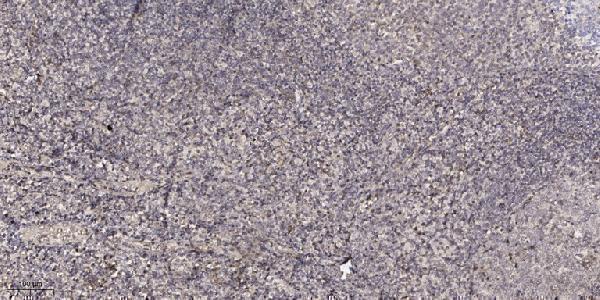
A13898 lists human and mouse reactivity (catalog: reactivity) and has a real IHC image from paraffin-embedded human tonsil (A13898 image caption). IF is listed without an image (catalog: applications, IF image alts).

Real IHC data Immunohistochemical analysis of paraffin-embedded human tonsil. 1, Antibody was diluted at 1:200 (4° overnight). 2, Tris-EDTA, pH9.0 was used for antigen retrieval. 3, Secondary antibody was diluted at 1:200 (room temperature, 30min).
Anti-Protein FAM3D FAM3D Antibody
Cat # A13898

A13898 will render with an IHC image of paraffin-embedded human tonsil (A13898 image caption). It lists IHC and IF applications and human and mouse reactivity; no IF image is supplied (catalog: applications, reactivity, IF image alts).

Which to pick: For tissue IHC, choose A13898: its own caption documents paraffin-embedded human tonsil, Tris-EDTA retrieval at pH 9.0, and primary antibody at 1:200 overnight at 4°C; the fixative is unreported (A13898 image caption). For IF/ICC, A13898 lists IF at 1:50, but supplies no IF image or ICC application (catalog: applications, IF dilution, IF image alts). For cross-species work, A13898 lists human and mouse reactivity, while its pictured IHC sample is human only; clonality is unreported.

Each figure is that product's own IHC / IF validation image from its datasheet.

References

  1. UniProt Consortium. UniProt entry Q96BQ1 (FAM3D_HUMAN, Protein FAM3D).
  2. Human Protein Atlas. FAM3D tissue IHC expression (reliability: Approved).
  3. Human Protein Atlas. FAM3D subcellular location (ICC-IF): Secreted.
  4. Human Protein Atlas. FAM3D antibody validation summary (1 antibodies).
  5. Deficiency of FAM3D (Family With Sequence Similarity 3, Member D), A Novel Chemokine, Attenuates Neutrophil Recruitment and Ameliorates Abdominal Aortic Aneurysm Development. Arteriosclerosis, thrombosis, and vascular biology 2018 — PMC6039426.
  6. FAM3D as a Prognostic Indicator of Head and Neck Squamous Cell Carcinoma Is Associated with Immune Infiltration. Computational and mathematical methods in medicine 2022 — PMC9741545.
  7. FAM3D is essential for colon homeostasis and host defense against inflammation associated carcinogenesis. Nature communications 2020 — PMC7679402.
  8. PubMed PMID:12160727 — UniProt-cited evidence.
  9. PubMed PMID:12975309 — UniProt-cited evidence.
  10. PubMed PMID:14702039 — UniProt-cited evidence.