This website uses cookies to ensure you get the best experience on our website.


- Table of Contents
Plan FGF1 chromogenic IHC in paraffin sections around the selective endothelial cytoplasmic pattern reported in tissue (HPA tissue IHC). This guide covers fixation consistency, interpretation of secreted FGF1, and epitope uncertainty across 2 isoforms (standard IHC practice; UniProt).

Expected localisation, validated protocols, controls and antibodies — the at-a-glance facts below, then the full design guide.
| Expected localisation | Endothelial cytoplasm (HPA tissue IHC) | |
| Staining pattern | Selective cytoplasmic staining in endothelial cells (HPA tissue IHC) | |
| Antigen retrieval | EDTA pH 8.0 HIER, heat-mediated (datasheet PB9944) | |
| Positive control | Kidney | |
| Negative control | Adipose tissue+4 more · see all |
| Fixation | Keep fixation consistent across sections. (standard IHC practice; not target-specific) | |
| Caveat | Secretion may separate FGF1 staining from its source cells (UniProt) | |
| Regulation | Expression regulation not established (UniProt) | |
| Isoform / epitope | 2 isoforms; antibody epitope coverage is unknown (UniProt) |
The catalog antibody’s paraffin IHC protocol (datasheet: PB9944) is followed by published FGF1 IHC protocols for rat brain sections (PMC1831852) and gastric adenocarcinoma (PMC4360790).
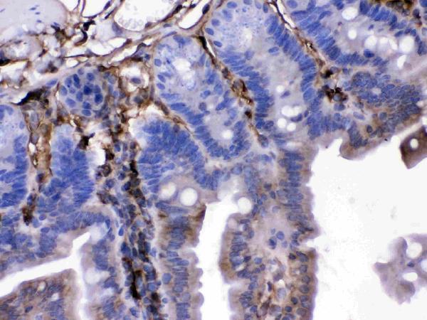
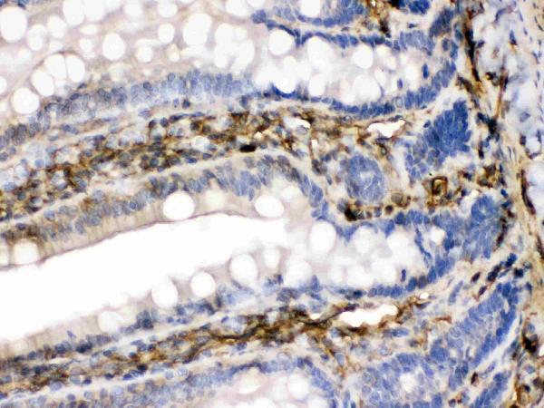
| Sample | Paraffin-embedded mouse intestine tissue; fixative not specified (datasheet PB9944) |
| Fixation | Image fixative and duration unreported (datasheet PB9944); verify before use. |
| Sectioning | 4–5 µm sections on charged slides (standard) |
| Deparaffinisation | Xylene, graded ethanol series to water (standard) |
| Antigen retrieval | Heat retrieval: EDTA pH 8.0 (datasheet PB9944); 20 min, 95–100 °C (standard) |
| Peroxidase block | 3% H2O2, 10 min, room temperature (standard) |
| Blocking | 10% goat serum (datasheet PB9944) |
| Primary antibody | Rabbit anti-FGF1, 0.5-1μg/ml (datasheet PB9944) |
| Primary incubation | Overnight at 4 °C (datasheet PB9944) |
| Detection | Streptavidin-biotin complex (SABC), DAB chromogen (datasheet PB9944) |
| Counterstain | Hematoxylin, blue, dehydrate and mount (standard) |
| Expected result | FGF1-positive staining in cells in glomeruli of kidney (HPA tissue IHC: High). HPA tissue profile: Highly selective cytoplasmic expression in endothelial cells. No signal in the no-primary control. |
FGF1 can occupy the cytoplasm, cell cortex, cytosol and nucleus, and can be secreted without a cleavable signal sequence (UniProt P05230). For paraffin-section IHC, expect selective cytoplasmic staining associated with endothelial cells (HPA: tissue IHC profile). Kidney glomerular cells provide a reported high-staining reference, although HPA does not identify every positive glomerular cell subtype (HPA: kidney, High). HPA rates its tissue IHC evidence Enhanced and notes medium agreement between staining and RNA expression (HPA: reliability description).
| Selective cytoplasmic chromogen in endothelial cells, with strong staining in cells of kidney glomeruli (HPA: tissue IHC profile; kidney, High). | This matches the reported tissue pattern. Identify the stained cell type from tissue morphology before assigning it to endothelium; the kidney entry names glomerular cells without resolving every subtype (HPA: tissue IHC profile; kidney, High). |
| Predominantly nuclear staining in a paraffin section, with little of the reported endothelial cytoplasmic pattern (HPA: tissue IHC profile). | Treat the mismatch as a pattern needing review, rather than proof of an artefact: UniProt includes nuclear FGF1, and HPA reports supported nucleoplasmic ICC-IF localization (UniProt P05230; HPA: subcellular ICC-IF). Check morphology and controls before interpreting the IHC result (general IHC practice). |
| Strong chromogen in adipocytes or respiratory epithelial cells, where HPA reports no detection (HPA: adipose tissue; bronchus). | These are unexpected cell-type results for the cited tissue entries. Check whether staining follows recognizable cells and whether controls show background; cross-reactivity or endogenous detection activity are possible explanations, not diagnoses from the slide alone (HPA: tissue IHC; general IHC practice). |
| Diffuse chromogen across cells and cell-free areas, without a selective cellular pattern (HPA: tissue IHC profile). | This does not reproduce HPA's highly selective cytoplasmic profile. Inspect the negative control and detection reagents for background, then reassess antibody concentration and wash conditions as general IHC variables (HPA: tissue IHC profile; general IHC practice). |
| No visible staining in kidney glomerular cells used as a positive reference (HPA: kidney, High). | A negative reference section leaves the run uninterpretable for tissue negatives. Verify that the positive control, primary antibody, detection system and counterstain were processed as intended before calling another specimen FGF1-negative (HPA: kidney, High; general IHC practice). |
| Tissue and cell-type reference (HPA: tissue IHC). | Kidney glomerular cells are High; adipocytes in adipose tissue and respiratory epithelial cells in bronchus are Not detected. Compare like cell types within the named tissues, since these entries do not classify every cell in either section (HPA: kidney; adipose tissue; bronchus). |
| RNA and protein location (HPA: reliability description). | HPA calls RNA tissue-enriched in brain yet describes selective endothelial cytoplasmic protein staining; it also reports no detection in cerebral-cortex endothelial cells. Because FGF1 can be secreted, RNA location alone is an unreliable slide-level positive-control choice (HPA: RNA specificity; tissue IHC; reliability description). |
| Intracellular and secreted pools (UniProt P05230). | FGF1 lacks a cleavable signal sequence and can move from cytoplasm toward the membrane for non-classical secretion. UniProt also reports nuclear localization. These annotations support checking compartment and morphology together; they do not establish the amount or appearance of extracellular chromogen in a particular paraffin section (UniProt P05230). |
| Antibody validation by application (HPA: antibody validation). | CAB017519 has Enhanced IHC validation, whereas HPA003265 has Supported ICC validation and no IHC status in the supplied record. An ICC localization result therefore cannot establish the expected performance of either antibody in paraffin-section IHC (HPA: antibody validation; subcellular ICC-IF). |
| Processing and isoforms (UniProt P05230). | UniProt lists a propeptide at residues 2–15, a chain at 16–155 and two isoforms. The supplied record gives no antibody epitope, so it cannot predict which molecular forms an IHC stain detects or whether processing changes the observed pattern (UniProt P05230). |
| Antigen retrieval and detection settings (general IHC practice). | Retrieval conditions and detection controls are routine run variables to check when staining fails or background rises. No FGF1-specific retrieval condition, dilution or fixation sensitivity is supplied here; do not infer one from HPA staining levels or UniProt localization (general IHC practice; HPA: tissue IHC; UniProt P05230). |
| Situation | Likely cause | Next action |
|---|---|---|
| Kidney reference section is blank where glomerular cells should stain (HPA: kidney, High). | A failed IHC run or unsuitable control section is possible; absence of chromogen alone does not identify which step failed (general IHC practice). | Repeat with a suitable kidney positive section and inspect primary-antibody, retrieval and detection steps using the antibody's validated IHC instructions (HPA: kidney, High; general IHC practice). |
| Negative-control slide also shows diffuse or cell-free chromogen (general IHC practice). | Background from detection reagents, endogenous activity or nonspecific binding is possible (general IHC practice). | Inspect the negative control alongside the test section; review blocking, washes and detection controls before scoring cell-associated staining (general IHC practice). |
| Unexpected strong staining appears in adipocytes or bronchial respiratory epithelium (HPA: adipose tissue and bronchus, Not detected). | Possible cross-reactivity or endogenous detection activity; the HPA entries support an unexpected-pattern flag, not a unique cause (HPA: tissue IHC; general IHC practice). | Confirm cell identity by morphology, compare control slides and check whether the expected endothelial cytoplasmic pattern is present (HPA: tissue IHC profile; general IHC practice). |
| The section shows nuclear staining but little selective endothelial cytoplasmic staining (HPA: tissue IHC profile). | The tissue IHC pattern differs from HPA's summary, although nuclear FGF1 is biologically plausible and HPA supports nucleoplasmic ICC-IF localization (UniProt P05230; HPA: subcellular ICC-IF). | Check the tissue positive reference, controls and cell morphology; report nuclear staining separately rather than treating it as confirmation of the expected tissue IHC pattern (HPA: tissue IHC; general IHC practice). |
| A brain section is negative despite brain-enriched RNA or UniProt tissue specificity (HPA: RNA specificity; UniProt P05230). | RNA abundance and tissue-wide expression do not guarantee staining in the selected cells; HPA reports cerebral-cortex endothelial cells as Not detected (HPA: reliability description; cerebral cortex). | Use the reported kidney glomerular-cell staining as the IHC reference and describe the exact brain region and cell type examined (HPA: kidney, High; cerebral cortex, Not detected). |
| Why might IF/ICC show nucleoplasmic FGF1 while tissue IHC favors cytoplasmic endothelial staining? (HPA: subcellular ICC-IF; tissue IHC). | HPA's supported nucleoplasmic result comes from ICC-IF, while its selective cytoplasmic profile comes from tissue IHC; UniProt includes both nuclear and cytoplasmic locations (HPA: subcellular ICC-IF; tissue IHC; UniProt P05230). | Interpret each image against its own application and cell context; use the separate IF/ICC guide for that application (HPA: antibody validation; general IHC/IF practice). |
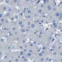
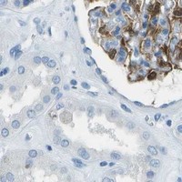


Comprehensive Human Protein Atlas IHC scoring per tissue (reliability: Enhanced — Medium consistency between antibody staining and RNA expression data. At least one protein variant secreted, tissue location of RNA and protein might differ and correlation is complex.). Rows are taken directly from the HPA tissue chart — click any row's HPA link to view the source.
| Tissue | Cell type | Level | Evidence | Source |
|---|---|---|---|---|
| Kidney | Cells in glomeruli | High | Protein (IHC) | HPA → |
| Tissue | Cell type | Level | Evidence | Source |
|---|---|---|---|---|
| Adipose tissue | Adipocytes | Not detected | Protein (IHC) | HPA → |
| Adrenal gland | Glandular cells | Not detected | Protein (IHC) | HPA → |
| Appendix | Glandular cells | Not detected | Protein (IHC) | HPA → |
| Bone marrow | Hematopoietic cells | Not detected | Protein (IHC) | HPA → |
| Bronchus | Respiratory epithelial cells | Not detected | Protein (IHC) | HPA → |
Use matched controls and cell-level scoring to troubleshoot FGF1 staining in paraffin sections; interpret IF observations separately.
The catalog lists anti-FGF1 antibodies for human, mouse and rat IHC (catalog: applications/reactivity). Its only IHC images are labeled GADD45A, leaving FGF1 staining unverified (PB9944 image captions).
M00206-1 is listed for human, mouse and rat IHC, and RP1005 for human IHC; neither has an IHC image (catalog: applications/reactivity/image alts). PB9944 is listed for human, mouse and rat IHC, but its paraffin-section images are labeled GADD45A in mouse and rat intestine and human placenta (PB9944 image captions).
Which to pick: For cross-species tissue IHC, M00206-1 is a rabbit monoclonal listed for human, mouse and rat, though it has no IHC image (catalog: host/clone/applications/reactivity/image alts). RP1005 is listed for human IHC; PB9944's caption shows paraffin sections but names GADD45A and does not report the fixative, so it cannot establish FGF1 staining or fixation (catalog: RP1005 applications/reactivity; PB9944 image captions). None of these SKUs lists IF/ICC, so there is no supported IF/ICC pick here (catalog: applications). The selected PB9944 tissue-IHC caption documents paraffin sections, but does not specify the fixative (selected-SKU IHC image PB9944).