FGF3 / Fibroblast growth factor 3 · IHC design guide

Design Immunohistochemistry for FGF3

Use cerebellar granular-layer processes as the tissue IHC reference (HPA tissue IHC). For paraffin sections, start the catalog antibody at 2–5 μg/ml (datasheet A03316-2) and include a negative tissue control.

Evidence assembled Oct 2026 · For research use; verify linked source records and product datasheet before use
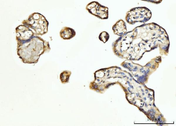
Immunohistochemistry protocol sheet for FGF3 (IHC for FGF3): expected localisation Secreted (UniProt); granular-layer processes stain (HPA tissue IHC), antibody A03316-2, validated IHC image, and IHC protocol steps
Printable FGF3 IHC protocol sheet — expected localisation Secreted (UniProt); granular-layer processes stain (HPA tissue IHC), antibody A03316-2, controls and protocol steps. Open the full FGF3 IHC guide →

FGF3 Immunohistochemistry Experimental Design Guide

Expected localisation, validated protocols, controls and antibodies — the at-a-glance facts below, then the full design guide.

Must know before staining
Expected localisation Secreted (UniProt); granular-layer processes stain (HPA tissue IHC)
Staining pattern High staining in cerebellar granular-layer processes (HPA tissue IHC)
Antigen retrieval EDTA pH 8.0 HIER, heat-mediated (datasheet A03316-2)
Positive control ⓘ Cerebellum
Negative control ⓘ Adipose tissue+4 more · see all
Important caveats
Reasons your staining may differ from the expected pattern.
Fixation Keep fixation consistent across paraffin sections (standard IHC practice; not target-specific); Matched tissue-IHC evidence does not establish the fixation claim. Validate the specimen-specific method before use. (selected-SKU IHC image A03316-2)
Caveat Secreted FGF3 may stain away from RNA-positive cells (HPA tissue IHC)
Regulation Brain-enriched RNA expression (HPA tissue RNA)
Isoform / epitope No isoforms annotated; mature chain spans aa 18–239 (UniProt)
Section 1

Recommended FGF3 IHC & IF Protocols

The catalog antibody’s IHC-P protocol is paired with one published mouse oral tissue FGF3 protocol (PMC5658092).

Recommended immunohistochemistry (IHC-P) protocol parameters
SampleParaffin-embedded human placenta tissue; fixative not specified (datasheet A03316-2)
FixationImage fixative and duration unreported (datasheet A03316-2); verify before use.
Sectioning4–5 µm sections on charged slides (standard)
DeparaffinisationXylene, graded ethanol series to water (standard)
Antigen retrievalHeat retrieval: EDTA pH 8.0 (datasheet A03316-2); 20 min, 95–100 °C (standard)
Peroxidase block3% H2O2, 10 min, room temperature (standard)
Blocking10% goat serum (datasheet A03316-2)
Primary antibodyRabbit anti-FGF3, 2-5 μg/ml (datasheet A03316-2)
Primary incubationOvernight at 4 °C (datasheet A03316-2)
DetectionStreptavidin-biotin complex (SABC), DAB chromogen (datasheet A03316-2)
CounterstainHematoxylin, blue, dehydrate and mount (standard)
Expected resultFGF3-positive staining in processes in granular layer of cerebellum (HPA tissue IHC: High). HPA tissue profile: Expressed in cerebellum. No signal in the no-primary control.
💡Decision noteStart with heat-mediated EDTA pH 8.0 retrieval (datasheet: A03316-2); the published protocol used microwave retrieval without specifying a buffer (PMC5658092).
Section 2

What Is the Expected FGF3 Staining Pattern?

FGF3 is a secreted protein with a signal peptide and no transmembrane segment (UniProt P11487 topology). In tissue IHC, the supported positive pattern is high staining of processes in the cerebellar granular layer (HPA: High in processes in granular layer). HPA rates its tissue staining “Enhanced,” while noting medium consistency with RNA and disregarded presumed off-target binding (HPA: reliability description).

What am I looking at on my slide?
High staining follows processes in the cerebellar granular layer (HPA: High in processes in granular layer).This matches the reported positive tissue pattern (HPA: cerebellum IHC). Assess whether staining traces processes rather than assigning it to a cell body solely from its position; the HPA record identifies processes, not their cell of origin (HPA: processes in granular layer).
Strong, isolated nuclear staining appears without the reported process pattern.A nuclear-only result does not match the reported tissue pattern or secreted annotation (HPA: cerebellum IHC; UniProt P11487: secreted). Treat it as suspect and review morphology, background and controls before interpreting it as FGF3.
Prominent staining appears in cells reported as undetected, such as adipocytes in adipose tissue (HPA: Not detected in adipocytes).That distribution conflicts with the listed HPA tissue result (HPA: adipose tissue IHC). Consider antibody cross-reactivity or endogenous chromogenic activity, and compare with a no-primary control (general IHC practice); the HPA result alone cannot identify the cause.
Diffuse color covers multiple structures and obscures process boundaries.The slide cannot be scored confidently against the process-specific reference pattern (HPA: High in processes in granular layer). Review the no-primary control, blocking, washes and detection conditions for nonspecific background (general IHC practice).
No convincing signal appears in the cerebellar granular layer.This disagrees with the reported high process staining (HPA: cerebellum IHC), but does not by itself establish absent FGF3. Check tissue identity, section quality, antibody and detection controls, and the validated IHC-P workflow (general IHC practice).
💡Expected FGF3 appearanceCall a section positive when high staining delineates processes in the cerebellar granular layer (HPA: cerebellum IHC); isolated nuclear or widespread nonspecific color is a suspect result, not the reported pattern (HPA: processes in granular layer; general IHC practice).
How each factor affects the staining
Secretion and topologyFGF3 is secreted and lacks a transmembrane segment (UniProt P11487 topology). Do not require a continuous cell-membrane outline to call the reported process-associated tissue pattern (HPA: cerebellum IHC); these annotations do not identify the producing cells.
Signal peptide and mature chainUniProt annotates a signal peptide at residues 1–17 and a chain at 18–239 (UniProt P11487 processing). Antibody epitope information is not supplied, so these annotations cannot predict whether processing affects staining.
Tissue evidence and validationHPA marks tissue IHC “Enhanced” and reports high cerebellar process staining (HPA: reliability; cerebellum IHC). It also notes medium agreement with RNA and presumed off-target binding that was disregarded; interpret unexpected staining with controls (HPA: reliability description; general IHC practice).
IF/ICC: what localisation is established?HPA lists FGF3 as secreted, gives no main subcellular location and supplies no cell-line ICC-IF images (HPA: subcellular). The cerebellar IHC pattern cannot establish an IF/ICC cell-compartment pattern (HPA: cerebellum IHC; HPA: subcellular).
Why is my staining missing, weak or wrong?
SituationLikely causeNext action
Cerebellar processes are unstained or unexpectedly faint.The result conflicts with the HPA positive reference, but the cause is undetermined (HPA: High in processes in granular layer).Confirm the granular layer on the section, then check primary-antibody, detection and counterstain steps against the IHC-P workflow (general IHC practice). Do not infer FGF3-specific fixation sensitivity; none is supplied.
Color develops in the no-primary control.Signal without primary antibody points to background from the detection system or endogenous tissue activity (general IHC practice).Review the detection reagent, appropriate endogenous-activity blocking and wash steps (general IHC practice). Reassess the full stain only after control background is resolved.
Color is widespread and process outlines are unclear.Broad background prevents comparison with the reported restricted process pattern (HPA: cerebellum IHC).Compare the no-primary control and review blocking, antibody concentration and washes (general IHC practice). Score only structures whose boundaries remain interpretable.
Strong nuclear staining dominates the section.A nuclear-only pattern is unsupported by the secreted annotation and reported tissue staining (UniProt P11487: secreted; HPA: cerebellum IHC).Inspect morphology and controls, and repeat with an IHC-validated antibody if needed (general IHC practice). Do not assign nuclear FGF3 from this pattern alone.
Adipocytes stain strongly in adipose tissue.HPA reports FGF3 as not detected in those cells (HPA: adipose tissue IHC); cross-reactivity or detection background are possibilities, not established causes.Compare the no-primary control and the cerebellar reference pattern, then assess whether staining follows the intended structures (HPA: cerebellum IHC; general IHC practice).
The stained area seems inconsistent with local RNA expression.FGF3 is secreted, and HPA says RNA and protein tissue locations are expected to differ (UniProt P11487: secreted; HPA: reliability description).Judge the protein stain against the reported cerebellar process pattern and staining controls (HPA: cerebellum IHC; general IHC practice). Do not use RNA location alone to reject it.

Sample controls for FGF3 IHC & IF

🧪Run cerebellum first and look for staining in processes in the granular layer (HPA: High in processes in granular layer). Use adipose tissue as a negative tissue, with adipocytes expected to be unstained (HPA: Not detected in adipocytes); cells outside the stained processes can provide a background reference on the cerebellum slide, but HPA does not identify a specific internal negative cell type.
Positive control tissue: Cerebellum (Processes in granular layer, HPA High)
Negative control tissue: Adipose tissue (HPA Not detected)
ICC-IF cell lines (HPA subcellular resource): HPA carries no ICC-IF cell line for FGF3; derive a cell-line control from the positive tissue's cell type (Processes in granular layer) and confirm it by RNA or western blot first.
Technical controls: Include a no-primary (secondary-only) control, an isotype control matched to the primary antibody’s host species and clonality, and a biological specificity control using FGF3 knockout material or peptide competition where available. Check for endogenous peroxidase or biotin signal in chromogenic IHC, and for tissue autofluorescence if examining cerebellum by IF (standard IHC/IF practice).
⚠️Feasibility: A target-specific fixation window and retrieval requirement are unreported in the supplied FGF3 evidence; the selected-SKU paraffin caption describes CD16/Fcgr3, and its fixative is unreported (selected-SKU caption). There is no basis here to conclude that frozen sections or IF are easier; HPA lists no FGF3 ICC-IF image cell lines (HPA subcellular). Because FGF3 is secreted (UniProt P11487), assess signal in the reported cerebellar processes against local background rather than assuming cell-bound staining.

HPA tissue IHC evidence for FGF3

Comprehensive Human Protein Atlas IHC scoring per tissue (reliability: Enhanced — Medium consistency between antibody staining and RNA expression data. Secreted protein, tissue location of RNA and protein is expected to differ. Presumed off target binding observed and disregarded. External characterization data supports antibody staining.). Rows are taken directly from the HPA tissue chart — click any row's HPA link to view the source.

Positive expression · recommended positive controls

TissueCell typeLevelEvidenceSource
Cerebellum Processes in granular layer High Protein (IHC) HPA →

Undetected expression · recommended negative controls

TissueCell typeLevelEvidenceSource
Adipose tissue Adipocytes Not detected Protein (IHC) HPA →
Adrenal gland Glandular cells Not detected Protein (IHC) HPA →
Appendix Glandular cells Not detected Protein (IHC) HPA →
Bone marrow Hematopoietic cells Not detected Protein (IHC) HPA →
Breast Adipocytes Not detected Protein (IHC) HPA →
Section 3

Advanced FGF3 IHC Tips

Troubleshoot FGF3 staining in paraffin sections by checking the antibody identity, retrieval conditions, tissue distribution and extracellular signal before scoring (UniProt P11487; HPA tissue IHC).

What retrieval conditions should I start with for FGF3 staining in paraffin sections?
Start with heat-mediated retrieval in EDTA at pH 8.0 (datasheet A03316-2). The supplied caption for A03316-2 identifies its target as CD16/Fcgr3, so that retrieval condition has no demonstrated FGF3 specificity in the supplied evidence (A03316-2 caption). Before interpreting FGF3 staining, confirm that the antibody and its antigen documentation actually match FGF3 (A03316-2 caption; UniProt P11487). If signal is weak after that check, compare retrieval heating times on adjacent sections while holding detection conditions constant (standard IHC practice). Examine tissue structure and background alongside signal, since excessive retrieval can impair section quality (standard IHC practice).
Could fixation explain weak or uneven FGF3 staining?
FGF3-specific fixation sensitivity is unknown from the supplied evidence; the selected tissue caption states no fixative (A03316-2 caption). Record the actual fixative, fixation duration and tissue processing history for every section before comparing staining intensity (standard IHC practice). If archival sections vary, test matched sections with the same retrieval and detection conditions, then inspect both morphology and signal (standard IHC practice). FGF3 is annotated as secreted, but that annotation does not establish how fixation affects its detection (UniProt P11487). Treat an apparent fixation effect as a finding from your controlled comparison, rather than an established property of FGF3 (standard IHC practice).
Where should convincing FGF3 staining appear in a chromogenic section?
FGF3 is a secreted protein with no transmembrane segment, so assess staining in the tissue context rather than demanding a membrane outline (UniProt P11487 topology and subcellular annotation). HPA reports high staining in processes in the cerebellar granular layer and describes FGF3 expression in cerebellum (HPA tissue IHC). Its reliability note cautions that RNA and protein locations may differ for a secreted protein and that presumed off-target staining was disregarded (HPA tissue IHC). Compare the stained structures with a matched negative control and the expected anatomy at the same magnification (standard IHC practice). Diffuse chromogen alone is insufficient to assign FGF3 to a particular cell type (HPA tissue IHC; standard IHC practice).
How can the FGF3 epitope affect staining interpretation?
First verify the antibody's stated target and immunogen: the supplied A03316-2 tissue caption names CD16/Fcgr3, not FGF3 (A03316-2 caption). The FGF3 record lists a 239-amino-acid precursor, signal peptide at residues 1–17, mature chain at 18–239, and one glycosylation site at residue 65 (UniProt P11487). It lists no isoforms, but provides no antibody epitope for this guide (UniProt P11487; supplied evidence). Ask whether the documented epitope lies in the signal peptide or mature chain before interpreting tissue staining (UniProt P11487; standard IHC practice). Epitope identity and independent FGF3 validation are necessary to resolve an unexpected pattern (standard IHC practice).
How should I investigate FGF3 localisation with multiplex immunofluorescence?
Use a validated marker for the cells or processes being examined in the cerebellar granular layer, where HPA reports high FGF3 staining (HPA tissue IHC). Choose spectrally separated fluorophores and inspect unstained tissue in each channel to distinguish signal from autofluorescence (standard IF practice). FGF3 is secreted and lacks a transmembrane segment, but the supplied evidence does not identify this antibody's epitope (UniProt P11487; A03316-2 caption). Use permeabilisation when a confirmed epitope requires intracellular access; assess staining without it when testing accessibility of an extracellular epitope (standard IF practice). The supplied evidence provides no FGF3 ICC/IF images or IF-specific fixation conditions, so establish those conditions experimentally (HPA subcellular; supplied evidence).
What should I check when FGF3 chromogenic staining has high background?
Check a no-primary control and a matched negative tissue section to separate detection-system background from candidate FGF3 signal (standard IHC practice). Block endogenous peroxidase before DAB development, and assess endogenous biotin if using a biotin-based detection system (standard IHC practice). The supplied A03316-2 caption describes serum blocking and biotin-based DAB detection, but identifies CD16/Fcgr3 as its target (A03316-2 caption). Optimise primary concentration and wash stringency only after confirming the antibody's FGF3 identity and validation (A03316-2 caption; standard IHC practice). HPA notes presumed off-target staining in its FGF3 assessment, making morphology and controls especially important (HPA tissue IHC).
How should I quantify FGF3 staining across paraffin sections? ⚠ ANSWER MARKED FOR VERIFICATION
Define the anatomical region and analysis area before examining treatment groups, using consistent section thickness, imaging and DAB settings (standard IHC practice). For discrete cells, report percentage positive and an intensity-weighted H-score; for the reported cerebellar granular-layer processes, measure positive area or signal density per mm² (HPA tissue IHC; standard IHC practice). Normalise process measurements to the sampled granular-layer area, and cell scores to the counted eligible cells (standard IHC practice). Set thresholds using negative controls and apply them consistently across sections (standard IHC practice). Confirm antibody identity first, because A03316-2's supplied tissue caption documents CD16/Fcgr3 rather than FGF3 (A03316-2 caption).
How do I distinguish true FGF3 staining from artefact?
Look for reproducible staining in anatomically plausible structures; HPA reports high FGF3 staining in processes in the cerebellar granular layer (HPA tissue IHC). Because FGF3 is secreted, signal need not coincide precisely with the cells producing its RNA (UniProt P11487; HPA tissue IHC). Examine unexpected cell-restricted staining, tissue edges and necrotic areas against morphology and negative controls before assigning specificity (standard IHC practice). Exclude endogenous peroxidase or detection-system signal with an appropriate no-primary control (standard IHC practice). Finally, the supplied A03316-2 image identifies CD16/Fcgr3, so its staining cannot validate an FGF3 interpretation (A03316-2 caption).
Boster reagents

Best FGF3 / Fibroblast growth factor 3 IHC Antibodies

The catalog lists A03316-2 for human IHC, but its sole IHC image caption identifies CD16/Fcgr3 rather than FGF3; FGF3-specific image validation is unresolved (catalog: A03316-2; image caption).

Real IHC data IHC analysis of CD16/Fcgr3 using anti-CD16/Fcgr3 antibody (A03316-2). CD16/Fcgr3 was detected in a paraffin-embedded section of human placenta tissue. Heat mediated antigen retrieval was performed in EDTA buffer (pH 8.0, epitope retrieval solution). The tissue section was blocked with 10% goat serum. The tissue section was then incubated with 2 μg/ml rabbit anti-CD16/Fcgr3 Antibody (A03316-2) overnight at 4°C. Biotinylated goat anti-rabbit IgG was used as secondary antibody and incubated for 30 minutes at 37°C. The tissue section was developed using Strepavidin-Biotin-Complex (SABC) (Catalog # SA1022) with DAB as the chromogen.
Anti-FGF3 Antibody
Cat # A03316-2

A03316-2 is listed as a human-reactive rabbit polyclonal anti-FGF3 antibody for IHC and ELISA (catalog: A03316-2). Its IHC caption describes CD16/Fcgr3 staining in a paraffin-embedded human placenta section; no IF image is supplied (image caption; catalog: IF images).

Which to pick: A03316-2 is the only listed tissue IHC option, at 2–5 μg/ml for human paraffin sections, but the CD16/Fcgr3 caption does not establish FGF3 staining (catalog: A03316-2 IHC dilution; image caption). No IF/ICC application or nonhuman reactivity is listed, so there is no supported IF/ICC or cross-species pick (catalog: A03316-2 applications and reactivity). The paraffin-section caption does not report a fixative (image caption: A03316-2).

Each figure is that product's own IHC / IF validation image from its datasheet.

References

  1. UniProt Consortium. UniProt entry P11487 (FGF3_HUMAN, Fibroblast growth factor 3).
  2. Human Protein Atlas. FGF3 tissue IHC expression (reliability: Enhanced).
  3. Human Protein Atlas. FGF3 subcellular location (ICC-IF): Secreted.
  4. Human Protein Atlas. FGF3 antibody validation summary (1 antibodies).
  5. Hypomethylated Fgf3 is a potential biomarker for early detection of oral cancer in mice treated with the tobacco carcinogen dibenzo[def,p]chrysene. PloS one 2017 — PMC5658092.
  6. Tbx1 controls the morphogenesis of pharyngeal pouch epithelia through mesodermal Wnt11r and Fgf8a. Development (Cambridge, England) 2014 — PMC4197720.
  7. Functional screening of amplification outlier oncogenes in organoid models of early tumorigenesis. Cell reports 2023 — PMC10841581.
  8. Fgf3 is crucial for the generation of monoaminergic cerebrospinal fluid contacting cells in zebrafish. Biology open 2019 — PMC6602327.
  9. PubMed PMID:2470007 — UniProt-cited evidence.
  10. PubMed PMID:15489334 — UniProt-cited evidence.
  11. PubMed PMID:8663044 — UniProt-cited evidence.