FLCN / Folliculin · IHC design guide

Design Immunohistochemistry for FLCN

Plan FLCN IHC in paraffin sections using its widespread cytoplasmic tissue profile as a reference (HPA tissue IHC). Compare high staining in bronchial respiratory epithelium with low staining in hippocampal glia (HPA tissue IHC).

Evidence assembled Oct 2026 · For research use; verify linked source records and product datasheet before use
Immunohistochemistry protocol sheet for FLCN (IHC for FLCN): expected localisation Cytoplasmic staining across tissues (HPA tissue IHC), antibody A00718-1, validated IHC image, and IHC protocol steps
Printable FLCN IHC protocol sheet — expected localisation Cytoplasmic staining across tissues (HPA tissue IHC), antibody A00718-1, controls and protocol steps. Open the full FLCN IHC guide →

FLCN Immunohistochemistry Experimental Design Guide

Expected localisation, validated protocols, controls and antibodies — the at-a-glance facts below, then the full design guide.

Must know before staining
Expected localisation Cytoplasmic staining across tissues (HPA tissue IHC)
Staining pattern Cytoplasmic staining in many tissue cell types (HPA tissue IHC)
Antigen retrieval EDTA pH 8.0 HIER, heat-mediated (datasheet A00718-1)
Positive control ⓘ Bronchus+4 more · see all
Negative control ⓘ None in HPA (detected in all 45 tissues); use no-primary + isotype controls
Important caveats
Reasons your staining may differ from the expected pattern.
Fixation Matched tissue-IHC evidence does not establish the fixation claim. Validate the specimen-specific method before use. (selected-SKU IHC image A00718-1)
Caveat Antibody staining has medium consistency with RNA data (HPA tissue IHC)
Regulation Refeeding shifts FLCN to cytosol (UniProt)
Isoform / epitope 3 isoforms; verify epitope coverage for each (UniProt)
Section 1

Recommended FLCN IHC & IF Protocols

The catalog antibody protocol uses EDTA pH 8.0 retrieval (datasheet: A00718-1). Two published FLCN IHC protocols provide additional paraffin-section conditions (PMC12267975; PMC4376441).

Recommended immunohistochemistry (IHC-P) protocol parameters
SampleParaffin-embedded human bladder cancer tissue; fixative not specified (datasheet A00718-1)
FixationImage fixative and duration unreported (datasheet A00718-1); verify before use.
Sectioning4–5 µm sections on charged slides (standard)
DeparaffinisationXylene, graded ethanol series to water (standard)
Antigen retrievalHeat retrieval: EDTA pH 8.0 (datasheet A00718-1); 20 min, 95–100 °C (standard)
Peroxidase block3% H2O2, 10 min, room temperature (standard)
Blocking10% goat serum (datasheet A00718-1)
Primary antibodyRabbit anti-FLCN, 0.5-1μg/ml (datasheet A00718-1)
Primary incubationOvernight at 4 °C (datasheet A00718-1)
DetectionStreptavidin-biotin complex (SABC), DAB chromogen (datasheet A00718-1)
CounterstainHematoxylin, blue, dehydrate and mount (standard)
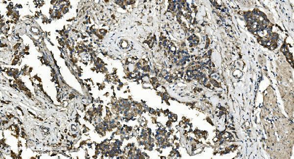
Expected resultFLCN-positive staining in respiratory epithelial cells of bronchus (HPA tissue IHC: High). HPA tissue profile: Ubiquitous cytoplasmic expression. No signal in the no-primary control.
💡Decision noteStart with heat-mediated EDTA pH 8.0 for the catalog antibody (datasheet: A00718-1); optimize retrieval for the tissue and antibody used (standard IHC practice).
Section 2

What Is the Expected FLCN Staining Pattern?

FLCN should appear mainly in the cytoplasm of many cell types, with strong IHC staining reported in respiratory epithelial cells and elongated or late spermatids (HPA tissue IHC: ubiquitous cytoplasmic expression; High in those cells). HPA rates the tissue pattern Supported, with medium consistency between staining and RNA data (HPA tissue IHC). Cytosolic and lysosome membrane localization is reported, but FLCN has no transmembrane segment (UniProt Q8NFG4: subcellular location; topology).

What am I looking at on my slide?
Cytoplasmic staining is strong in bronchial or nasopharyngeal respiratory epithelium, or in elongated or late spermatids (HPA tissue IHC: High in these cells).This matches the reported IHC pattern. Compare signal within the named cell population and its neighbors; widespread cytoplasmic expression means staining outside these examples can also be genuine (HPA tissue IHC: ubiquitous cytoplasmic expression; standard IHC interpretation).
Staining appears exclusively as a sharp cell border or uniformly strong nuclei, with little cytoplasmic signal (HPA tissue IHC: cytoplasmic profile).Treat this as discordant with the predominant IHC pattern and check controls. Nuclear, lysosomal and other locations are reported, so a secondary compartment alone does not prove artefact (UniProt Q8NFG4: subcellular location; HPA subcellular: plasma membrane uncertain).
A low-staining population, such as hippocampal glia or soft-tissue fibroblasts, stains much more strongly than neighboring expected-positive cells (HPA tissue IHC: Low in these populations).Consider cross-reactivity or endogenous chromogen-generating activity, then check antibody-omission controls and cell identity (standard IHC practice). Low HPA staining is not evidence that every cell in that population must be negative (HPA tissue IHC: Low).
Diffuse color covers stroma, lumens or much of the section without clear cellular boundaries (standard IHC interpretation).The distribution is difficult to score as FLCN because the reported pattern is cellular and mainly cytoplasmic (HPA tissue IHC: ubiquitous cytoplasmic expression). Check the detection control and background-reduction steps before assigning positivity (standard IHC practice).
No signal appears in an intact bronchial or nasopharyngeal respiratory epithelium on the same run (HPA tissue IHC: High in respiratory epithelial cells).First suspect an assay or detection failure, including antibody, retrieval or chromogen conditions (standard IHC practice). HPA provides a useful expected-positive reference, but its Supported rating reflects medium agreement with RNA data, not guaranteed staining in every specimen (HPA tissue IHC: reliability description).
💡Expected FLCN appearanceCall a section positive when identifiable respiratory epithelial cells or elongated or late spermatids show clear cytoplasmic staining, typically strong in these HPA examples; isolated uniform nuclear, edge-only or diffuse noncellular color warrants review (HPA tissue IHC: High and ubiquitous cytoplasmic expression; UniProt Q8NFG4: nuclear localization; standard IHC interpretation).
How each factor affects the staining
Tissue and cell selection (HPA tissue IHC: High, Medium and Low cell-level entries).Bronchial or nasopharyngeal respiratory epithelium and elongated or late spermatids offer strong reference patterns; adipocytes and several glandular or neuronal populations are Medium, while listed glia, fibroblasts and hematopoietic cells are Low (HPA tissue IHC). Score the named cells, not the whole tissue (standard IHC practice).
Subcellular distribution and nutrient state (UniProt Q8NFG4: subcellular location).FLCN can associate with lysosome membranes during amino acid depletion and move toward the cytosol after refeeding (UniProt Q8NFG4: subcellular location). Routine tissue IHC chiefly supports a cytoplasmic call; it does not establish the specimen's nutrient state (HPA tissue IHC: cytoplasmic profile; standard IHC interpretation).
Topology and secondary locations (UniProt Q8NFG4: topology and subcellular location).FLCN has no transmembrane segment, although lysosome membrane, nucleus, cilium, centrosome and spindle locations are reported (UniProt Q8NFG4). A membrane-like or nuclear accent needs context; HPA's plasma membrane IF assignment is uncertain (HPA subcellular).
Antibody evidence (HPA antibodies: HPA028760 and CAB015158).Both listed antibodies have Supported IHC status; HPA028760 also has Supported ICC status (HPA antibodies). The supplied record does not state an Enhanced IHC designation or establish that every antibody reproduces the same intensity (HPA antibodies; HPA tissue IHC: Supported).
Why is my staining missing, weak or wrong?
SituationLikely causeNext action
Expected-positive respiratory epithelium is blank (HPA tissue IHC: High in bronchus and nasopharynx).Assay failure or insufficient staining conditions are possible; the tissue result alone does not identify which step failed (standard IHC practice).Confirm section integrity and controls, then review retrieval, antibody dilution, detection reagent and chromogen steps against the catalog antibody's IHC-P procedure (standard IHC practice).
Color is diffuse across cells and empty spaces (standard IHC interpretation).Nonspecific binding or excess chromogen/background is possible (standard IHC practice).Compare an antibody-omission control; review blocking, washes, dilution and development time before scoring cytoplasmic cells (standard IHC practice).
Strong nuclear-only staining dominates a section (HPA tissue IHC: cytoplasmic profile).This differs from the principal tissue IHC pattern, though nuclear localization is listed for FLCN (UniProt Q8NFG4: nucleus).Check whether cytoplasmic signal is present in expected-positive cells and compare controls; avoid calling every nuclear signal false solely from its compartment (HPA tissue IHC; standard IHC practice).
Low-listed cells stain strongly while expected-positive cells do not (HPA tissue IHC: Low and High cell-level entries).Cell misidentification, cross-reactivity or endogenous detection activity may explain the mismatch (standard IHC practice).Recheck morphology and an antibody-omission control, then compare with a listed Supported IHC antibody if available (HPA antibodies: IHC Supported; standard IHC practice).
Puncta or cell-edge color appears without a clear cytoplasmic pattern (HPA tissue IHC: cytoplasmic profile).Lysosome association is biologically plausible, while HPA rates plasma membrane IF localization uncertain (UniProt Q8NFG4: lysosome membrane; HPA subcellular).Interpret the whole cell and compare expected-positive tissue and controls before assigning FLCN; record puncta separately from diffuse cytoplasmic staining (standard IHC interpretation).
IF/ICC: where should FLCN appear (HPA subcellular: ICC-IF)?HPA reports mainly cytosolic signal, with vesicles as an additional approved location and plasma membrane as uncertain (HPA subcellular).Use cytosol as the main localization reference; assess vesicles as secondary and interpret apparent plasma membrane signal cautiously (HPA subcellular). Consult the separate IF/ICC guide for assay setup.

Sample controls for FLCN IHC & IF

🧪Run bronchus first and score respiratory epithelial cells for FLCN staining (HPA: High in bronchus respiratory epithelial cells). HPA detects FLCN in all 45 scored tissues, so there is no HPA-supported negative tissue; use no-primary and isotype controls to define background, and treat cells without visible staining on the positive slide as internal background comparators rather than proven FLCN-negative cells (HPA: no negative rows; FLCN detected in all 45 scored tissues).
Positive control tissue: Bronchus (Respiratory epithelial cells, HPA High)
Negative control tissue: None in HPA: FLCN is detected in all 45 scored tissues. Use a no-primary (secondary-only) and an isotype control instead.
ICC-IF cell lines (HPA subcellular resource): HPA ICC-IF images show FLCN in A-431, U-251MG, U2OS, RPTEC/TERT1, hTERT-RPE1 (serum starved), KOLF2.1J, with annotated localisation: Cytosol (supported) (HPA subcellular).
Technical controls: Include a no-primary, secondary-only slide and a concentration-matched rabbit IgG isotype slide alongside the rabbit catalog antibody, and use FLCN knockout material as a biological specificity control if available (catalog caption: rabbit anti-FLCN antibody). For chromogenic detection in bronchus, quench endogenous peroxidase and check for background from endogenous biotin when using the caption’s biotinylated secondary and streptavidin–biotin detection (catalog caption: biotinylated goat anti-rabbit IgG, SABC and DAB).
⚠️Feasibility: A target-specific fixation window or fixation effect is unreported in the supplied evidence; the selected-SKU paraffin-section caption does not state the fixative (catalog caption: fixative not stated). The documented IHC example uses heat retrieval in EDTA at pH 8.0 and 1 μg/ml primary antibody, but it does not establish whether FLCN staining depends on that retrieval condition (catalog caption: EDTA retrieval and antibody concentration). There is no matched evidence that frozen sections or IF are easier; for bronchus, check blood and inflammatory cells for peroxidase background when interpreting DAB staining (HPA: High in bronchus respiratory epithelial cells; catalog caption: DAB detection).

HPA tissue IHC evidence for FLCN

Comprehensive Human Protein Atlas IHC scoring per tissue (reliability: Supported — Medium consistency between antibody staining and RNA expression data.). Rows are taken directly from the HPA tissue chart — click any row's HPA link to view the source.

Positive expression · recommended positive controls

TissueCell typeLevelEvidenceSource
Bronchus Respiratory epithelial cells High Protein (IHC) HPA →
Nasopharynx Respiratory epithelial cells High Protein (IHC) HPA →
Testis Elongated or late spermatids High Protein (IHC) HPA →
Adipose tissue Adipocytes Medium Protein (IHC) HPA →
Adrenal gland Glandular cells Medium Protein (IHC) HPA →

Undetected expression · recommended negative controls

TissueCell typeLevelEvidenceSource
None in HPA: FLCN is detected in all 45 scored tissues. Use a no-primary (secondary-only) and an isotype control instead.
Section 3

Advanced FLCN IHC Tips

Troubleshoot FLCN staining by separating tissue processing effects from its variable cytosolic, lysosomal and nuclear distribution (UniProt Q8NFG4).

Which retrieval conditions should I try first when FLCN staining is weak?
Start with heat-mediated antigen retrieval in EDTA at pH 8.0 for paraffin sections (datasheet A00718-1). The selected tissue image used that retrieval, followed by 1 μg/ml primary antibody overnight at 4°C (datasheet A00718-1). If staining remains weak, compare retrieval duration on adjacent sections while keeping antibody concentration, detection and development time constant (standard IHC practice). Score respiratory epithelial cells as a positive reference, since high staining is reported in bronchus and nasopharynx (HPA tissue IHC). Check whether signal stays predominantly cytoplasmic; stronger background throughout the section does not establish improved FLCN detection (HPA tissue IHC; standard IHC practice).
Could fixation explain inconsistent FLCN staining between paraffin blocks?
Target-specific FLCN sensitivity to fixation is unknown: the selected paraffin-section caption does not report a fixative (datasheet A00718-1). Record each block’s fixative and fixation interval, then compare matched sections using the same EDTA pH 8.0 retrieval and 1 μg/ml primary antibody conditions (datasheet A00718-1; standard IHC practice). Assess preservation of cell outlines alongside staining, because poor morphology can make compartment scoring unreliable (standard IHC practice). Include a section with a known staining pattern in the same run, and avoid attributing a block difference to fixation until retrieval, detection and tissue composition have been checked (HPA tissue IHC; standard IHC practice).
How should I assess punctate or nuclear FLCN staining in tissue?
Expect broadly cytoplasmic staining across tissues as the starting pattern for chromogenic IHC (HPA tissue IHC). Puncta may warrant closer examination because FLCN localizes to the lysosome membrane during amino acid depletion and shifts toward cytosol after refeeding (UniProt Q8NFG4). Nuclear, ciliary, centrosomal and spindle localization are also reported, so a signal outside diffuse cytoplasm needs context rather than automatic rejection (UniProt Q8NFG4). Compare adjacent sections processed with the same EDTA pH 8.0 retrieval and 1 μg/ml antibody incubation, then document which cells and compartments stain (datasheet A00718-1; standard IHC practice).
Can absent staining be explained by an FLCN isoform or masked epitope?
FLCN has 3 reported isoforms, but the supplied tissue-IHC caption does not identify this antibody’s epitope or isoform coverage (UniProt Q8NFG4; datasheet A00718-1). Do not assign an unstained cell population to a particular isoform without epitope mapping and independent expression evidence (standard IHC practice). FLCN contains uDENN and cDENN regions and several reported phosphorylation sites; these annotations alone do not establish whether tissue processing masks this antibody’s epitope (UniProt Q8NFG4). First repeat the documented EDTA pH 8.0 retrieval and 1 μg/ml overnight incubation, then compare a separately validated antibody if the question remains (datasheet A00718-1; standard IHC practice).
How can IF help resolve an ambiguous chromogenic FLCN pattern?
Use IF as a separate validation experiment, since the supplied 1 μg/ml overnight protocol documents paraffin-section chromogenic IHC, not IF performance (datasheet A00718-1). Multiplex FLCN with a marker for the expected cell type, such as a respiratory epithelial marker when examining bronchus, where those cells show high FLCN staining (HPA tissue IHC; standard IF practice). Choose a fluorophore channel after checking unstained tissue for autofluorescence, and include single-label controls to assess channel spillover (standard IF practice). Because FLCN has no transmembrane segment and can associate with lysosomes, optimize permeabilisation for intracellular epitope access without assuming a membrane-facing epitope position (UniProt Q8NFG4; standard IF practice).
How do I distinguish widespread FLCN expression from nonspecific brown staining?
Widespread cytoplasmic signal can be plausible because tissue IHC reports ubiquitous cytoplasmic FLCN expression, with only medium consistency between antibody staining and RNA data (HPA tissue IHC). Run a no-primary control through secondary antibody, detection reagent and DAB steps to reveal reagent-associated staining (standard IHC practice). The selected image used 10% goat serum blocking, a biotinylated secondary antibody and DAB development; match those steps when troubleshooting its pattern (datasheet A00718-1). Compare staining within intact cells against blank spaces, necrotic areas and section edges, and shorten chromogen development if diffuse deposit obscures cellular boundaries (standard IHC practice).
What is a defensible way to quantify FLCN across tissue sections? ⚠ ANSWER MARKED FOR VERIFICATION
Define the cell population and cytoplasmic compartment before scoring, because the reported tissue-IHC profile is ubiquitous and cytoplasmic (HPA tissue IHC). Record the percentage of positive eligible cells and an H-score calculated from intensity categories 0–3, giving a possible range of 0–300 (standard IHC practice). If counting positive cells per mm² instead, normalize to evaluable tissue area and report cell density separately when tissue composition differs (standard IHC practice). Keep retrieval at EDTA pH 8.0 and apply the same detection and DAB timing across compared sections; exclude damaged or necrotic regions consistently (datasheet A00718-1; standard IHC practice).
Which FLCN staining features suggest a true signal rather than artefact?
A convincing result identifies stained, intact cells and a reproducible cellular pattern, with cytoplasm as the main tissue-IHC reference compartment (HPA tissue IHC; standard IHC practice). High staining in bronchial or nasopharyngeal respiratory epithelial cells is a useful comparison, while some other cell populations have low reported staining (HPA tissue IHC). Nuclear or punctate signal is not automatically false because FLCN has reported nuclear and lysosome-associated localization; assess it against matched controls and tissue context (UniProt Q8NFG4; standard IHC practice). Reject conclusions based solely on section-edge color, necrotic deposits or signal reproduced by a no-primary control, which can reveal endogenous enzyme or detection background (standard IHC practice).
Boster reagents

Best FLCN / Folliculin IHC Antibodies

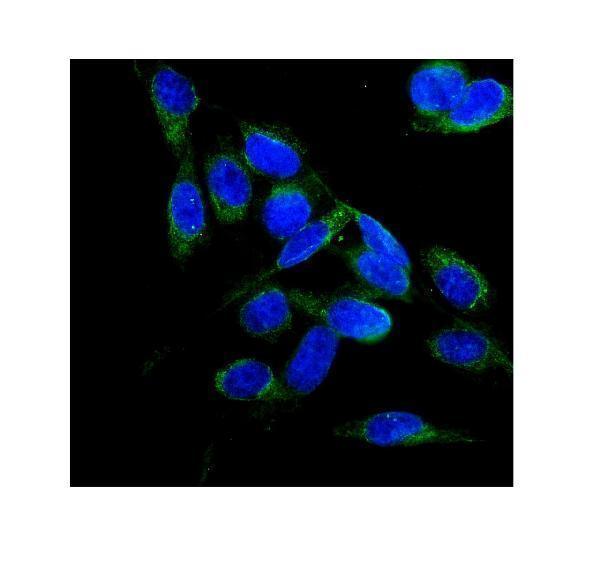
A00718-1 has pictured IHC staining in human paraffin tissue and IF staining in U20S cells (catalog image captions); M00718 lists IHC and IF/ICC for human, mouse, and rat (catalog applications/reactivity).

Real IHC data IHC analysis of FLCN using anti-FLCN antibody (A00718-1). FLCN was detected in paraffin-embedded section of human bladder cancer tissue. Heat mediated antigen retrieval was performed in EDTA buffer (pH8.0, epitope retrieval solution). The tissue section was blocked with 10% goat serum. The tissue section was then incubated with 1μg/ml rabbit anti-FLCN Antibody (A00718-1) overnight at 4°C. Biotinylated goat anti-rabbit IgG was used as secondary antibody and incubated for 30 minutes at 37°C. The tissue section was developed using Strepavidin-Biotin-Complex (SABC) (Catalog # SA1022) with DAB as the chromogen.
Anti-FLCN ® Antibody
Cat # A00718-1

A00718-1 has pictured IHC results in human bladder cancer, appendicitis, and liver cancer paraffin sections, plus IF in U20S cells (catalog image captions). M00718 lists IHC and IF/ICC applications and human, mouse, and rat reactivity, but has no supplied IHC or IF image (catalog applications/reactivity and image fields).

Which to pick: For tissue IHC, choose A00718-1: its pictured human bladder cancer paraffin section used EDTA pH 8 retrieval and DAB detection; the fixative is unreported (catalog IHC image caption). For IF/ICC, A00718-1 has a pictured U20S cell result, while M00718 is a rabbit monoclonal, clone 32F42, with listed IF/ICC use (catalog IF image caption; catalog clone/applications). For mouse or rat work, both list reactivity, but the supplied images document human material only (catalog reactivity and image captions).

Each figure is that product's own IHC / IF validation image from its datasheet.

References

  1. UniProt Consortium. UniProt entry Q8NFG4 (FLCN_HUMAN, Folliculin).
  2. Human Protein Atlas. FLCN tissue IHC expression (reliability: Supported).
  3. Human Protein Atlas. FLCN subcellular location (ICC-IF): Mainly localized to the cytosol. In addition localized to the plasma membrane and vesicles..
  4. Human Protein Atlas. FLCN antibody validation summary (2 antibodies).
  5. Parotid Oncocytomas Express the Glycoprotein Non-metastatic Melanoma Protein B (GPNMB): A Potential Link with Birt-Hogg-Dubé Syndrome. Head and neck pathology 2026 — PMC13269580.
  6. Oncocytoma-like renal tumor with transformation toward high-grade oncocytic carcinoma: a unique case with morphologic, immunohistochemical, and genomic characterization. Medicine 2014 — PMC4616290.
  7. Clinic Examination and Gene Diagnosis for a Birt-Hogg-Dubé Syndrome Family With a Novel flcn Frameshift Mutation Causing Nonsense-Mediated mRNA Degradation. Human mutation 2025 — PMC12267975.
  8. Distinctive expression patterns of glycoprotein non-metastatic B and folliculin in renal tumors in patients with Birt-Hogg-Dubé syndrome. Cancer science 2015 — PMC4376441.
  9. PubMed PMID:12204536 — UniProt-cited evidence.
  10. PubMed PMID:14702039 — UniProt-cited evidence.
  11. PubMed PMID:16625196 — UniProt-cited evidence.