This website uses cookies to ensure you get the best experience on our website.


- Table of Contents
Plan paraffin-section IHC for FOLH1 using prostate glands, kidney proximal tubules and intestinal enterocytes as positive tissues (HPA tissue IHC). Interpret the observed cytoplasmic staining (HPA tissue IHC) alongside the annotated membrane topology and isoforms (UniProt).

Expected localisation, validated protocols, controls and antibodies — the at-a-glance facts below, then the full design guide.
| Expected localisation | Cytoplasmic staining in tissue sections (HPA tissue IHC) | |
| Staining pattern | Cytoplasm of prostate glands, kidney tubules and enterocytes (HPA tissue IHC) | |
| Antigen retrieval | Citrate pH 6.0 HIER, 95–98 °C, 20 min (rule: cytoplasmic / membrane antigen) | |
| Positive control | Duodenum+3 more · see all | |
| Negative control | Adipose tissue+4 more · see all |
| Fixation | Keep fixation consistent across sections (standard IHC practice; not target-specific). Selected-image fixative and duration unreported (datasheet M02846-9); verify before use. | |
| Caveat | Normal and primary tumor prostate favor different isoforms (UniProt) | |
| Regulation | Primary prostate tumors favor PSMA-1 (UniProt) | |
| Isoform / epitope | 8 isoforms; check whether the epitope is extracellular or cytoplasmic (UniProt) |
The catalog antibody protocol and the published ileal IHC protocol provide starting points for FOLH1 staining in paraffin sections (datasheet; PMC3351422 methods).
| Sample | Tissue sections; selected-image fixative not specified (standard IHC workflow) |
| Fixation | Image fixative and duration unreported (datasheet M02846-9); verify before use. |
| Sectioning | 4–5 µm sections on charged slides (standard) |
| Deparaffinisation | Xylene, graded ethanol series to water (standard) |
| Antigen retrieval | Heat-induced epitope retrieval in citrate buffer, pH 6.0, 20 min at 95–98 °C (standard rule: cytoplasmic / membrane antigen) |
| Peroxidase block | 3% H2O2, 10 min, room temperature (standard) |
| Blocking | 10% normal serum of the secondary host, 30 min, room temperature (standard) |
| Primary antibody | Rabbit anti-FOLH1, 1:200-1:1000 (datasheet M02846-9) |
| Primary incubation | Overnight at 4 °C (standard) |
| Detection | HRP-polymer secondary, DAB chromogen 5–10 min (standard) |
| Counterstain | Hematoxylin, blue, dehydrate and mount (standard) |
| Expected result | FOLH1-positive staining in enterocytes of duodenum (HPA tissue IHC: High). HPA tissue profile: Selective cytoplasmic expression in prostate, kidney tubules, duodenum and small intestine. No signal in the no-primary control. |
FOLH1 is a membrane protein with an extracellular region and a short cytoplasmic tail (UniProt Q04609 topology). In paraffin-section IHC, expect strong staining in prostate glandular cells, kidney proximal-tubule microvilli, and duodenal and small-intestinal enterocytes (HPA tissue IHC: High). HPA also describes selective cytoplasmic expression in these tissues and rates the tissue staining profile Enhanced for consistency with RNA expression (HPA tissue IHC).
| Strong staining in prostate glandular cells, proximal-tubule microvilli, or intestinal enterocytes. | This matches the reported high-expression cell types (HPA tissue IHC: High). Record the stained cell type and whether signal appears at an apical edge, membrane, or within cytoplasm; HPA reports cytoplasmic IHC staining, while UniProt assigns FOLH1 to the membrane and cytoplasm (HPA tissue IHC; UniProt Q04609 subcellular location). |
| Predominantly nuclear staining in the expected positive cells. | Nuclear localisation falls outside the reported membrane and cytoplasmic locations (UniProt Q04609 subcellular location; HPA subcellular: Membrane). Treat a nuclear-only pattern as suspect and check antibody specificity and detection controls before scoring it as FOLH1 (general IHC practice). |
| Strong staining in a cell type reported as undetected, such as adipocytes. | Adipocytes are reported as Not detected (HPA tissue IHC: adipocytes). Consider cross-reactivity or endogenous detection activity, then compare with a no-primary control and an IHC-validated antibody (general IHC practice; HPA antibodies: IHC Enhanced). A single unexpected stain does not establish new FOLH1 expression. |
| Uniform colour across cells, stroma, or tissue edges, obscuring cell boundaries. | This is less interpretable than the selective cell pattern reported by HPA (HPA tissue IHC). Assess nonspecific background with a no-primary control; review blocking, wash steps, and chromogen development under the chosen IHC workflow (general IHC practice). |
| No staining in prostate glandular cells or the other reported high-expression cells. | A blank known-positive section conflicts with the expected high staining (HPA tissue IHC: prostate, kidney, duodenum, small intestine). Check section integrity and run conditions before calling the specimen negative; a failed positive control cannot establish FOLH1 absence (general IHC practice). |
| Which compartment should be scored? | FOLH1 has a cytoplasmic segment at residues 1–19, a transmembrane segment at 20–43, and an extracellular region at 44–750 (UniProt Q04609 topology). HPA calls the tissue IHC pattern cytoplasmic and the subcellular summary membrane (HPA tissue IHC; HPA subcellular). Assess compartment together with cell identity. |
| Which tissues provide comparison sections? | Prostate glandular cells, kidney proximal-tubule microvilli, and duodenal or small-intestinal enterocytes are High (HPA tissue IHC). Adipocytes are Not detected, while salivary glandular cells and hepatocytes are Low (HPA tissue IHC). Use the same staining run for comparisons (general IHC practice). |
| Can a tissue labelled negative rule out all expression? | No. Adrenal glandular cells are Not detected by HPA tissue IHC, while UniProt reports FOLH1 protein detected in adrenal gland (HPA tissue IHC; UniProt Q04609 tissue specificity). Interpret an adrenal result in its assay and cell-type context; these sources do not give a target-specific fixation explanation. |
| Do isoforms change the expected IHC call? | UniProt lists 8 isoforms and reports PSMA' as most abundant in normal prostate, PSMA-1 in primary prostate tumors, and PSMA-9 specifically in prostate cancer (UniProt Q04609 isoforms; tissue specificity). The payload does not identify this antibody's epitope or establish isoform-specific IHC staining. |
| How strong is the supplied antibody evidence? | Both listed antibodies, HPA010593 and CAB001451, have IHC Enhanced status (HPA antibodies). This supports the reported IHC pattern through independent antibodies or orthogonal data (HPA antibodies: IHC Enhanced); it does not validate every specimen or identify the catalog antibody's epitope. |
| What should the IF/ICC result look like? | HPA summarizes FOLH1 as Membrane in its subcellular entry, but provides no main location or ICC-IF image cell lines here (HPA subcellular). UniProt also lists cytoplasm (UniProt Q04609 subcellular location). Treat this as localisation context for the separate IF/ICC guide, without inferring an IF protocol or validated image pattern. |
| Situation | Likely cause | Next action |
|---|---|---|
| The prostate positive control is blank. | The section or staining run may have failed; HPA reports High staining in prostate glandular cells (HPA tissue IHC). | Repeat with a suitable positive section and verify antibody dilution, retrieval, detection reagents, and counterstain against the chosen IHC protocol (general IHC practice). Do not score test sections as negative until the positive control works. |
| Staining appears mainly in nuclei. | A nuclear-only pattern conflicts with the reported membrane and cytoplasmic locations (UniProt Q04609 subcellular location; HPA subcellular). | Inspect the no-primary control, review chromogen deposits and counterstain, and compare the result with an IHC-validated antibody if available (general IHC practice; HPA antibodies: IHC Enhanced). |
| Many unrelated cells stain as strongly as the expected cells. | The broad pattern differs from HPA's selective cytoplasmic tissue profile and may reflect nonspecific staining or endogenous detection activity (HPA tissue IHC; general IHC practice). | Compare positive and reported undetected cell types on the same run; check a no-primary control, blocking, and detection steps before assigning FOLH1 positivity (HPA tissue IHC; general IHC practice). |
| Weak, widespread colour obscures proximal-tubule or enterocyte staining. | Background can mask the reported High signal in proximal-tubule microvilli and enterocytes (HPA tissue IHC; general IHC practice). | Use the no-primary control to locate background; review washes, antibody dilution, and chromogen development under the chosen IHC protocol (general IHC practice). Reassess the expected cell boundaries afterward (HPA tissue IHC). |
| Adrenal glandular cells stain despite an HPA Not detected entry. | The sources differ in scope: HPA reports those cells Not detected by tissue IHC, while UniProt reports adrenal protein detection (HPA tissue IHC; UniProt Q04609 tissue specificity). | Document the cell type and controls, then compare with a reported High tissue and an independent IHC-validated antibody where available (HPA tissue IHC; HPA antibodies: IHC Enhanced). Avoid assigning a fixation cause from these records. |
| An IF/ICC image seems cytoplasmic rather than sharply membranous. | The supplied IF record has a Membrane summary but no ICC-IF image cell lines or main-location detail; UniProt also lists cytoplasm (HPA subcellular; UniProt Q04609 subcellular location). | Use the separate IF/ICC guide to assess its own controls and imaging conditions (general IF practice). Do not treat the paraffin IHC tissue pattern or IHC antibody status as direct IF validation (HPA tissue IHC; HPA antibodies). |
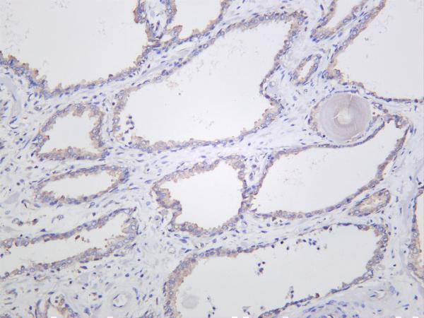
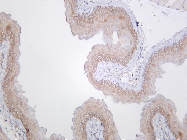


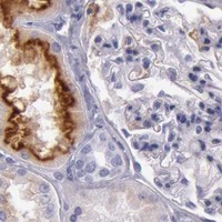
Comprehensive Human Protein Atlas IHC scoring per tissue (reliability: Enhanced — High consistency between antibody staining and RNA expression data.). Rows are taken directly from the HPA tissue chart — click any row's HPA link to view the source.
| Tissue | Cell type | Level | Evidence | Source |
|---|---|---|---|---|
| Duodenum | Enterocytes | High | Protein (IHC) | HPA → |
| Kidney | Proximal tubules (microvilli) | High | Protein (IHC) | HPA → |
| Prostate | Glandular cells | High | Protein (IHC) | HPA → |
| Small intestine | Enterocytes | High | Protein (IHC) | HPA → |
| Tissue | Cell type | Level | Evidence | Source |
|---|---|---|---|---|
| Adipose tissue | Adipocytes | Not detected | Protein (IHC) | HPA → |
| Adrenal gland | Glandular cells | Not detected | Protein (IHC) | HPA → |
| Appendix | Endocrine cells | Not detected | Protein (IHC) | HPA → |
| Bone marrow | Hematopoietic cells | Not detected | Protein (IHC) | HPA → |
| Bronchus | Respiratory epithelial cells | Not detected | Protein (IHC) | HPA → |
These checks help troubleshoot FOLH1 staining in paraffin sections while keeping observed tissue patterns distinct from antibody validation and processing evidence.
Catalog anti-FOLH1 antibodies have human prostate IHC images (catalog IHC captions); M02846-9 also lists IF and human, mouse, and rat reactivity (catalog applications/reactivity).
M02846-9 will render with an IHC image of human prostate; its listed applications include IHC and IF, with human, mouse, and rat reactivity (catalog IHC caption; catalog applications/reactivity). M02846 will render with an IHC image of paraffin-embedded human prostate carcinoma; it lists IHC and human reactivity (catalog IHC caption; catalog applications/reactivity).
Which to pick: For paraffin-section tissue IHC, choose M02846: its own image caption specifies paraffin-embedded human prostate carcinoma, while the fixative is unreported (catalog IHC caption); its listed IHC dilution is 1:50 (catalog dilution). For IF/ICC, choose M02846-9 because IF is a listed application at 1:200–1:1000 (catalog applications/dilution); its own IHC caption says human prostate but does not report section processing or fixative (catalog IHC caption). For cross-species work, M02846-9 lists human, mouse, and rat reactivity and is described as a recombinant rabbit monoclonal antibody (catalog reactivity/description).