This website uses cookies to ensure you get the best experience on our website.


- Table of Contents
Plan chromogenic FUS IHC in paraffin sections, from fixation and antigen retrieval through nuclear scoring. Expect widespread nuclear staining across tissue cell types (HPA tissue IHC).

Expected localisation, validated protocols, controls and antibodies — the at-a-glance facts below, then the full design guide.
| Expected localisation | Nuclear staining (HPA tissue IHC) | |
| Staining pattern | Widespread nuclear staining across cell types (HPA tissue IHC) | |
| Antigen retrieval | EDTA pH 8.0 HIER, heat-mediated (datasheet A00771-3) | |
| Positive control | Adipose tissue+4 more · see all | |
| Negative control | None in HPA (detected in all 45 tissues); use no-primary + isotype controls |
| Fixation | Keep fixation conditions consistent across sections (standard IHC practice; not target-specific) | |
| Caveat | Widespread nuclear signal can complicate cell-specific scoring (HPA tissue IHC) | |
| Regulation | Low tissue specificity (HPA tissue RNA) | |
| Isoform / epitope | Long and Short isoforms; check epitope coverage (UniProt) |
The catalog antibody protocol is paired with published FUS IHC methods for post-mortem CNS tissue and paraffin sections (PMC12407188; PMC2864899; PMC3319073).
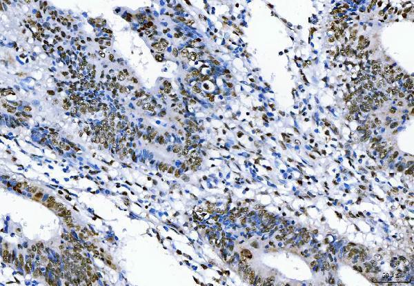
| Sample | Paraffin-embedded human rectal cancer tissue; fixative not specified (datasheet A00771-3) |
| Fixation | Image fixative and duration unreported (datasheet A00771-3); verify before use. |
| Sectioning | 4–5 µm sections on charged slides (standard) |
| Deparaffinisation | Xylene, graded ethanol series to water (standard) |
| Antigen retrieval | Heat retrieval: EDTA pH 8.0 (datasheet A00771-3); 20 min, 95–100 °C (standard) |
| Peroxidase block | 3% H2O2, 10 min, room temperature (standard) |
| Blocking | 10% goat serum (datasheet A00771-3) |
| Primary antibody | Rabbit anti-FUS, 2 μg/ml (datasheet A00771-3) |
| Primary incubation | Overnight at 4 °C (datasheet A00771-3) |
| Detection | HRP-conjugated secondary, DAB chromogen (datasheet A00771-3) |
| Counterstain | Hematoxylin, blue, dehydrate and mount (standard) |
| Expected result | FUS-positive staining in adipocytes of adipose tissue (HPA tissue IHC: High). HPA tissue profile: Ubiquitous nuclear expression. No signal in the no-primary control. |
FUS should stain predominantly in nuclei across many cell types in paraffin sections: HPA describes ubiquitous nuclear expression with supported tissue IHC reliability and low tissue RNA specificity (HPA: tissue IHC). UniProt places FUS in the nucleus, where it can appear punctate and is excluded from nucleoli (UniProt P35637: subcellular location). FUS has no transmembrane segment, so membrane staining is unexpected (UniProt P35637: topology).
| Nuclear chromogen in adipocytes, marrow hematopoietic cells, or brain glial cells. | This matches reported high staining in those cells (HPA: tissue IHC). Assess the fraction of stained nuclei and their intensity against an appropriate reference section; HPA's 'High' category does not set a numerical scoring cutoff. |
| Predominantly membranous or cytoplasmic staining, with little nuclear signal. | The compartment conflicts with HPA's ubiquitous nuclear IHC profile and UniProt's nuclear location and lack of a transmembrane segment (HPA: tissue IHC; UniProt P35637: subcellular location, topology). Check morphology and detection controls before calling it FUS. |
| Strong staining in a cell population absent from the matched reference pattern. | Cross-reactivity or endogenous detection activity is possible; verify with a reagent control and a second validated antibody. FUS is broadly expressed, and HPA lists no negative tissues, so an unfamiliar positive cell type alone cannot establish a false positive (HPA: tissue IHC, antibodies). |
| Hazy chromogen across nuclei, cytoplasm, and surrounding tissue. | Poor compartment definition prevents confident scoring against the expected nuclear pattern (HPA: tissue IHC). General IHC practice: inspect reagent controls and review blocking, washes, antibody concentration, and detection conditions to locate background. |
| No nuclear signal in a known-positive reference section. | An absent signal conflicts with high staining reported in selected reference cells, such as marrow hematopoietic cells or cerebellar granular-layer cells (HPA: tissue IHC). First check reference morphology and run controls; general IHC practice is to review retrieval, antibody dilution, and detection. |
| Cellular location and topology | HPA reports ubiquitous nuclear IHC staining, while UniProt describes punctate nuclear FUS that excludes nucleoli and has no transmembrane segment (HPA: tissue IHC; UniProt P35637: subcellular location, topology). Use nuclear location as the primary interpretation criterion; fine puncta may be difficult to resolve with chromogenic IHC. |
| Tissue and cell context | HPA reports high staining in adipocytes, adrenal glandular cells, bronchial respiratory epithelial cells, caudate glial cells, and other listed populations (HPA: tissue IHC). Its positive examples guide reference selection; the supplied HPA record lists no negative tissue or cell population, so do not use one as a required negative control. |
| Antibody validation | HPA lists three antibodies with supported IHC status: HPA008784, CAB033036, and CAB058691 (HPA: antibodies). That supports checking the nuclear pattern with an independently validated reagent when a result is disputed. The supplied validation summary does not establish that every catalog antibody recognizes the same epitope. |
| Isoforms and sequence features | UniProt lists long and short FUS isoforms, one protein chain spanning residues 1–526, and an RNA-recognition motif at residues 285–371 (UniProt P35637: isoforms, processing, domains). Without an antibody epitope in the supplied record, these features do not predict isoform-specific staining or justify an antigen-retrieval setting. |
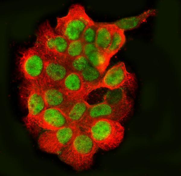
| IF/ICC Q&A: where should signal appear? | In IF/ICC, expect nucleoplasmic FUS; HPA supports that location and lists U2OS, A-431, and U-251MG image examples (HPA: subcellular ICC-IF). UniProt additionally describes punctate nuclear distribution with nucleolar exclusion (UniProt P35637: subcellular location). Interpret that localization in the separate IF/ICC guide. |
| Situation | Likely cause | Next action |
|---|---|---|
| A reference section shows weak or absent nuclear chromogen. | The result is inconsistent with HPA's high staining in the chosen listed cell population (HPA: tissue IHC); the assay step responsible is not identifiable from that observation alone. | Confirm that the reference contains the expected cells. As general IHC checks, review retrieval, antibody dilution, detection reagents, and a working positive control. |
| Cytoplasmic or membrane signal dominates. | That distribution conflicts with the reported nuclear pattern and absence of a transmembrane segment (HPA: tissue IHC; UniProt P35637: subcellular location, topology). | Compare the stained section with morphology and a reagent control; repeat with an independently IHC-supported antibody if the compartment discrepancy persists (HPA: antibodies). |
| Broad staining obscures nuclear boundaries. | The signal cannot be assigned reliably to the expected nuclear compartment (HPA: tissue IHC). General IHC causes to investigate include concentrated antibody or nonspecific detection background. | Use reagent controls to locate the background; review antibody dilution, blocking, washes, and detection conditions as general IHC practice. |
| An unexpected cell population stains strongly. | Cross-reactivity or endogenous detection activity may contribute, but HPA reports ubiquitous nuclear expression and supplies no negative population (HPA: tissue IHC). | Check whether staining remains nuclear, inspect a detection control, and compare with a second IHC-supported antibody (HPA: antibodies) before rejecting the cell population. |
| Two antibodies produce different staining patterns. | HPA supports IHC use for HPA008784, CAB033036, and CAB058691, but the supplied record gives no shared epitope or isoform coverage (HPA: antibodies; UniProt P35637: isoforms). | Compare the same reference tissue and nuclear compartment under controlled conditions; document each reagent's result without assigning the difference to a specific isoform. |
| A proposed retrieval change appears to restore signal. | The supplied HPA and UniProt records do not establish FUS-specific fixation sensitivity or an optimal retrieval condition. | Treat retrieval as a general IHC variable: compare sections with matched positive and reagent controls, then score nuclear localization against the HPA pattern (HPA: tissue IHC). |


Comprehensive Human Protein Atlas IHC scoring per tissue (reliability: Supported — High consistency between antibody staining and RNA expression data.). Rows are taken directly from the HPA tissue chart — click any row's HPA link to view the source.
| Tissue | Cell type | Level | Evidence | Source |
|---|---|---|---|---|
| Adipose tissue | Adipocytes | High | Protein (IHC) | HPA → |
| Adrenal gland | Glandular cells | High | Protein (IHC) | HPA → |
| Appendix | Endocrine cells | High | Protein (IHC) | HPA → |
| Bone marrow | Hematopoietic cells | High | Protein (IHC) | HPA → |
| Breast | Adipocytes | High | Protein (IHC) | HPA → |
| Tissue | Cell type | Level | Evidence | Source |
|---|---|---|---|---|
| None in HPA: FUS is detected in all 45 scored tissues. Use a no-primary (secondary-only) and an isotype control instead. | ||||
Troubleshoot FUS staining in paraffin section IHC by checking retrieval, nuclear localisation, epitope access, controls and scoring before interpreting changes in expression.
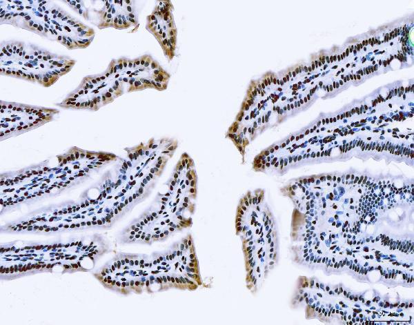
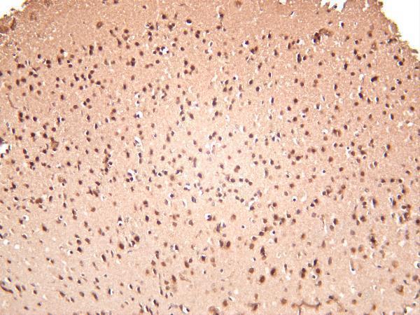
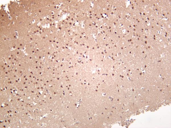
IHC images show human rectal cancer and mouse and rat colon paraffin sections, plus mouse and rat brain; an IF/ICC image shows A431 cells (catalog image captions).
A00771-3 has IHC images from human rectal cancer and mouse and rat colon paraffin sections, plus an IF/ICC image from A431 cells (A00771-3 image captions). M00771-2 has mouse and rat brain IHC images and lists human, mouse, and rat reactivity for IHC and IF (M00771-2 image captions; catalog).
Which to pick: Choose A00771-3 for paraffin-section IHC when a matching tissue image matters; its captions show human rectal cancer and mouse and rat colon, but do not report the fixative (A00771-3 IHC captions). For IF/ICC, A00771-3 has an A431-cell image, while M00771-2 lists IF without an IF image (A00771-3 IF caption; M00771-2 catalog). For cross-species IHC, A00771-3 has images from all three listed species; M00771-2 is a rabbit monoclonal with mouse and rat brain images and listed human reactivity (A00771-3 IHC captions; M00771-2 IHC captions and catalog).