GABRD / Gamma-aminobutyric acid receptor subunit delta · IHC design guide

Design Immunohistochemistry for GABRD

Plan chromogenic GABRD IHC on paraffin sections using cerebellar granule cells as a high-staining reference (HPA tissue IHC). This guide covers fixation consistency, the catalog antibody’s 1:100–1:300 IHC range (datasheet A30688), and interpretation of neuronal cytoplasmic and synaptic staining (HPA tissue IHC).

Evidence assembled Oct 2026 · For research use; verify linked source records and product datasheet before use
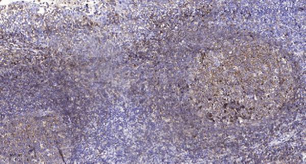
Immunohistochemistry protocol sheet for GABRD (IHC for GABRD): expected localisation Neuronal cytoplasm and synapses (HPA tissue IHC); cell membrane expected (UniProt), antibody A30688, validated IHC image, and IHC protocol steps
Printable GABRD IHC protocol sheet — expected localisation Neuronal cytoplasm and synapses (HPA tissue IHC); cell membrane expected (UniProt), antibody A30688, controls and protocol steps. Open the full GABRD IHC guide →

GABRD Immunohistochemistry Experimental Design Guide

Expected localisation, validated protocols, controls and antibodies — the at-a-glance facts below, then the full design guide.

Must know before staining
Expected localisation Neuronal cytoplasm and synapses (HPA tissue IHC); cell membrane expected (UniProt)
Staining pattern Neuronal cytoplasm and synapses; cerebellar granule cell membrane (HPA tissue IHC)
Antigen retrieval Tris-EDTA pH 9.0 HIER, heat-mediated (datasheet A30688)
Positive control ⓘ Cerebellum+4 more · see all
Negative control ⓘ Adipose tissue+4 more · see all
Important caveats
Reasons your staining may differ from the expected pattern.
Fixation Keep fixation conditions consistent across sections. (standard IHC practice; not target-specific)
Caveat Presumed off-target binding can confound staining (HPA tissue IHC)
Regulation No expression regulation annotated (UniProt)
Isoform / epitope No isoforms annotated; extracellular versus cytoplasmic epitope matters (UniProt)
Section 1

Recommended GABRD IHC & IF Protocols

The catalog antibody uses Tris-EDTA pH 9.0 retrieval (datasheet A30688); the published HCC protocol uses citrate pH 6.0 (PMC13470470).

Recommended immunohistochemistry (IHC-P) protocol parameters
SampleParaffin-embedded human tonsil tissue; fixative not specified (datasheet A30688)
FixationImage fixative and duration unreported (datasheet A30688); verify before use.
Sectioning4–5 µm sections on charged slides (standard)
DeparaffinisationXylene, graded ethanol series to water (standard)
Antigen retrievalHeat retrieval: Tris-EDTA pH 9.0 (datasheet A30688); 20 min, 95–100 °C (standard)
Peroxidase block3% H2O2, 10 min, room temperature (standard)
Blocking10% normal serum of the secondary host, 30 min, room temperature (standard)
Primary antibodyRabbit anti-GABRD, 1:100 - 1:300 (datasheet A30688)
Primary incubationOvernight at 4 °C (standard)
DetectionHRP-polymer secondary, DAB chromogen 5–10 min (standard)
CounterstainHematoxylin, blue, dehydrate and mount (standard)
Expected resultGABRD-positive staining in granular cells - cytoplasm/membrane of cerebellum (HPA tissue IHC: High). HPA tissue profile: Cytoplasmic expression in neuronal cells and synapses. No signal in the no-primary control.
💡Decision noteStart with Tris-EDTA pH 9.0 for the catalog antibody (datasheet A30688). For FFPE HCC sections, compare citrate pH 6.0 (PMC13470470).
Section 2

What Is the Expected GABRD Staining Pattern?

GABRD is a 4-pass cell membrane receptor subunit (UniProt O14764 topology). In paraffin-section IHC, expect the strongest staining in cerebellar granular cells, at the cytoplasm/membrane, with neuronal and synaptic expression elsewhere (HPA: tissue IHC). HPA rates the IHC evidence Enhanced, while reporting medium agreement with RNA data and presumed off-target staining that was disregarded (HPA: tissue IHC reliability).

What am I looking at on my slide?
Strong cytoplasmic/membrane staining in cerebellar granular cells.This matches the clearest supplied positive reference: cerebellar granular cells are High (HPA: tissue IHC). Judge the pattern within the identified cells, since HPA also describes neuronal and synaptic expression (HPA: tissue IHC profile).
Predominantly nuclear staining, without a neuronal cytoplasmic/membrane pattern.A nuclear pattern lacks support from the cell membrane topology and observed tissue profile (UniProt O14764 topology; HPA: tissue IHC). Treat it as suspect until controls and tissue morphology establish that the signal is specific.
Strong staining concentrated in an unexpected cell population.Cell identity matters: HPA reports cerebellar granular cells and selected neuronal cells, but also endocrine cells in colon, duodenum and rectum (HPA: tissue IHC). Outside those reported contexts, investigate cross-reactivity or endogenous detection activity before calling it GABRD.
Broad, even chromogen deposits obscure cells and tissue boundaries.This is difficult to score as the localized neuronal pattern reported by HPA (HPA: tissue IHC). Diffuse background can reflect nonspecific detection or inadequate background control (general IHC practice); compare a no-primary control and tissue morphology.
No convincing signal in cerebellar granular cells.That conflicts with the High cerebellar reference pattern (HPA: tissue IHC). Review section quality, detection controls and the IHC workflow before interpreting a test tissue as negative (general IHC practice).
💡Expected GABRD appearanceCall a result positive when cerebellar granular cells show strong cytoplasmic/membrane staining, with weaker context-dependent neuronal staining; isolated nuclear or widespread cell-independent deposits warrant investigation (HPA: tissue IHC; UniProt O14764 topology).
How each factor affects the staining
Cellular location and topologyGABRD has 4 transmembrane segments and a cell membrane assignment (UniProt O14764 topology). Interpret membrane-associated signal alongside HPA's observed cytoplasmic neuronal and synaptic staining; the latter does not make an exclusively nuclear pattern expected (HPA: tissue IHC).
Tissue and cell distributionCerebellar granular cells are High; caudate and hippocampal neuronal cells and selected intestinal endocrine cells are Medium (HPA: tissue IHC). Cerebral cortical neuronal cells are Low, so a uniformly strong signal across brain regions is not the supplied reference pattern (HPA: tissue IHC).
Validation limitsThe listed antibody HPA044371 is IHC Enhanced (HPA: antibody validation). HPA nevertheless reports medium staining/RNA consistency and presumed off-target binding that was disregarded; use cell type, location and controls together rather than treating intensity alone as proof (HPA: tissue IHC reliability).
Mature protein and epitope positionUniProt annotates a signal peptide at residues 1–24 and the mature chain at 25–452 (UniProt O14764 processing). Epitope position is not supplied, so these annotations cannot predict how this catalog antibody will stain a paraffin section.
Glycosylation and retrievalUniProt annotates glycosylation sites at residues 103 and 106 (UniProt O14764). Their effect on IHC staining is unreported here; antigen retrieval may be assessed as general paraffin-section practice, without assuming a GABRD-specific retrieval requirement (general IHC practice).
Why is my staining missing, weak or wrong?
SituationLikely causeNext action
Cerebellar granular cells are blank while the counterstain is visible.The result conflicts with the reported High granular-cell staining (HPA: tissue IHC); counterstaining alone does not verify antigen detection (general IHC practice).Check tissue identity and preservation, the positive-control section, primary-antibody application and detection steps; review retrieval as a general IHC variable without claiming a GABRD-specific fixation effect (general IHC practice).
Signal is nuclear rather than cytoplasmic/membranous.Nuclear localization is unsupported by the supplied membrane topology and tissue observations (UniProt O14764 topology; HPA: tissue IHC).Compare the staining with cell morphology and a no-primary control; reassess specificity if nuclei remain the dominant positive structure (general IHC practice).
Many unrelated cell types show similarly strong staining.The pattern may reflect nonspecific binding or endogenous detection activity, especially where it exceeds HPA's reported cell distribution (HPA: tissue IHC; general IHC practice).Compare a no-primary control and a tissue/cell type reported as not detected, then review blocking and detection chemistry (HPA: tissue IHC; general IHC practice).
The section has diffuse staining that masks cell boundaries.Widespread deposits cannot be assigned confidently to the neuronal and synaptic profile (HPA: tissue IHC); background from staining reagents is possible (general IHC practice).Check the no-primary control, background blocking and chromogen development; score only signal that remains localizable to identifiable cells (general IHC practice).
A negative-control tissue shows focal staining.HPA reports no detection in adipocytes of adipose tissue and several other listed cell populations (HPA: tissue IHC); focal signal may be background or an unverified cell-specific finding.Identify the stained cell type, compare its no-primary control and avoid calling it GABRD solely from a focal deposit (general IHC practice).
Does an ICC-IF Golgi or vesicle image define the IHC-P positive pattern?HPA lists Golgi apparatus and vesicles as uncertain ICC-IF locations, and rates HPA044371 ICC as Uncertain (HPA: subcellular; HPA: antibody validation).Use the tissue IHC neuronal/cytoplasmic-membrane observations to interpret paraffin sections; treat ICC-IF localization as uncertain and consult its separate guide for that application (HPA: tissue IHC; HPA: subcellular).

Sample controls for GABRD IHC & IF

🧪Run cerebellum first: granular cells should stain (HPA: High, granular cells—cytoplasm/membrane). Use adipose tissue as the negative tissue because adipocytes are listed as not detected (HPA: Not detected, adipocytes); on the cerebellar slide, compare granular cells with adjacent cells outside that population and treat any staining there as background only after checking those cells’ identity and expression.
Positive control tissue: Cerebellum (Granular cells - cytoplasm/membrane, HPA High)
Negative control tissue: Adipose tissue (HPA Not detected)
ICC-IF cell lines (HPA subcellular resource): HPA ICC-IF images show GABRD in MCF-7, SH-SY5Y, U2OS, with annotated localisation: Golgi apparatus (uncertain), Vesicles (uncertain) (HPA subcellular).
Technical controls: Include a no-primary (secondary-only) control, an isotype control matched to the primary antibody’s host species and clonality, and a GABRD knockout biological control where available (standard IHC practice). For chromogenic cerebellar sections, quench endogenous peroxidase and inspect background staining before scoring (standard IHC practice).
⚠️Feasibility: A target-specific fixation window or fixation effect is unreported, and the selected A30688 paraffin-section caption does not state a fixative (selected-SKU caption: fixative unreported). The caption uses Tris–EDTA at pH 9.0 for antigen retrieval, but does not establish whether retrieval is required for all specimens (selected-SKU caption: Tris–Tris-EDTA, pH 9.0). Frozen sections and IF cannot be judged easier from the supplied evidence; interpret cerebellar neuronal staining against the stated granular-cell pattern, and account for autofluorescence if using IF (HPA: High, cerebellar granular cells; standard IF practice).

HPA tissue IHC evidence for GABRD

Comprehensive Human Protein Atlas IHC scoring per tissue (reliability: Enhanced — Medium consistency between antibody staining and RNA expression data. Presumed off target binding observed and disregarded.). Rows are taken directly from the HPA tissue chart — click any row's HPA link to view the source.

Positive expression · recommended positive controls

TissueCell typeLevelEvidenceSource
Cerebellum Granular cells - cytoplasm/membrane High Protein (IHC) HPA →
Caudate Neuronal cells Medium Protein (IHC) HPA →
Colon Endocrine cells Medium Protein (IHC) HPA →
Duodenum Endocrine cells Medium Protein (IHC) HPA →
Hippocampus Neuronal cells Medium Protein (IHC) HPA →

Undetected expression · recommended negative controls

TissueCell typeLevelEvidenceSource
Adipose tissue Adipocytes Not detected Protein (IHC) HPA →
Adrenal gland Glandular cells Not detected Protein (IHC) HPA →
Appendix Endocrine cells Not detected Protein (IHC) HPA →
Bone marrow Hematopoietic cells Not detected Protein (IHC) HPA →
Breast Adipocytes Not detected Protein (IHC) HPA →
Section 3

Advanced GABRD IHC Tips

Use the catalog antibody’s paraffin-section evidence as a starting point, then assess GABRD staining against cell type, compartment and controls.

Which retrieval condition should I start with for GABRD paraffin sections?
Start with heat-mediated Tris-EDTA retrieval at pH 9.0 (datasheet A30688). The selected paraffin-section image used that buffer before staining human tonsil with antibody at 1:200 overnight at 4°C (datasheet A30688). Keep heating, cooling and section handling consistent across the run, then compare signal and tissue morphology with a no-primary control (standard IHC practice). If staining is weak, test an alternative retrieval condition on adjacent sections as a fallback and assess whether it improves specific staining without increasing background (standard IHC practice). Record each condition separately because the caption reports the buffer and pH, but no heating duration (datasheet A30688).
How should I assess fixation-related loss of GABRD staining?
Target-specific sensitivity to fixation is unknown from the supplied evidence; the catalog image identifies paraffin-embedded human tonsil but does not state its fixative (datasheet A30688). Record the fixative, fixation interval and processing history for each specimen, and compare sections processed together (standard IHC practice). If signal differs between specimens, first check retrieval, section preservation and staining controls before attributing the difference to fixation (standard IHC practice). An antibody incubation of 1:200 overnight at 4°C is documented for the selected image, but it does not establish a fixation condition (datasheet A30688). Report any fixation effect as an observation from your own controlled comparison.
What staining pattern is credible for GABRD in tissue sections?
Expect GABRD interpretation to center on neuronal membranes and nearby processes: it is a cell-membrane receptor subunit with 4 transmembrane segments (UniProt O14764 topology). Tissue IHC reports high cytoplasm/membrane staining in cerebellar granular cells and medium staining in hippocampal neurons (HPA tissue IHC). HPA also describes cytoplasmic staining in neuronal cells and synapses, so chromogenic signal need not trace a crisp cell outline (HPA tissue IHC). Its Golgi and vesicle assignments are uncertain and come from subcellular imaging, so treat them as context rather than a required paraffin-section pattern (HPA subcellular). Compare cellular and neuropil staining against matched controls before scoring (standard IHC practice).
How can epitope position change interpretation of GABRD staining?
Check the antibody’s stated immunogen or mapped epitope before interpreting a negative result; none is specified in the supplied catalog caption (datasheet A30688). GABRD has an extracellular region at residues 25–251, a cytoplasmic region at 332–426, and 4 transmembrane segments (UniProt O14764 topology). Glycosylation sites at 103 and 106 and phosphoserine at 390 are annotated, but their effects on this antibody’s staining are unknown (UniProt O14764 processing and modifications). No isoforms are annotated, so do not explain discordant tissue staining by invoking a documented alternative isoform (UniProt O14764 isoforms). Compare retrieval conditions and control tissues before proposing epitope masking (standard IHC practice).
How should I design a complementary IF experiment for GABRD?
For complementary IF, multiplex GABRD with a validated neuronal marker when examining cerebellar granular cells or hippocampal neurons (HPA tissue IHC; standard IF practice). Select fluorophores after inspecting unstained sections for autofluorescence, and include single-label controls to check channel bleed-through (standard IF practice). Choose permeabilisation according to the mapped epitope: GABRD includes extracellular residues 25–251 and cytoplasmic residues 332–426, but this antibody’s epitope is unspecified (UniProt O14764 topology; datasheet A30688). Avoid assuming that the catalog paraffin-section retrieval and 1:200 dilution transfer to IF (datasheet A30688). Compare signal with no-primary controls and the expected neuronal distribution before calling colocalisation (standard IF practice; HPA tissue IHC).
How can I reduce diffuse or misleading chromogenic background?
First inspect a no-primary control and tissue edges; widespread colour in both suggests a detection or processing problem (standard IHC practice). Apply a peroxidase block before chromogenic detection, use an appropriate protein block, and rinse consistently between incubations (standard IHC practice). The selected image used primary antibody at 1:200 overnight at 4°C and secondary antibody at 1:200 for 45 minutes at room temperature; these are starting conditions for that image, not proven background limits (datasheet A30688). If background persists, titrate the primary and secondary separately while retaining the same retrieval and controls (standard IHC practice). HPA reports presumed off-target binding, so require a credible cell and compartment pattern (HPA tissue IHC).
How should I quantify GABRD staining across sections? ⚠ ANSWER MARKED FOR VERIFICATION
Define the region and cell population before scoring; cerebellar granular cells show high cytoplasm/membrane staining, while hippocampal neurons are reported at medium levels (HPA tissue IHC). For cell-based scoring, report the percentage of positive neurons and an intensity-weighted H-score using fixed thresholds across the compared sections (standard IHC practice). For process-rich regions, measure positive stain area or optical density per mm² of intact annotated tissue and report background subtraction (standard IHC practice). Normalize cell-based results to the number of evaluable target cells, rather than all nuclei in a mixed tissue field (standard IHC practice). Keep retrieval, detection and imaging settings consistent (standard IHC practice).
When should a GABRD-positive IHC result be treated cautiously?
Give greatest weight to staining that aligns with neuronal cells or processes in supported regions: HPA reports high cerebellar granular-cell and medium hippocampal neuronal staining (HPA tissue IHC). Diffuse staining in an unexpected compartment or cell population needs independent validation, particularly because presumed off-target binding was observed in the HPA assessment (HPA tissue IHC). Check section edges, damaged or necrotic areas, no-primary controls and endogenous peroxidase before assigning brown reaction product to GABRD (standard IHC practice). The receptor is membrane-associated, while tissue IHC can also show neuronal cytoplasmic and synaptic signal; compartment alone is insufficient to establish specificity (UniProt O14764 subcellular location; HPA tissue IHC). Record discordant patterns separately from confidently scored cells (standard IHC practice).
Boster reagents

Best GABRD / Gamma-aminobutyric acid receptor subunit delta IHC Antibodies

A30688 has real IHC data from paraffin-embedded human tonsil (IHC image caption). IF/ICC are listed applications, with human, mouse, and rat reactivity (catalog applications/reactivity).

Real IHC data Immunohistochemical analysis of paraffin-embedded human tonsil. 1, Antibody was diluted at 1:200(4° overnight). 2, Tris-EDTA,pH9.0 was used for antigen retrieval. 3,Secondary antibody was diluted at 1:200(room temperature, 45min).
Anti-GABRD/Gaba A Receptor Delta Antibody
Cat # A30688

A30688 will render with an IHC figure from paraffin-embedded human tonsil (IHC image caption). A30688 lists IHC, IF, and ICC applications and human, mouse, and rat reactivity; no IF figure is supplied (catalog applications/reactivity; IF image alts).

Which to pick: Choose A30688 for paraffin-section tissue IHC: its own caption reports human tonsil, Tris-EDTA pH 9.0 retrieval, and a 1:200 primary dilution overnight at 4°C (IHC image caption). For IF/ICC, A30688 lists both applications, although no IF figure is supplied (catalog applications; IF image alts). It is the listed cross-species option because the polyclonal antibody lists human, mouse, and rat reactivity; its paraffin-only caption does not report the fixative (catalog dilution_raw/reactivity; IHC image caption).

Each figure is that product's own IHC / IF validation image from its datasheet.

References

  1. UniProt Consortium. UniProt entry O14764 (GBRD_HUMAN, Gamma-aminobutyric acid receptor subunit delta).
  2. Human Protein Atlas. GABRD tissue IHC expression (reliability: Enhanced).
  3. Human Protein Atlas. GABRD subcellular location (ICC-IF): Localized to the Golgi apparatus and vesicles..
  4. Human Protein Atlas. GABRD antibody validation summary (1 antibodies).
  5. GABRD Accelerates Tumour Progression via Regulating CCND1 Signalling Pathway in Gastric Cancer. Journal of cellular and molecular medicine 2025 — PMC11947670.
  6. Neurotransmitter receptor-related gene signature as potential prognostic and therapeutic biomarkers in colorectal cancer. Frontiers in cell and developmental biology 2023 — PMC10720326.
  7. GABRD promotes hepatocellular carcinoma progression via the IL-10RA/JAK2-STAT3 signaling axis. Cell death & disease 2026 — PMC13470470.
  8. Allopregnanolone Mediates Affective Switching Through Modulation of Oscillatory States in the Basolateral Amygdala. Biological psychiatry 2022 — PMC8714669.
  9. PubMed PMID:16710414 — UniProt-cited evidence.
  10. PubMed PMID:15489334 — UniProt-cited evidence.
  11. PubMed PMID:35355020 — UniProt-cited evidence.