This website uses cookies to ensure you get the best experience on our website.


- Table of Contents
Plan GORAB paraffin-section IHC around its general cytoplasmic tissue staining (HPA tissue IHC). Colon glandular cells show high staining, while heart cardiomyocytes have no detected staining (HPA tissue IHC).

Expected localisation, validated protocols, controls and antibodies — the at-a-glance facts below, then the full design guide.
| Expected localisation | Cytoplasmic in tissue (HPA tissue IHC); Golgi/TGN expected molecularly (UniProt) | |
| Staining pattern | General cytoplasmic staining in tissue cells (HPA tissue IHC) | |
| Antigen retrieval | Tris-EDTA pH 9.0 HIER, heat-mediated (datasheet A09417) | |
| Positive control | Cerebellum+4 more · see all | |
| Negative control | Adipose tissue+4 more · see all |
| Fixation | Keep fixation consistent across sections (standard IHC practice; not target-specific) | |
| Caveat | Medium consistency between staining and RNA expression (HPA tissue IHC) | |
| Regulation | Expression regulation unreported (UniProt) | |
| Isoform / epitope | Two isoforms; epitope coverage is unknown (UniProt) |
The catalog antibody protocol uses paraffin sections (datasheet A09417). One published mouse bone immunostaining protocol provides an additional IHC reference (PMC5880397).
| Sample | Paraffin-embedded human Colon cancer tissue; fixative not specified (datasheet A09417) |
| Fixation | Image fixative and duration unreported (datasheet A09417); verify before use. |
| Sectioning | 4–5 µm sections on charged slides (standard) |
| Deparaffinisation | Xylene, graded ethanol series to water (standard) |
| Antigen retrieval | Heat retrieval: Tris-EDTA pH 9.0 (datasheet A09417); 20 min, 95–100 °C (standard) |
| Peroxidase block | 3% H2O2, 10 min, room temperature (standard) |
| Blocking | 10% normal serum of the secondary host, 30 min, room temperature (standard) |
| Primary antibody | Rabbit anti-GORAB, 1:50 recommended; image 1:200 (datasheet A09417) |
| Primary incubation | Overnight at 4 °C (standard) |
| Detection | HRP-polymer secondary, DAB chromogen 5–10 min (standard) |
| Counterstain | Hematoxylin, blue, dehydrate and mount (standard) |
| Expected result | GORAB-positive staining in cells in granular layer of cerebellum (HPA tissue IHC: High). HPA tissue profile: General cytoplasmic expression. No signal in the no-primary control. |
GORAB localizes to the cytoplasm and trans-Golgi network (UniProt Q5T7V8). In tissue IHC, HPA reports general cytoplasmic staining, with high staining in selected neuronal, epithelial, glomerular and decidual cells (HPA tissue IHC). GORAB has no transmembrane segment (UniProt Q5T7V8 topology). Treat this as a working pattern: HPA rates its tissue IHC evidence Approved, with medium agreement between staining and RNA data, pending external verification (HPA tissue IHC).
| Cytoplasmic staining, sometimes concentrated near the nucleus, in the expected cell population. | Consistent with HPA's general cytoplasmic tissue pattern and UniProt's Golgi localization (HPA tissue IHC; UniProt Q5T7V8). A Golgi-like focus is plausible, but tissue IHC alone does not prove organelle identity. |
| Predominantly nuclear staining, or staining confined to cell borders or extracellular material. | Cell-border or extracellular staining is discordant with the reported cytoplasmic and Golgi pattern (HPA tissue IHC; UniProt Q5T7V8). Nuclear signal needs caution: HPA also reports nucleoplasm and nucleoli in ICC-IF, so nuclear staining alone does not establish an artefact (HPA subcellular ICC-IF). |
| Strong staining in cells expected to be unstained, such as adipocytes or cardiomyocytes. | These cells were not detected in HPA tissue IHC (HPA tissue IHC). Consider antibody cross-reactivity or endogenous chromogen activity; review morphology and detection controls before calling the cells GORAB-positive (general IHC practice). |
| Diffuse color across cells, stroma and empty spaces, with little cellular definition. | A broad, poorly localized signal cannot establish the reported cellular pattern (HPA tissue IHC). In chromogenic IHC, nonspecific binding, endogenous enzyme activity or detection background can produce this appearance (general IHC practice). |
| No staining in an expected positive cell population. | Absence in a high-staining HPA population, such as colon glandular cells, calls for a run-level check (HPA tissue IHC). Assess tissue preservation, retrieval and detection performance before interpreting the result as biological absence (general IHC practice). |
| Tissue and cell selection | HPA reports high staining in colon and duodenal glandular cells, cerebral cortical neurons, cerebellar granular-layer cells, esophageal squamous epithelium, gallbladder glandular cells, glomerular cells and placental decidual cells (HPA tissue IHC). Compare the named cell population, not the whole section. |
| Negative comparison | HPA reports no detection in adipocytes, cardiomyocytes, skeletal myocytes, smooth muscle cells and splenic red-pulp cells (HPA tissue IHC). These are useful contextual comparisons, although absence in one sample cannot validate antibody specificity by itself (general IHC practice). |
| Localization evidence across applications | UniProt lists cytoplasm, Golgi apparatus and trans-Golgi network; HPA tissue IHC describes general cytoplasmic expression (UniProt Q5T7V8; HPA tissue IHC). HPA ICC-IF additionally reports nucleoplasm and nucleoli, so IF compartment calls should be assessed on its separate guide page (HPA subcellular ICC-IF). |
| Validation strength | HPA027250 is Approved for IHC, whereas the listed HPA027746 entry has no IHC status; both have Supported ICC status (HPA antibodies). HPA rates the tissue pattern Approved, with medium RNA agreement and external verification pending (HPA tissue IHC). |
| Protein features and epitope uncertainty | UniProt lists two isoforms, one 1–369 chain, no signal peptide, no propeptide and no annotated glycosylation sites or modified residues (UniProt Q5T7V8). The supplied record gives no antibody epitope, so isoform recognition and retrieval requirements cannot be inferred. |
| Situation | Likely cause | Next action |
|---|---|---|
| A high-staining reference population is blank. | The run may have inadequate retrieval, antibody binding or detection; HPA reports high staining in colon glandular cells (HPA tissue IHC; general IHC practice). | Check the reference slide, reagent sequence and chromogen performance; optimize retrieval against the antibody's validated IHC instructions (general IHC practice). |
| Expected cells stain, but the entire section has diffuse color. | Nonspecific binding or chromogenic detection background can obscure the cellular pattern (general IHC practice). | Review blocking, washes, detection timing and a no-primary control; score only cell-associated staining with clear morphology (general IHC practice). |
| Adipocytes or muscle cells appear strongly positive. | This conflicts with HPA's not-detected calls for adipocytes, cardiomyocytes, skeletal myocytes and smooth muscle cells (HPA tissue IHC). Cross-reactivity or endogenous activity is possible (general IHC practice). | Check cell identification and a no-primary control; reassess antibody and detection conditions before treating the signal as GORAB (general IHC practice). |
| Staining is mainly at cell borders or in extracellular material. | That distribution does not match the reported cytoplasmic and Golgi locations; UniProt reports no transmembrane segment (HPA tissue IHC; UniProt Q5T7V8 topology). | Compare with a reference population and detection control, then reassess staining localization before scoring it as positive (general IHC practice). |
| A predominantly nuclear signal conflicts with the expected tissue IHC pattern. | HPA tissue IHC describes general cytoplasmic staining, while HPA ICC-IF reports nucleoplasm as a main location (HPA tissue IHC; HPA subcellular ICC-IF). | Record nuclear and cytoplasmic staining separately. Confirm the tissue pattern with controls; use the separate IF/ICC guide for nuclear localization questions. |
| Different tissues or runs give inconsistent intensity. | HPA's Approved tissue pattern has medium agreement with RNA data and awaits external verification; intensity also depends on IHC run conditions (HPA tissue IHC; general IHC practice). | Compare the same named cell populations with a shared reference section and consistent scoring criteria across runs (HPA tissue IHC; general IHC practice). |


Comprehensive Human Protein Atlas IHC scoring per tissue (reliability: Approved — Medium consistency between antibody staining and RNA expression data. Pending external verification.). Rows are taken directly from the HPA tissue chart — click any row's HPA link to view the source.
| Tissue | Cell type | Level | Evidence | Source |
|---|---|---|---|---|
| Cerebellum | Cells in granular layer | High | Protein (IHC) | HPA → |
| Cerebral cortex | Neuronal cells | High | Protein (IHC) | HPA → |
| Colon | Glandular cells | High | Protein (IHC) | HPA → |
| Duodenum | Glandular cells | High | Protein (IHC) | HPA → |
| Esophagus | Squamous epithelial cells | High | Protein (IHC) | HPA → |
| Tissue | Cell type | Level | Evidence | Source |
|---|---|---|---|---|
| Adipose tissue | Adipocytes | Not detected | Protein (IHC) | HPA → |
| Heart muscle | Cardiomyocytes | Not detected | Protein (IHC) | HPA → |
| Skeletal muscle | Myocytes | Not detected | Protein (IHC) | HPA → |
| Smooth muscle | Smooth muscle cells | Not detected | Protein (IHC) | HPA → |
| Spleen | Cells in red pulp | Not detected | Protein (IHC) | HPA → |
Troubleshoot GORAB staining in paraffin sections by checking retrieval, cell type, compartment and controls before comparing signal intensity (datasheet A09417; HPA tissue IHC).
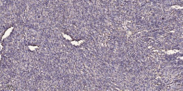
A09417 has a real IHC image from paraffin-embedded human colon cancer (catalog image caption). Human, mouse, and rat reactivity is listed (catalog reactivity); no IF image is supplied (catalog images).
A09417 is the only card and shows IHC on paraffin-embedded human colon cancer, with 1:200 primary antibody dilution and Tris-EDTA retrieval at pH 9.0 (catalog image caption). Its listed applications include IHC, and its listed reactivity covers human, mouse, and rat (catalog applications/reactivity).
Which to pick: Choose A09417, a rabbit polyclonal, for paraffin-section tissue IHC because its own image documents that preparation; the fixative is unreported (catalog dilution data; catalog image caption). No SKU has listed IF/ICC application or IF image data here (catalog applications/images). For mouse or rat IHC, A09417 lists those species as reactive, but its supplied IHC image shows human tissue only (catalog reactivity; catalog image caption).