HGH1 / Co-chaperone protein HGH1 homolog · IHC design guide

Design Immunohistochemistry for HGH1

Plan HGH1 staining in paraffin sections with a human-reactive IHC antibody at 1:100–1:300 (datasheet: A30576). Compare staining in glandular cells with undetected adipocytes, and interpret results in light of the low consistency between staining and RNA expression (HPA tissue IHC).

Evidence assembled Oct 2026 · For research use; verify linked source records and product datasheet before use
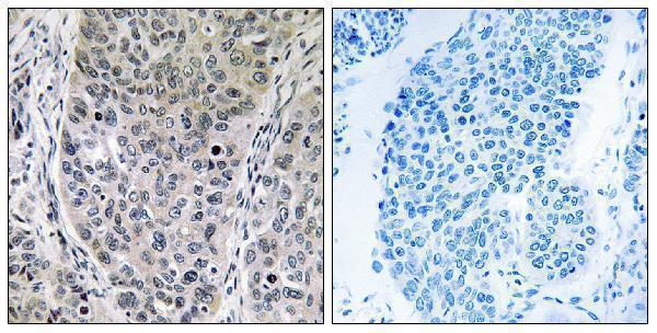
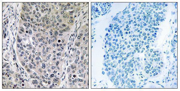
Immunohistochemistry protocol sheet for HGH1 (IHC for HGH1): expected localisation Cytoplasmic and nuclear tissue staining (HPA tissue IHC), antibody A13420-1, validated IHC image, and IHC protocol steps
Printable HGH1 IHC protocol sheet — expected localisation Cytoplasmic and nuclear tissue staining (HPA tissue IHC), antibody A13420-1, controls and protocol steps. Open the full HGH1 IHC guide →

HGH1 Immunohistochemistry Experimental Design Guide

Expected localisation, validated protocols, controls and antibodies — the at-a-glance facts below, then the full design guide.

Must know before staining
Expected localisation Cytoplasmic and nuclear tissue staining (HPA tissue IHC)
Staining pattern Glandular and other cells show cytoplasmic/nuclear staining (HPA tissue IHC)
Antigen retrieval Citrate pH 6.0 HIER, 95–98 °C, 20 min (rule: cytoplasmic / membrane antigen)
Positive control ⓘ Adrenal gland+4 more · see all
Negative control ⓘ Adipose tissue+4 more · see all
Important caveats
Reasons your staining may differ from the expected pattern.
Fixation Matched tissue-IHC evidence does not establish the fixation claim. Validate the specimen-specific method before use. (selected-SKU IHC image A13420-1)
Caveat Staining shows low consistency with RNA expression (HPA tissue IHC)
Regulation Expression regulation is unannotated (UniProt)
Isoform / epitope No isoforms annotated; chain spans residues 2–390 (UniProt)
Section 1

Recommended HGH1 IHC & IF Protocols

The catalog antibody’s IHC-P protocol is accompanied by one published HGH1 paraffin-section workflow (PMC11155144).

Recommended immunohistochemistry (IHC-P) protocol parameters
SampleParaffin-embedded human lung carcinoma tissue; fixative not specified (datasheet A13420-1)
FixationImage fixative and duration unreported (datasheet A13420-1); verify before use.
Sectioning4–5 µm sections on charged slides (standard)
DeparaffinisationXylene, graded ethanol series to water (standard)
Antigen retrievalHeat-induced epitope retrieval in citrate buffer, pH 6.0, 20 min at 95–98 °C (standard rule: cytoplasmic / membrane antigen)
Peroxidase block3% H2O2, 10 min, room temperature (standard)
Blocking10% normal serum of the secondary host, 30 min, room temperature (standard)
Primary antibodyRabbit anti-HGH1, 1:100-1:300 (datasheet A13420-1)
Primary incubationOvernight at 4 °C (standard)
DetectionHRP-polymer secondary, DAB chromogen 5–10 min (standard)
CounterstainHematoxylin, blue, dehydrate and mount (standard)
Expected resultHGH1-positive staining in glandular cells of adrenal gland (HPA tissue IHC: Medium). HPA tissue profile: Cytoplasmic and nuclear expression in most tissues. No signal in the no-primary control.
💡Decision noteStart with citrate pH 6.0 retrieval at 95–98 °C for 20 min (page retrieval rule; UniProt Q9BTY7: cytoplasm).
Section 2

What Is the Expected HGH1 Staining Pattern?

HGH1 is cytoplasmic by UniProt and has no transmembrane segment (UniProt Q9BTY7). In tissue IHC, expect cytoplasmic and sometimes nuclear staining across cell types, including medium staining in several glandular cell populations (HPA tissue IHC). Treat this as a provisional reference pattern: the HPA antibody is Approved, but staining has low consistency with RNA expression and awaits external verification (HPA tissue IHC).

What am I looking at on my slide?
Cytoplasmic staining in adrenal gland or appendix glandular cells, with or without nuclear staining.This fits the reported medium staining in those cell populations and the broader cytoplasmic and nuclear tissue profile (HPA tissue IHC). Score the cells and compartments separately; nuclear signal alone is less persuasive because UniProt places HGH1 in the cytoplasm (UniProt Q9BTY7).
Predominantly membrane outlining, or signal confined to extracellular material.That distribution conflicts with the reported cytoplasmic tissue pattern and a protein with no transmembrane segment or signal peptide (HPA tissue IHC; UniProt Q9BTY7). Consider artefact or antibody cross-reactivity, then compare a positive control and the no-primary control (general IHC practice).
Strong staining in adipocytes or caudate glial cells while expected positive cells remain faint.Those particular cells are reported as not detected, whereas several glandular cell populations are medium (HPA tissue IHC). Cross-reactivity or endogenous detection activity is possible; first check tissue identity, cell assignment, and controls (general IHC practice).
A uniform haze covers tissue, stroma, and empty regions, obscuring cell boundaries.A field-wide deposit is difficult to reconcile with a cell-associated cytoplasmic and nuclear profile (HPA tissue IHC). Check the no-primary control and detection reagents, and review blocking and washes before assigning HGH1 positivity (general IHC practice).
No staining in adrenal gland or appendix glandular cells.These are medium-staining examples, so an entirely blank run warrants a technical check (HPA tissue IHC). Review tissue preservation, retrieval, primary antibody application, detection, and counterstain using routine IHC controls (general IHC practice). A blank section alone cannot establish absent HGH1.
💡Expected HGH1 appearanceCall a section positive when identifiable cells show cytoplasmic, sometimes nuclear, staining—for example medium signal in adrenal gland or appendix glandular cells (HPA tissue IHC); membrane-only outlines or uniform field haze warrant investigation rather than a positive call (UniProt Q9BTY7 topology; general IHC practice).
How each factor affects the staining
Tissue and cell choiceUse the named cell population when judging a control: adrenal gland, appendix, breast, and duodenum glandular cells are Medium, while adipocytes are Not detected (HPA tissue IHC). A tissue name alone does not define every cell within its section (general IHC practice).
Compartment and topologyUniProt assigns HGH1 to cytoplasm and reports no transmembrane segment or signal peptide (UniProt Q9BTY7). HPA tissue IHC also describes nuclear staining; assess that observation alongside cytoplasmic signal rather than treating all nuclear staining as automatically specific (HPA tissue IHC).
Strength of the reference patternThe tissue antibody is Approved, yet its staining shows low consistency with RNA expression and awaits external verification (HPA tissue IHC). The reported positive and negative cells are useful comparators, but neither a match nor a mismatch independently proves antibody specificity.
Expression breadthHPA describes low tissue specificity at the RNA level and cytoplasmic and nuclear protein expression in most tissues (HPA tissue IHC). Compare the specified cell populations and their reported levels; a broadly stained section is harder to interpret than a clearly localized signal (general IHC practice).
IF/ICC Q: Should nuclear bodies be expected in IHC?A: HPA ICC-IF places HGH1 mainly in approved nuclear bodies and cytosol, with additional approved nucleoplasmic localization (HPA subcellular ICC-IF). That supports an IF compartment comparison; it does not establish that nuclear bodies will resolve as distinct structures in chromogenic tissue IHC.
Why is my staining missing, weak or wrong?
SituationLikely causeNext action
Positive control is blank.The selected glandular cells may be absent from the section, or an IHC step may have failed (HPA tissue IHC; general IHC practice).Confirm cell identity in the counterstain, then verify retrieval, primary antibody application, detection reagents, and a routine run control (general IHC practice).
Only membrane edges stain.Membrane-restricted signal conflicts with cytoplasmic localization and the lack of a transmembrane segment (UniProt Q9BTY7).Compare cytoplasmic staining in a reported medium cell population and inspect the no-primary control; reassess specificity if the edge pattern persists (HPA tissue IHC; general IHC practice).
Unexpected cells stain strongly.Cell assignment may be mistaken, or staining may reflect cross-reactivity or endogenous detection activity (general IHC practice).Compare the exact cell type with HPA, including Not detected adipocytes or caudate glia; check no-primary and detection controls before scoring (HPA tissue IHC; general IHC practice).
Diffuse brown background obscures cells.Excess detection deposit, insufficient blocking, or incomplete washing can produce nonspecific background (general IHC practice).Inspect the no-primary control; review blocking, washes, and detection exposure, then score only signal that remains cell associated (general IHC practice).
Nuclear signal appears without clear cytoplasm.HPA reports nuclear tissue staining, but UniProt assigns cytoplasmic localization, so the isolated pattern has limited interpretive support (HPA tissue IHC; UniProt Q9BTY7).Compare another reported positive cell population and controls; record nuclear and cytoplasmic scores separately and avoid declaring specificity from nuclear signal alone (HPA tissue IHC; general IHC practice).
A negative comparator shows a few positive cells.A Not detected label applies to the listed cell population, not necessarily every neighboring cell; tissue IHC also has an external-verification caveat (HPA tissue IHC).Identify the stained cells before calling a contradiction, compare a reported positive population, and document the discrepancy for independent confirmation (HPA tissue IHC; general IHC practice).

Sample controls for HGH1 IHC & IF

🧪Run bronchus first: respiratory epithelial cells show medium HGH1 staining (HPA: bronchus, respiratory epithelial cells). Use adipose tissue as the negative comparator because adipocytes are not detected (HPA: adipose tissue, adipocytes); within the bronchus slide, cells judged negative should show background-level chromogen, but the supplied HPA row does not identify a specific negative cell type there.
Positive control tissue: Adrenal gland (Glandular cells, HPA Medium)
Negative control tissue: Adipose tissue (HPA Not detected)
ICC-IF cell lines (HPA subcellular resource): HPA ICC-IF images show HGH1 in HEK293, PC-3, U2OS, with annotated localisation: Nuclear bodies (approved), Cytosol (approved) (HPA subcellular).
Technical controls: Include a no-primary, secondary-only slide and a host- and clonality-matched isotype control; use a validated HGH1 knockout sample or the peptide block shown for the catalog antibody as a biological specificity control (catalog antibody A13420-1 tissue-IHC caption: peptide block). Quench endogenous peroxidase for chromogenic detection and check any inflammatory cells in bronchus for residual signal (standard IHC practice).
⚠️Feasibility: No target-specific fixation window or fixation effect is reported in the supplied evidence; the paraffin-section caption does not state a fixative (catalog antibody A13420-1 tissue-IHC caption). HGH1-specific retrieval dependence is unreported, so optimize antigen retrieval empirically for the paraffin-section assay (standard IHC practice). ICC-IF images exist in HEK293, PC-3 and U2OS (HPA: subcellular), but the evidence does not establish whether IF or frozen sections are easier than paraffin IHC; inflammatory-cell peroxidase in bronchus can complicate chromogenic interpretation (standard IHC practice).

HPA tissue IHC evidence for HGH1

Comprehensive Human Protein Atlas IHC scoring per tissue (reliability: Approved — Low consistency between antibody staining and RNA expression data. Pending external verification.). Rows are taken directly from the HPA tissue chart — click any row's HPA link to view the source.

Positive expression · recommended positive controls

TissueCell typeLevelEvidenceSource
Adrenal gland Glandular cells Medium Protein (IHC) HPA →
Appendix Glandular cells Medium Protein (IHC) HPA →
Bone marrow Hematopoietic cells Medium Protein (IHC) HPA →
Breast Glandular cells Medium Protein (IHC) HPA →
Bronchus Respiratory epithelial cells Medium Protein (IHC) HPA →

Undetected expression · recommended negative controls

TissueCell typeLevelEvidenceSource
Adipose tissue Adipocytes Not detected Protein (IHC) HPA →
Caudate Glial cells Not detected Protein (IHC) HPA →
Cerebellum Cells in granular layer Not detected Protein (IHC) HPA →
Cerebral cortex Endothelial cells Not detected Protein (IHC) HPA →
Cervix Glandular cells Not detected Protein (IHC) HPA →
Section 3

Advanced HGH1 IHC Tips

Troubleshoot HGH1 staining in paraffin sections by checking retrieval, compartment patterns, controls, and cell-level scoring before interpreting chromogenic signal.

What retrieval should I try first when HGH1 staining is weak?
Use citrate buffer at pH 6.0 for heat-induced retrieval at 95–98 °C for 20 minutes (page retrieval setting). If staining remains weak, compare a modestly longer heating step on matched sections, while watching for tissue damage and increased background (standard IHC practice). Keep section thickness, cooling, antibody incubation, and chromogen development consistent so the comparison isolates retrieval (standard IHC practice). Because the selected image shows peptide-blocked staining in paraffin-embedded lung carcinoma but reports no fixative, it cannot establish a fixation-specific retrieval requirement (selected tissue-IHC caption, A13420-1). Evaluate any gain in cytoplasmic signal alongside nuclear staining, which has also been reported in tissue IHC (UniProt Q9BTY7 subcellular; HPA tissue IHC).
How can I troubleshoot variable HGH1 staining across differently fixed blocks?
Record each block’s fixative, fixation duration, and processing history before comparing staining, because these variables can change epitope accessibility in paraffin sections (standard IHC practice). Target-specific fixation sensitivity for HGH1 is unknown; the selected paraffin-section caption does not state a fixative (selected tissue-IHC caption, A13420-1). Do not infer a preferred fixative from tissue expression patterns or the protein’s cytoplasmic annotation and modified residues (HPA tissue IHC; UniProt Q9BTY7 subcellular and modified residues). Stain matched sections in one run with citrate at pH 6.0, 95–98 °C for 20 minutes, then compare morphology, background, and cellular signal (page retrieval setting; standard IHC practice).
How should I assess HGH1 signal in cytoplasm and nuclei?
Start by scoring cytoplasmic staining separately from nuclear staining: UniProt annotates HGH1 as cytoplasmic, while tissue IHC reports both compartments in most tissues (UniProt Q9BTY7 subcellular; HPA tissue IHC). Nuclear signal deserves review rather than automatic rejection, because approved subcellular imaging places HGH1 in nuclear bodies and cytosol, with additional nucleoplasmic localisation (HPA subcellular). Compare staining with cell boundaries and a counterstain at the same magnification, and include a no-primary control to reveal detection background (standard IHC practice). A crisp, reproducible cellular pattern that exceeds control staining is more persuasive than diffuse color over damaged tissue (standard IHC practice).
Could isoforms or epitope masking explain discordant HGH1 staining?
The supplied UniProt record lists 0 isoforms, so an isoform-specific explanation is unsupported by this evidence (UniProt Q9BTY7 isoforms). HGH1 has no annotated transmembrane segment, signal peptide, glycosylation sites, or domains; those annotations do not identify the catalog antibody’s epitope (UniProt Q9BTY7 topology, processing, glycosylation, and domains). Its recorded modifications include N-terminal acetylation and phosphorylation at residues 17, 214, and 388, but their effects on this antibody’s staining are unknown (UniProt Q9BTY7 modified residues). If signal changes with retrieval, compare adjacent sections and seek an epitope map before attributing the change to a modification (standard IHC practice). The selected peptide-blocked image supports a competition check for that image, not precise epitope assignment (selected tissue-IHC caption, A13420-1).
How should I follow up an ambiguous IHC pattern with IF?
Use IF/ICC as a separate localisation check, pairing HGH1 with a marker for the expected cell type and retaining single-stain controls for channel assessment (standard IF practice). Choose fluorophores after inspecting unstained tissue autofluorescence, and favor a channel with clear separation from that background (standard IF practice). HGH1 has no transmembrane segment and is annotated as cytoplasmic, so permeabilisation should allow intracellular antibody access; optimise detergent exposure to preserve nuclear structures (UniProt Q9BTY7 topology and subcellular; standard IF practice). Compare cytosolic and nuclear-body patterns with the reported IF localisation, while recognising that IF and chromogenic tissue IHC can differ in preparation and detection (HPA subcellular; standard IHC/IF practice).
What should I check when HGH1 staining looks widespread or muddy?
Run no-primary and detection-only controls to identify nonspecific reagent signal, then inspect whether color concentrates at section edges, folds, or damaged regions (standard IHC practice). Include a peroxidase-blocking step before chromogenic detection, and check endogenous pigment or enzyme activity if brown deposits remain in controls (standard chromogenic IHC practice). Titrate the catalog antibody against background using matched sections and keep DAB development time constant across conditions (standard IHC practice). Widespread staining alone does not prove background: HPA reports cytoplasmic and nuclear expression in most tissues, though its tissue-IHC reliability notes low agreement with RNA data (HPA tissue IHC).
How should I quantify HGH1 staining across samples? ⚠ ANSWER MARKED FOR VERIFICATION
Define the cell population and compartment before scoring, then record cytoplasmic and nuclear HGH1 signal separately because both occur in reported tissue staining (HPA tissue IHC). For chromogenic IHC, use a consistent 0–300 H-score from intensity categories and percent positive cells, or report percent positive cells alone (standard IHC scoring practice). For scattered positive cells, density per mm² can be useful when the sampled tissue area is measured consistently (standard IHC scoring practice). Normalise cell-based scores to the number of eligible cells and area-based counts to viable tissue area, excluding folds and necrosis by predefined rules (standard IHC scoring practice). Keep exposure, threshold, and observer criteria fixed across the comparison (standard IHC scoring practice).
When is an apparent HGH1-positive pattern convincing?
Look for reproducible cellular staining that exceeds no-primary controls and follows plausible compartments: cytoplasm is annotated by UniProt, while HPA also reports nuclear tissue staining (UniProt Q9BTY7 subcellular; HPA tissue IHC). Check the stained cell type against the tissue context instead of treating every brown deposit as HGH1, since HPA reports different positive cell populations across tissues (HPA tissue IHC). Discount signal confined to section edges, folds, necrosis, or areas with endogenous peroxidase activity unless controls resolve those artefacts (standard IHC practice). The selected lung carcinoma image includes peptide competition, but that single image does not establish specificity in every tissue or fixation condition (selected tissue-IHC caption, A13420-1; standard IHC interpretation).
Boster reagents

Best HGH1 / Co-chaperone protein HGH1 homolog IHC Antibodies

The supplied antibodies are labeled BRP16, not HGH1 (catalog titles: Anti-Brp16 Antibody; Anti-BRP16 Antibody). Both have paraffin-section IHC images from human lung carcinoma; no IF images are supplied (catalog image captions).

Real IHC data Immunohistochemistry analysis of paraffin-embedded human lung carcinoma tissue, using BRP16 Antibody. The picture on the right is blocked with the synthesized peptide.
Anti-Brp16 Antibody
Cat # A13420-1
Real IHC data Immunohistochemistry analysis of paraffin-embedded human lung carcinoma tissue, using BRP16 Antibody. The picture on the right is blocked with the synthesized peptide.
Anti-BRP16 Antibody
Cat # A30576

A13420-1 lists IHC and IF for human, mouse and rat, with an IHC image from paraffin-embedded human lung carcinoma tissue (A13420-1 catalog applications, reactivity and image caption). A30576 lists IHC, IF and ICC for the same species, with its own IHC image from paraffin-embedded human lung carcinoma tissue (A30576 catalog applications, reactivity and image caption).

Which to pick: For tissue IHC, either rabbit polyclonal SKU has its own paraffin-section image; the fixative is unreported for both (A13420-1 and A30576 catalog host, dilution_raw and image captions). For IF/ICC, A30576 lists both applications, while A13420-1 lists IF only; neither has a supplied IF image (catalog applications and if_image_alts). Both list human, mouse and rat reactivity, but the supplied IHC images show human tissue only (catalog reactivity and image captions).

Each figure is that product's own IHC / IF validation image from its datasheet.

References

  1. UniProt Consortium. UniProt entry Q9BTY7 (HGH1_HUMAN, Co-chaperone protein HGH1 homolog).
  2. Human Protein Atlas. HGH1 tissue IHC expression (reliability: Approved).
  3. Human Protein Atlas. HGH1 subcellular location (ICC-IF): Mainly localized to the nuclear bodies and cytosol. In addition localized to the nucleoplasm..
  4. Human Protein Atlas. HGH1 antibody validation summary (1 antibodies).
  5. Knockdown of HGH1 in breast cancer cell lines can inhibit the viability, invasion and migration of tumor cells. Cell adhesion & migration 2025 — PMC11657051.
  6. HGH1 Promotes Breast Cancer Progression Through the PI3K/AKT/NF-κB Signaling Pathway: Potential Role for Prognosis and Targeted Therapy. Breast cancer (Dove Medical Press) 2025 — PMC12007510.
  7. The role of HGH1 in breast cancer prognosis: a study on immune response and cell cycle. BMC cancer 2024 — PMC11382511.
  8. NSUN2/YBX1 promotes the progression of breast cancer by enhancing HGH1 mRNA stability through m(5)C methylation. Breast cancer research : BCR 2024 — PMC11155144.
  9. PubMed PMID:16421571 — UniProt-cited evidence.
  10. PubMed PMID:15489334 — UniProt-cited evidence.
  11. PubMed PMID:18669648 — UniProt-cited evidence.