This website uses cookies to ensure you get the best experience on our website.


- Table of Contents
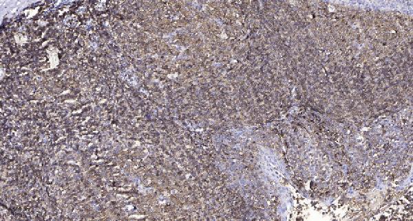
Plan chromogenic IHC for HTR3A in paraffin sections using the reported immune-cell staining pattern (HPA tissue IHC). The catalog antibody was shown on human tonsil at 1:200 with overnight primary incubation at 4°C (datasheet A02391-1).

Expected localisation, validated protocols, controls and antibodies — the at-a-glance facts below, then the full design guide.
| Expected localisation | Immune-cell cytoplasm and membrane (HPA tissue IHC) | |
| Staining pattern | Cytoplasmic and membranous staining mainly in immune cells (HPA tissue IHC) | |
| Antigen retrieval | Tris-EDTA pH 9.0 HIER, heat-mediated (datasheet A02391-1) | |
| Positive control | Lymph node+1 more · see all | |
| Negative control | Adipose tissue+4 more · see all |
| Fixation | Keep fixation consistent across paraffin sections. (standard IHC practice; not target-specific) | |
| Caveat | Whole-tissue scoring can obscure cell-specific staining (HPA tissue IHC) | |
| Regulation | Expression regulation is unreported (UniProt) | |
| Isoform / epitope | 5 isoforms; check whether the epitope is extracellular or cytoplasmic (UniProt) |
The catalog antibody’s IHC-P protocol is accompanied by published HTR3A IHC methods for lung adenocarcinoma, human brain, and rat intestine (PMC7540989; PMC10233771; PMC11250020).
| Sample | Paraffin-embedded human tonsil tissue; fixative not specified (datasheet A02391-1) |
| Fixation | Image fixative and duration unreported (datasheet A02391-1); verify before use. |
| Sectioning | 4–5 µm sections on charged slides (standard) |
| Deparaffinisation | Xylene, graded ethanol series to water (standard) |
| Antigen retrieval | Heat retrieval: Tris-EDTA pH 9.0 (datasheet A02391-1); 20 min, 95–100 °C (standard) |
| Peroxidase block | 3% H2O2, 10 min, room temperature (standard) |
| Blocking | 10% normal serum of the secondary host, 30 min, room temperature (standard) |
| Primary antibody | Rabbit anti-HTR3A, 1:100-1:300 (datasheet A02391-1) |
| Primary incubation | Overnight at 4 °C (standard) |
| Detection | HRP-polymer secondary, DAB chromogen 5–10 min (standard) |
| Counterstain | Hematoxylin, blue, dehydrate and mount (standard) |
| Expected result | HTR3A-positive staining in non-germinal center cells of lymph node (HPA tissue IHC: Medium). HPA tissue profile: Cytoplasmic and membranous expression mainly in immune cells. No signal in the no-primary control. |
HTR3A is a cell membrane receptor with 4 transmembrane segments and a postsynaptic location (UniProt P46098 topology; UniProt P46098 subcellular location). In paraffin tissue sections, expect cytoplasmic and membranous staining mainly in immune cells, especially non-germinal center cells in lymph node and tonsil (HPA tissue IHC). HPA rates the tissue pattern Supported, with medium consistency between staining and RNA expression (HPA tissue IHC).
| Membranous staining, with some cytoplasmic signal, in non-germinal center cells of lymph node or tonsil. | This matches HPA's immune-cell profile and its Medium staining level in those cells (HPA tissue IHC). Membrane signal also fits the receptor's topology (UniProt P46098 topology). Cytoplasmic signal can fit the observed HPA pattern, so its presence alone is not grounds to reject a slide (HPA tissue IHC). |
| Predominantly nuclear staining, with little staining at the cell edge. | A nuclear-dominant pattern conflicts with the reported membrane and cytoplasmic tissue pattern and membrane topology (HPA tissue IHC; UniProt P46098 topology). Treat it as suspect and compare it with a no-primary control and the expected cells on the same slide (general IHC practice). |
| Strong staining in cells HPA lists as not detected, such as adipocytes in adipose tissue. | That distribution conflicts with HPA's cell-specific observation (HPA tissue IHC). Cross-reactivity or endogenous detection activity is possible; check a no-primary control and, if available, an independent antibody before assigning the signal to HTR3A (general IHC practice). A negative HPA call applies to the named cell type, not automatically to every cell in that tissue (HPA tissue IHC). |
| Broad, diffuse color obscures cell boundaries across the section. | This prevents a reliable comparison with HPA's cell-restricted cytoplasmic and membranous pattern (HPA tissue IHC). Check background in the no-primary control, then review blocking, washing and detection conditions as general IHC troubleshooting steps (general IHC practice). |
| No convincing staining in non-germinal center cells of lymph node or tonsil. | Those cells show Medium staining in the HPA tissue record, so a blank result merits a workflow check (HPA tissue IHC). Confirm that the expected cells are present and review the antibody, antigen retrieval and detection controls (general IHC practice). A single negative run does not overturn the HPA observation (HPA tissue IHC). |
| Topology and epitope location | HTR3A has extracellular and cytoplasmic regions around 4 transmembrane segments (UniProt P46098 topology). Interpret the cell pattern with that structure in mind; the supplied record gives no antibody epitope, so it cannot predict which region this stain detects. |
| Isoforms and processing | UniProt lists 5 isoforms and a signal peptide at residues 1–23, followed by the 24–478 chain (UniProt P46098 isoforms; UniProt P46098 processing). Without an antibody epitope, the record cannot establish isoform coverage or a processing-related staining difference. |
| Glycosylation | Four glycosylation sites are annotated at residues 28, 104, 170 and 186 (UniProt P46098 glycosylation). The supplied evidence does not show whether these modifications affect this antibody's staining or antigen retrieval. |
| Strength and scope of tissue evidence | HPA calls tissue IHC Supported and reports medium staining in specified lymph node and tonsil cells (HPA tissue IHC). The listed antibody, HPA069442, is IHC Supported (HPA antibodies); this does not establish the identity of every stained immune cell or validate an IF pattern. |
| Situation | Likely cause | Next action |
|---|---|---|
| The expected lymph node or tonsil cells appear negative. | A failed staining run or absent expected cells is possible; HPA reports Medium signal in non-germinal center cells there (HPA tissue IHC). | Verify the cells are present, inspect a run control, and review antigen retrieval, antibody dilution and detection steps (general IHC practice). |
| Nuclear staining dominates the slide. | That pattern disagrees with HPA's cytoplasmic and membranous profile and UniProt's membrane location (HPA tissue IHC; UniProt P46098 subcellular location). | Compare a no-primary control and the distribution in the expected cells before scoring it as HTR3A (general IHC practice). |
| Color appears in many cells across a nominally negative area. | Background or an off-target signal is possible; HPA's negative observations name particular cell types, including adipocytes in adipose tissue (HPA tissue IHC). | Compare the named cells and a no-primary control; review blocking, washing and endogenous detection activity (general IHC practice). |
| Membrane outlines are weak, but immune-cell cytoplasm stains. | HPA describes both cytoplasmic and membranous expression, mainly in immune cells (HPA tissue IHC). Cytoplasmic staining alone is therefore inconclusive. | Assess whether the signal remains cell restricted and whether lymph node or tonsil non-germinal center cells show the expected pattern (HPA tissue IHC). |
| Cerebral cortex appears negative in the cells being assessed. | UniProt reports expression in cerebral cortex, while HPA specifically lists cortical endothelial cells as not detected (UniProt P46098 tissue specificity; HPA tissue IHC). | Identify the cell type before interpreting the result; do not use endothelial-cell negativity to infer that every cortical cell is negative (HPA tissue IHC). |
| Can an IF/ICC image be scored against this IHC pattern? | HPA summarizes the subcellular location as membrane but provides no ICC-IF images or ICC validation for the listed antibody (HPA subcellular; HPA antibodies). | Use the membrane annotation as context, but assess IF/ICC with controls and validation appropriate to that assay (HPA subcellular; general IF practice). |


Comprehensive Human Protein Atlas IHC scoring per tissue (reliability: Supported — Medium consistency between antibody staining and RNA expression data.). Rows are taken directly from the HPA tissue chart — click any row's HPA link to view the source.
| Tissue | Cell type | Level | Evidence | Source |
|---|---|---|---|---|
| Lymph node | Non-germinal center cells | Medium | Protein (IHC) | HPA → |
| Tonsil | Non-germinal center cells | Medium | Protein (IHC) | HPA → |
| Tissue | Cell type | Level | Evidence | Source |
|---|---|---|---|---|
| Adipose tissue | Adipocytes | Not detected | Protein (IHC) | HPA → |
| Adrenal gland | Glandular cells | Not detected | Protein (IHC) | HPA → |
| Bone marrow | Hematopoietic cells | Not detected | Protein (IHC) | HPA → |
| Breast | Adipocytes | Not detected | Protein (IHC) | HPA → |
| Bronchus | Respiratory epithelial cells | Not detected | Protein (IHC) | HPA → |
Use the catalog antibody’s paraffin-section IHC conditions as a starting point, then judge staining against HTR3A localisation and tissue controls (datasheet A02391-1; UniProt P46098; HPA tissue IHC).
A02391-1 is a human-reactive HTR3A antibody listed for IHC and IF (catalog: applications and reactivity), with a paraffin-section IHC figure from human tonsil (catalog: IHC image caption).
A02391-1 will render with an IHC figure from paraffin-embedded human tonsil (catalog: IHC image caption). The catalog lists human reactivity and IHC and IF applications for A02391-1, but provides no IF figure (catalog: reactivity, applications and image captions).
Which to pick: Choose A02391-1 for human tissue IHC: its own figure shows paraffin-embedded tonsil, Tris-EDTA retrieval at pH 9.0, and primary antibody at 1:200 overnight at 4°C; the fixative is unreported (catalog: A02391-1 IHC image caption). A02391-1 is also listed for IF at 1:200–1:1000, but ICC validation is unreported (catalog: A02391-1 applications and dilution). No cross-species option is supported because A02391-1 lists human reactivity only (catalog: A02391-1 reactivity).