This website uses cookies to ensure you get the best experience on our website.


- Table of Contents
Plan IRF4 staining in paraffin sections using the selective nuclear pattern seen in immune cells (HPA tissue IHC). Use tonsil non-germinal center cells as a positive reference (HPA tissue IHC) and start the IHC-validated antibody at 0.5–1 µg/mL (datasheet: PB9222 IHC-P).

Expected localisation, validated protocols, controls and antibodies — the at-a-glance facts below, then the full design guide.
| Expected localisation | Selective nuclear staining in immune cells (HPA tissue IHC) | |
| Staining pattern | Nuclear staining in a subset of immune cells (HPA tissue IHC) | |
| Antigen retrieval | Tris-EDTA pH 9.0 HIER, 95–98 °C, 20 min (rule: nuclear antigen) | |
| Positive control | Appendix+4 more · see all | |
| Negative control | Adipose tissue+4 more · see all |
| Fixation | Matched tissue-IHC evidence does not establish the fixation claim. Validate the specimen-specific method before use. (selected-SKU IHC image PB9222) | |
| Caveat | Only a subset of immune cells may stain (HPA tissue IHC) | |
| Regulation | Lymphoid-cell enriched; regulation unreported (UniProt) | |
| Isoform / epitope | 2 isoforms; epitope differences unreported (UniProt) |
The catalog antibody’s IHC-P protocol is accompanied by one published IRF4 protocol for paraffin-embedded whole-tissue sections (PMC3122134).
| Sample | Tissue sections; selected-image fixative not specified (standard IHC workflow) |
| Fixation | Image fixative and duration unreported (datasheet PB9222); verify before use. |
| Sectioning | 4–5 µm sections on charged slides (standard) |
| Deparaffinisation | Xylene, graded ethanol series to water (standard) |
| Antigen retrieval | Heat-induced epitope retrieval in Tris-EDTA buffer, pH 9.0, 20 min at 95–98 °C (standard rule: nuclear antigen) |
| Peroxidase block | 3% H2O2, 10 min, room temperature (standard) |
| Blocking | 10% normal serum of the secondary host, 30 min, room temperature (standard) |
| Primary antibody | Rabbit anti-IRF4, 0.5-1μg/ml (datasheet PB9222) |
| Primary incubation | Overnight at 4 °C (standard) |
| Detection | HRP-polymer secondary, DAB chromogen 5–10 min (standard) |
| Counterstain | Hematoxylin, blue, dehydrate and mount (standard) |
| Expected result | IRF4-positive staining in non-germinal center cells of appendix (HPA tissue IHC: Medium). HPA tissue profile: Selective nuclear expression in a subset of immune cells in bone marrow, lymphoid tissues and gastrointestinal mucosa. No signal in the no-primary control. |
IRF4 (MUM1) is a transcriptional activator annotated in the nucleus and cytoplasm, with no transmembrane segment (UniProt Q15306). In paraffin-section IHC, expect selective nuclear staining in a subset of immune cells in bone marrow, lymphoid tissues and gastrointestinal mucosa (HPA tissue IHC). HPA rates the tissue pattern Enhanced, citing high consistency between antibody staining and RNA expression (HPA tissue IHC).
| Discrete nuclear staining in some, but not all, immune cells in a lymphoid section. | This fits HPA’s selective nuclear pattern; uniform staining of every cell is unnecessary (HPA tissue IHC). Appendix, lymph node and tonsil show medium staining in non-germinal center cells (HPA tissue IHC). Score the stained cell population and its nuclear location together, rather than judging the section as wholly positive or negative. |
| Strong, widespread cytoplasmic staining with little or no nuclear staining. | This does not match the predominant tissue IHC pattern (HPA tissue IHC). Cytoplasmic IRF4 is annotated by UniProt, but HPA’s additional cytosolic ICC-IF location is uncertain (UniProt Q15306; HPA subcellular). Review nuclear counterstain alignment, detection background and controls before calling the cytoplasmic signal specific. |
| Prominent staining in a cell population listed as negative by HPA, such as adipocytes or respiratory epithelium. | HPA reports no detection in adipocytes from adipose tissue and respiratory epithelial cells from bronchus (HPA tissue IHC). A discrepant signal can reflect cross-reactivity or endogenous detection activity; morphology alone cannot distinguish them. Check the compartment and compare the IHC-validated antibody with appropriate detection controls (general IHC practice). |
| Diffuse color across cells and surrounding tissue, obscuring individual nuclei. | This prevents assessment of HPA’s selective nuclear pattern (HPA tissue IHC). Uneven blocking, detection background or excessive chromogen development are general IHC possibilities, not established IRF4-specific effects (general IHC practice). Interpret the run only after cell boundaries and counterstained nuclei can be resolved against a low-background control. |
| No nuclear signal in bone marrow hematopoietic cells or lymph-node non-germinal center cells. | Both are reported at medium staining by HPA, so their absence warrants a run-level check (HPA tissue IHC). Because expression is selective, first confirm that the expected cells are present in the section. Then assess antibody validation, detection reagents and the control slide before treating the sample as IRF4-negative (general IHC practice). |
| Tissue and cell selection | Bone marrow hematopoietic cells, lymph-node and tonsil non-germinal center cells, appendix non-germinal center cells, and splenic red-pulp cells are reported at medium staining (HPA tissue IHC). Lung macrophages are listed as low, making them a less clear visual benchmark (HPA tissue IHC). |
| Compartment and topology | IRF4 has no transmembrane segment and is annotated in the nucleus and cytoplasm (UniProt Q15306). For tissue IHC, HPA describes selective nuclear expression (HPA tissue IHC). These records support a nuclear scoring focus but do not prove that every cytoplasmic signal is an artefact. |
| Antibody validation | HPA lists Enhanced IHC validation for HPA002038, HPA002698 and CAB013508 (HPA antibodies). That status supports the reported tissue pattern; it does not validate an unlisted catalog antibody or establish its working dilution. Interpret a new antibody against suitable positive and negative controls (general IHC practice). |
| Isoforms and target-specific limits | UniProt lists 2 IRF4 isoforms and one annotated chain spanning residues 1–451 (UniProt Q15306). The supplied records give no epitope mapping or isoform-specific IHC pattern. They also give no target-specific fixation or antigen-retrieval effect, so a failed signal cannot be assigned to either from these sources. |
| IF/ICC Q: where should signal appear? | A: HPA reports mainly nucleoplasmic localization, with additional cytosolic localization marked uncertain (HPA subcellular). This is ICC-IF evidence, not a paraffin-section IHC protocol or proof that cytosolic tissue staining is expected. Use the separate IF/ICC guide for that application. |
| Situation | Likely cause | Next action |
|---|---|---|
| Expected positive cells stain weakly or remain unstained. | The selected field may lack the relevant subset, or the IHC run may have inadequate signal; HPA reports selective expression rather than positivity in every cell (HPA tissue IHC). | Confirm the cell population on the counterstained section. Compare with a bone marrow or lymphoid positive control reported by HPA, then review the catalog antibody’s IHC-P instructions and detection controls (HPA tissue IHC; general IHC practice). |
| Only diffuse cytoplasmic color appears in the expected tissue. | The pattern departs from HPA’s selective nuclear tissue signal, although UniProt also annotates cytoplasm (HPA tissue IHC; UniProt Q15306). The image alone cannot identify the source. | Check whether color tracks counterstained nuclei. Review detection-only controls and repeat under the catalog antibody’s IHC-P conditions before scoring the run (general IHC practice). |
| Adipocytes or respiratory epithelial cells stain prominently. | Those cell populations are reported as not detected by HPA; cross-reactivity or endogenous chromogenic activity is possible (HPA tissue IHC; general IHC practice). | Compare a known-positive immune-cell population, a relevant negative tissue and a detection-only control. Record the stained cell type and compartment before attributing the signal to IRF4 (general IHC practice). |
| The whole section has brown haze or patchy background. | Background obscures the cell-restricted nuclear pattern reported by HPA (HPA tissue IHC). Blocking, reagent carryover or chromogen development are general IHC variables, with no IRF4-specific cause established here (general IHC practice). | Inspect the detection-only control, wash steps and chromogen development according to the assay instructions. Rescore only when individual nuclei and negative areas remain distinguishable (general IHC practice). |
| A low-signal lung field seems negative. | HPA lists lung macrophages as low, whereas several lymphoid and bone marrow populations are medium (HPA tissue IHC). A low-signal field gives limited evidence about run performance. | Use a reported medium-staining tissue as the primary positive comparison, and identify the relevant cells before scoring the lung field (HPA tissue IHC; general IHC practice). |
| IF/ICC shows nucleoplasmic signal plus faint cytosol. | Nucleoplasm is HPA’s supported main ICC-IF location; additional cytosol is marked uncertain (HPA subcellular). This observation does not establish an IHC tissue pattern. | Interpret the two applications separately: score selective nuclear cells in paraffin IHC (HPA tissue IHC), and consult the separate IF/ICC guide for fluorescence controls and interpretation. |

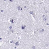
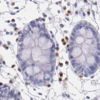


Comprehensive Human Protein Atlas IHC scoring per tissue (reliability: Enhanced — High consistency between antibody staining and RNA expression data.). Rows are taken directly from the HPA tissue chart — click any row's HPA link to view the source.
| Tissue | Cell type | Level | Evidence | Source |
|---|---|---|---|---|
| Appendix | Non-germinal center cells | Medium | Protein (IHC) | HPA → |
| Bone marrow | Hematopoietic cells | Medium | Protein (IHC) | HPA → |
| Lymph node | Non-germinal center cells | Medium | Protein (IHC) | HPA → |
| Spleen | Cells in red pulp | Medium | Protein (IHC) | HPA → |
| Tonsil | Non-germinal center cells | Medium | Protein (IHC) | HPA → |
| Tissue | Cell type | Level | Evidence | Source |
|---|---|---|---|---|
| Adipose tissue | Adipocytes | Not detected | Protein (IHC) | HPA → |
| Adrenal gland | Glandular cells | Not detected | Protein (IHC) | HPA → |
| Breast | Adipocytes | Not detected | Protein (IHC) | HPA → |
| Bronchus | Respiratory epithelial cells | Not detected | Protein (IHC) | HPA → |
| Caudate | Glial cells | Not detected | Protein (IHC) | HPA → |
Troubleshoot IRF4 chromogenic IHC by checking nuclear signal, cell context and section quality before comparing staining intensity across samples.
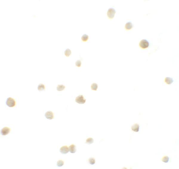
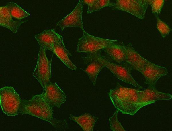
Anti-IRF4 figures show human tonsil paraffin-section IHC and Jurkat and A549 cell IF/ICC; catalog reactivity also includes Mouse and Rat (product image captions; A00401 catalog reactivity).
PB9222 shows human tonsil IHC(P) (PB9222 image caption), while A00401 shows Jurkat cell ICC at 10 μg/mL and lists IF/ICC applications (A00401 image caption; catalog applications). A00401-2 shows A549 cell IF/ICC at 5 μg/mL (A00401-2 image caption).
Which to pick: For tissue IHC, choose PB9222: its own human tonsil IHC(P) image supports paraffin sections; the fixative is unreported (PB9222 image caption). For IF/ICC, choose A00401-2 for its A549 cell image and polyclonal format (A00401-2 image caption; datasheet). For cross-species work, A00401 lists Human, Mouse and Rat reactivity and IF/ICC applications, though its shown cell image is from Jurkat cells (A00401 catalog reactivity and applications; image caption).