This website uses cookies to ensure you get the best experience on our website.


- Table of Contents
Plan chromogenic ITGA8 IHC on paraffin sections using kidney glomeruli as a high-staining reference (HPA tissue IHC) and the IHC-validated antibody at 1:50–1:100 (datasheet: A06636-1 IHC-P). Assess the observed vascular cytoplasmic staining (HPA tissue IHC) alongside ITGA8’s expected cell membrane location (UniProt).

Expected localisation, validated protocols, controls and antibodies — the at-a-glance facts below, then the full design guide.
| Expected localisation | Vascular cytoplasm observed (HPA tissue IHC); cell membrane expected (UniProt) | |
| Staining pattern | Cytoplasmic staining mainly in blood vessels; high in glomerular cells (HPA tissue IHC) | |
| Antigen retrieval | Citrate pH 6.0 HIER, 95–98 °C, 20 min (rule: cytoplasmic / membrane antigen) | |
| Positive control | Kidney | |
| Negative control | Adipose tissue+4 more · see all |
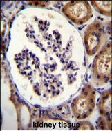
| Fixation | Formalin-fixed paraffin sections are documented (datasheet: A06636-1 IHC-P). (selected-SKU IHC image A06636-1) | |
| Caveat | Vascular staining may complicate glomerular scoring (HPA tissue IHC) | |
| Regulation | Expression regulation not specified (UniProt) | |
| Isoform / epitope | 0 isoforms; cleavage and extracellular vs cytoplasmic epitope matter (UniProt) |
The catalog antibody’s IHC-P protocol is accompanied by one published IHC workflow for human samples (PMC6975017: Methods).
| Sample | FFPE human kidney tissue (datasheet A06636-1) |
| Fixation | Image formalin-fixed; duration unreported (datasheet A06636-1); verify before use. |
| Sectioning | 4–5 µm sections on charged slides (standard) |
| Deparaffinisation | Xylene, graded ethanol series to water (standard) |
| Antigen retrieval | Heat-induced epitope retrieval in citrate buffer, pH 6.0, 20 min at 95–98 °C (standard rule: cytoplasmic / membrane antigen) |
| Peroxidase block | 3% H2O2, 10 min, room temperature (standard) |
| Blocking | 10% normal serum of the secondary host, 30 min, room temperature (standard) |
| Primary antibody | Rabbit anti-ITGA8, 1:50-1:100 (datasheet A06636-1) |
| Primary incubation | Overnight at 4 °C (standard) |
| Detection | HRP-polymer secondary, DAB chromogen 5–10 min (standard) |
| Counterstain | Hematoxylin, blue, dehydrate and mount (standard) |
| Expected result | ITGA8-positive staining in cells in glomeruli of kidney (HPA tissue IHC: High). HPA tissue profile: Cytoplasmic expression mainly in blood vessels. No signal in the no-primary control. |
ITGA8 is a cell membrane protein with an extracellular region and short cytoplasmic tail (UniProt P53708 topology). In tissue IHC, HPA reports mainly cytoplasmic staining in blood vessels and high staining in kidney glomerular cells (HPA tissue IHC, Enhanced reliability). Read the vessel-associated signal alongside the expected membrane localisation; HPA's tissue description does not establish that every cytoplasmic signal represents intracellular ITGA8.
| Vessel-associated staining and strong staining in kidney glomerular cells. | This matches HPA's reported distribution: mainly cytoplasmic expression in blood vessels and high staining in glomerular cells (HPA tissue IHC). Record which structures stain rather than scoring the section as uniformly positive (general IHC practice). |
| Staining outlines cells or lies close to their borders within a positive structure. | A peripheral pattern is consistent with ITGA8 at the cell membrane (UniProt P53708 topology; HPA ICC-IF: approved plasma membrane location). HPA describes its tissue IHC signal as mainly cytoplasmic, so compartment alone cannot decide whether an IHC result is valid (HPA tissue IHC). |
| Strong nuclear staining dominates, with little staining in vessels or glomerular cells. | Nuclear localisation conflicts with the reported membrane location (UniProt P53708 topology; HPA ICC-IF). Treat it as suspect, then compare with a reagent control and a kidney positive section before calling it ITGA8 (general IHC practice; HPA tissue IHC: high in glomerular cells). |
| Respiratory epithelium or adipocytes stain strongly while expected structures are weak. | HPA reports ITGA8 as not detected in bronchial respiratory epithelial cells and adipocytes (HPA tissue IHC). Consider off-target staining or endogenous detection activity; check cell identity and controls before interpreting those cells as positive (general IHC practice). |
| Broad, low-level colour covers cells and extracellular areas without a clear structural pattern. | This does not resemble HPA's vessel-focused tissue profile (HPA tissue IHC). Diffuse signal can reflect background from detection or insufficient blocking (general IHC practice); compare a reagent control and inspect whether glomerular cells remain distinguishable (HPA tissue IHC: high in glomerular cells). |
| Tissue and cell choice | Kidney glomerular cells offer an HPA-reported high reference, while the overall tissue profile is mainly vessel-associated (HPA tissue IHC). UniProt lists mesenchymal populations, including kidney mesangial and smooth muscle cells, but HPA's label 'cells in glomeruli' does not identify a precise cell subtype (UniProt P53708 tissue specificity; HPA tissue IHC). |
| Compartment and epitope orientation | The extracellular region spans residues 39–1012, the transmembrane segment 1013–1033, and the cytoplasmic tail 1034–1063 (UniProt P53708 topology). The antibody epitope is unspecified here; these coordinates cannot predict its staining pattern or retrieval needs. |
| Processing and glycosylation | UniProt describes disulfide-linked heavy and light chains and 16 glycosylation sites (UniProt P53708 processing and glycosylation). The supplied sources do not show how these features affect this antibody's IHC signal; do not infer shedding, loss of staining, or a required retrieval method. |
| Antibody evidence and negative comparators | HPA lists antibody HPA003432 as IHC Enhanced; its tissue profile has Enhanced reliability from staining and RNA consistency (HPA antibodies; HPA tissue IHC). HPA reports no detection in bronchial respiratory epithelial cells and adipocytes, useful cell-level comparators rather than proof that an entire section must be blank (HPA tissue IHC). |
| IF/ICC question: what localisation is expected? | Answer: plasma membrane is the approved ICC-IF location, with images listed for HEK293 and U2OS (HPA subcellular). This supports a localisation check in the separate IF/ICC guide; it does not prescribe an IF protocol or override the mainly cytoplasmic vessel description for tissue IHC (HPA tissue IHC). |
| Fixation and antigen retrieval | No target-specific fixation sensitivity or retrieval condition is supplied by UniProt or HPA. If optimisation is needed, compare retrieval conditions using the same positive tissue and controls; this is general IHC practice, not an ITGA8-specific finding. |
| Situation | Likely cause | Next action |
|---|---|---|
| No signal in kidney glomerular cells. | The section may be uninformative, or the IHC workflow may have failed; HPA reports high glomerular-cell staining (HPA tissue IHC). The supplied sources do not identify a target-specific failure mechanism. | Check tissue preservation and detection controls, then compare routine retrieval and antibody conditions on a kidney section (general IHC practice). Score the expected glomerular cells, not the kidney section as a whole (HPA tissue IHC). |
| Signal appears mainly nuclear. | Nuclear dominance conflicts with membrane localisation (UniProt P53708 topology; HPA ICC-IF). Its precise cause cannot be assigned from localisation alone. | Inspect a reagent control and the distribution in vessels and glomerular cells; review detection background before assigning the nuclear colour to ITGA8 (general IHC practice; HPA tissue IHC). |
| Strong staining appears in bronchial respiratory epithelial cells or adipocytes. | Those cells are listed as not detected by HPA tissue IHC (HPA tissue IHC); possible causes include off-target binding or endogenous detection activity (general IHC practice). | Confirm the cell type, compare a reagent control, and check whether the expected vessel or glomerular pattern is also present (general IHC practice; HPA tissue IHC). |
| Diffuse colour obscures vessel and glomerular-cell boundaries. | Non-specific reagent binding or detection background can reduce contrast (general IHC practice). HPA's tissue description calls for a mainly vessel-associated pattern, with high signal in glomerular cells (HPA tissue IHC). | Inspect reagent controls, blocking and wash steps, and detection exposure; adjust the general IHC workflow until structures can be distinguished (general IHC practice). |
| Vessels stain, but the signal looks cytoplasmic rather than sharply peripheral. | HPA explicitly describes tissue IHC staining as mainly cytoplasmic in blood vessels, despite approved plasma membrane localisation by ICC-IF and membrane topology (HPA tissue IHC; HPA ICC-IF; UniProt P53708 topology). | Judge the vessel-associated pattern together with controls and glomerular staining; document the compartment seen rather than discarding a tissue pattern solely because it lacks a crisp membrane outline (HPA tissue IHC; general IHC practice). |
| Different retrieval conditions give different staining intensity. | The supplied UniProt and HPA evidence does not establish an ITGA8-specific fixation or retrieval effect. Variation between preparation conditions must be assessed experimentally (general IHC practice). | Hold tissue, antibody and detection conditions constant while comparing retrieval conditions; retain the condition that gives interpretable glomerular and vessel signal with acceptable controls (HPA tissue IHC; general IHC practice). |
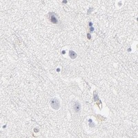


Comprehensive Human Protein Atlas IHC scoring per tissue (reliability: Enhanced — High consistency between antibody staining and RNA expression data. Antibody staining in cells/structures not annotated, view images.). Rows are taken directly from the HPA tissue chart — click any row's HPA link to view the source.
| Tissue | Cell type | Level | Evidence | Source |
|---|---|---|---|---|
| Kidney | Cells in glomeruli | High | Protein (IHC) | HPA → |
| Tissue | Cell type | Level | Evidence | Source |
|---|---|---|---|---|
| Adipose tissue | Adipocytes | Not detected | Protein (IHC) | HPA → |
| Adrenal gland | Glandular cells | Not detected | Protein (IHC) | HPA → |
| Appendix | Glandular cells | Not detected | Protein (IHC) | HPA → |
| Bone marrow | Hematopoietic cells | Not detected | Protein (IHC) | HPA → |
| Breast | Adipocytes | Not detected | Protein (IHC) | HPA → |
Troubleshoot ITGA8 staining in chromogenic IHC by checking retrieval, cell identity, compartment, controls, and scoring before interpreting signal.
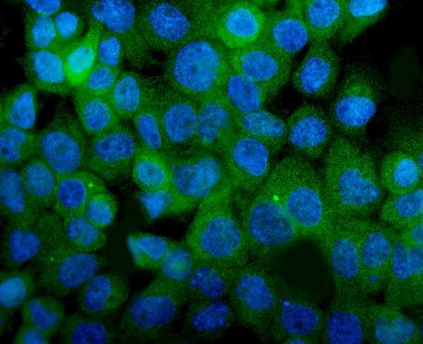
Two human-reactive ITGA8 antibodies have image data: one for chromogenic IHC in kidney tissue and one for IF in A431 cells (catalog reactivity; A06636-1 and A06636-2 image captions).
A06636-1 lists human IHC-P and shows DAB staining of formalin-fixed, paraffin-embedded human kidney (catalog applications/reactivity; A06636-1 image caption). A06636-2 lists human IF/ICC and shows IF staining of A431 cells (catalog applications/reactivity; A06636-2 image caption).
Which to pick: Choose A06636-1 for paraffin-section IHC: it is listed for IHC-P at 1:50–1:100 and has a formalin-fixed, paraffin-embedded kidney image (A06636-1 catalog dilution and image caption). Choose A06636-2 for IF/ICC at 5 μg/ml; its image shows IF in A431 cells, while its application list does not include IHC-P (A06636-2 catalog applications/dilution and image caption). Neither SKU has documented reactivity beyond human, so the payload does not support a cross-species choice (catalog reactivity).