This website uses cookies to ensure you get the best experience on our website.


- Table of Contents
Plan JMJD4 IHC-P around cytoplasmic staining reported in several tissues (HPA tissue IHC). Start with the catalog antibody at 2.5 μg/mL (datasheet: A16245) and assess glandular cells and kidney tubules as high-staining references (HPA tissue IHC).

Expected localisation, validated protocols, controls and antibodies — the at-a-glance facts below, then the full design guide.
| Expected localisation | Cytoplasmic (HPA tissue IHC) | |
| Staining pattern | Cytoplasmic staining across several tissues (HPA tissue IHC) | |
| Antigen retrieval | Citrate pH 6.0 HIER, 95–98 °C, 20 min (rule: cytoplasmic / membrane antigen) | |
| Positive control | Adrenal gland+4 more · see all | |
| Negative control | Adipose tissue+4 more · see all |
| Fixation | Keep fixation consistent across sections. (standard IHC practice; not target-specific) | |
| Caveat | Tissue staining awaits external verification (HPA tissue IHC) | |
| Regulation | No tissue-specific regulation annotated (UniProt) | |
| Isoform / epitope | 3 isoforms; epitope coverage unspecified (UniProt) |
The catalog antibody has an IHC protocol; the published JMJD4 protocol below adds details from paired kidney specimens (PMC10439757).
| Sample | Tissue sections; selected-image fixative not specified (standard IHC workflow) |
| Fixation | Image fixative and duration unreported (datasheet A16245); verify before use. |
| Sectioning | 4–5 µm sections on charged slides (standard) |
| Deparaffinisation | Xylene, graded ethanol series to water (standard) |
| Antigen retrieval | Heat-induced epitope retrieval in citrate buffer, pH 6.0, 20 min at 95–98 °C (standard rule: cytoplasmic / membrane antigen) |
| Peroxidase block | 3% H2O2, 10 min, room temperature (standard) |
| Blocking | 10% normal serum of the secondary host, 30 min, room temperature (standard) |
| Primary antibody | Rabbit anti-JMJD4, 2.5 μg/mL (datasheet A16245) |
| Primary incubation | Overnight at 4 °C (standard) |
| Detection | HRP-polymer secondary, DAB chromogen 5–10 min (standard) |
| Counterstain | Hematoxylin, blue, dehydrate and mount (standard) |
| Expected result | JMJD4-positive staining in glandular cells of adrenal gland (HPA tissue IHC: High). HPA tissue profile: Cytoplasmic expression in several tissues. No signal in the no-primary control. |
For paraffin-section IHC, expect predominantly cytoplasmic JMJD4 staining in the cell types reported as high by HPA, including kidney tubular cells, lung alveolar cells, heart cardiomyocytes, and glandular cells in several organs (UniProt Q9H9V9: cytoplasm; HPA tissue IHC: High in these cells). JMJD4 has no transmembrane segment (UniProt Q9H9V9: topology). Treat the pattern as provisional: HPA rates its tissue IHC profile Uncertain, pending external verification (HPA: tissue IHC reliability).
| Cytoplasmic signal in kidney tubular cells or lung alveolar cells, with discernible cell boundaries. | This fits the reported compartment and two reported high-staining cell populations (UniProt Q9H9V9: cytoplasm; HPA tissue IHC: High in kidney tubular and lung alveolar cells). Score signal in the named cells rather than assigning one intensity to the whole section (general IHC practice). The match supports interpretation but does not independently validate antibody specificity (HPA: IHC reliability Uncertain). |
| Signal is predominantly nuclear or outlines the plasma membrane, with little cytoplasmic staining. | For tissue IHC, this conflicts with the reported cytoplasmic pattern and warrants an artefact or specificity check (UniProt Q9H9V9: cytoplasm; HPA: cytoplasmic tissue expression). HPA separately reports mainly plasma-membrane and additional vesicular ICC-IF localization, both uncertain (HPA: subcellular ICC-IF). That discordant IF observation does not establish a membrane-positive tissue IHC pattern. |
| Strong staining appears in skin keratinocytes or skeletal-muscle myocytes. | HPA reports JMJD4 as Not detected in those cell types, so strong signal there is a warning for cross-reactivity or detection-system activity (HPA tissue IHC: Not detected in skin keratinocytes and skeletal-muscle myocytes; general IHC practice). Compare the suspect cells with a reported high-staining population and appropriate staining controls before calling the result JMJD4-specific (HPA tissue IHC: High in selected cells; general IHC practice). |
| Chromogen covers most of the section, including extracellular spaces or cells expected to be unstained. | A widespread deposit obscures the cell-specific pattern needed for interpretation (general IHC practice). Uneven blocking, excessive antibody or detection reagent, and endogenous enzyme activity are general IHC possibilities, not documented JMJD4-specific effects (general IHC practice). HPA's uncertain tissue profile cannot resolve whether broad staining represents target signal (HPA: tissue IHC reliability Uncertain). |
| No detectable signal appears in kidney tubules or heart cardiomyocytes. | Both are reported as High, so a blank result merits checking the staining run before interpreting JMJD4 as absent (HPA tissue IHC: High in kidney tubular cells and heart cardiomyocytes; general IHC practice). Verify that the positive-control section and detection controls worked, then review antibody dilution and retrieval conditions as general IHC variables (general IHC practice). HPA's uncertain reliability limits certainty about any single sample (HPA: tissue IHC reliability Uncertain). |
| Tissue and cell selection | HPA reports High staining in kidney tubular cells, heart cardiomyocytes, lung alveolar cells, and several glandular cell populations, but Not detected in skin keratinocytes and skeletal-muscle myocytes (HPA: tissue IHC). These are useful contrasts for reading a section; they are observations from an Uncertain profile, not certified positive and negative controls (HPA: tissue IHC reliability). |
| Compartment evidence across applications | UniProt assigns JMJD4 to the cytoplasm, and HPA describes cytoplasmic tissue expression (UniProt Q9H9V9: subcellular location; HPA: tissue IHC). HPA ICC-IF instead lists plasma membrane and vesicles, both uncertain (HPA: subcellular ICC-IF). Read the tissue IHC pattern on its own terms; the ICC-IF result does not define a paraffin-section scoring rule. |
| Protein features and isoforms | JMJD4 has no signal peptide, propeptide, transmembrane segment, or annotated glycosylation site; UniProt lists three isoforms and a JmjC domain at residues 142–301 (UniProt Q9H9V9: processing, topology, glycosylation, isoforms, domains). These annotations do not identify the antibody epitope or establish retrieval requirements or isoform-specific staining. |
| Strength of antibody evidence | HPA marks tissue IHC reliability Uncertain and lists IHC status as Uncertain for both HPA027260 and HPA052861 (HPA: tissue IHC reliability; HPA: antibody validation). A pattern match is therefore supportive rather than conclusive. Interpret compartment, cell type, and controls together, especially when a slide differs from the reported pattern (general IHC practice). |
| Situation | Likely cause | Next action |
|---|---|---|
| A reported high-staining cell population is blank. | The staining run may have failed, or the sample may differ from HPA's observed examples (general IHC practice; HPA tissue IHC: High in selected cells, reliability Uncertain). | Check a same-run positive-control section and detection controls, then review retrieval and antibody dilution as general IHC variables (general IHC practice). Do not infer a JMJD4-specific fixation effect; none is established by the supplied UniProt or HPA records. |
| Predominantly nuclear or sharp membrane-rim staining appears in tissue IHC. | The pattern differs from UniProt's cytoplasmic assignment and HPA's cytoplasmic tissue description (UniProt Q9H9V9: cytoplasm; HPA: tissue IHC). HPA's uncertain membrane ICC-IF observation cannot establish its cause in paraffin sections (HPA: subcellular ICC-IF). | Review morphology and compartment boundaries, compare a reported high-staining cell population, and inspect control staining before scoring the rim or nuclear signal as JMJD4 (HPA: tissue IHC; general IHC practice). |
| Strong signal appears in skin keratinocytes or skeletal-muscle myocytes. | Those populations are reported Not detected; cross-reactivity or detection-system activity is possible (HPA tissue IHC: Not detected; general IHC practice). | Check an appropriate primary-antibody omission control and detection controls, then compare cell-specific staining in a reported high-staining tissue (general IHC practice; HPA tissue IHC: High in selected cells). Treat HPA's negative observations as provisional because its IHC reliability is Uncertain (HPA: tissue IHC reliability). |
| Diffuse chromogen or extracellular staining obscures cells. | Background from antibody concentration, blocking, washing, or endogenous enzyme activity is possible in chromogenic IHC (general IHC practice). | Inspect omission and detection controls; adjust blocking, washes, dilution, or endogenous-enzyme quenching according to the detection system (general IHC practice). Reassess whether signal resolves into the reported cytoplasmic, cell-specific pattern (UniProt Q9H9V9: cytoplasm; HPA: tissue IHC). |
| Two IHC-validated antibodies give different tissue patterns. | HPA assigns IHC status Uncertain to both listed antibodies, so agreement with one image alone cannot settle specificity (HPA: HPA027260 and HPA052861 IHC validation; HPA: tissue IHC reliability Uncertain). | Compare the same named cell populations and subcellular compartment across sections, using matched staining controls (HPA: tissue IHC; UniProt Q9H9V9: cytoplasm; general IHC practice). Record disagreement rather than averaging it into an apparently definitive JMJD4 score. |
| IF/ICC shows membrane or vesicular signal. Does that change the IHC expectation? | HPA reports mainly plasma-membrane and additional vesicular ICC-IF localization, with both locations marked uncertain (HPA: subcellular ICC-IF). Tissue IHC instead reports cytoplasmic expression (HPA: tissue IHC). | Keep the tissue IHC interpretation anchored to the cytoplasmic tissue observation and UniProt localization (HPA: tissue IHC; UniProt Q9H9V9: cytoplasm). Treat the IF/ICC observation as unresolved application-specific evidence; it supplies no paraffin-section protocol or basis for calling a membrane-rim IHC result positive. |
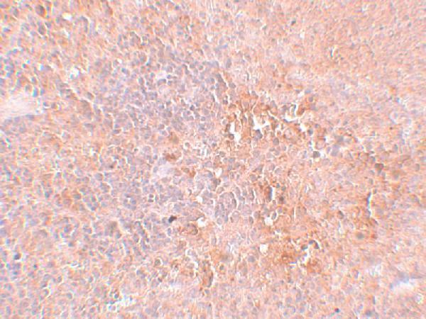
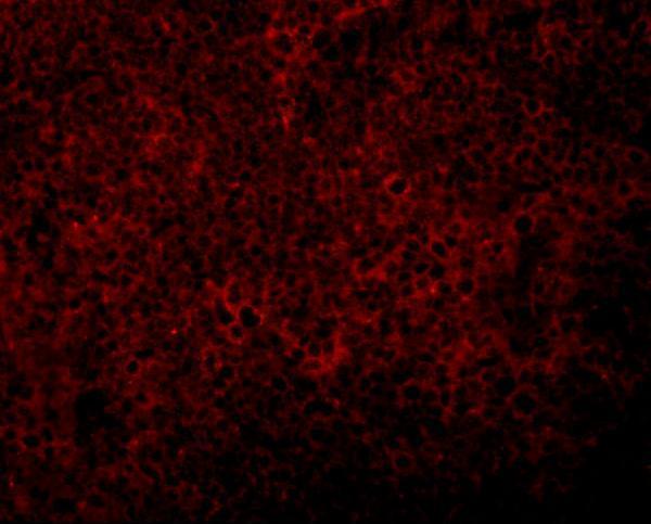
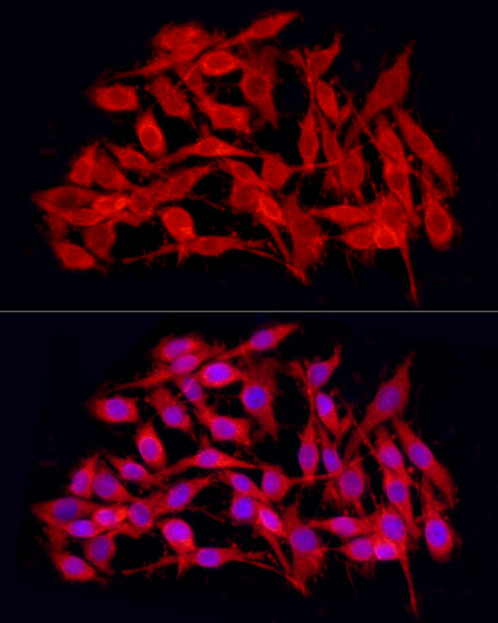


Comprehensive Human Protein Atlas IHC scoring per tissue (reliability: Uncertain — Pending external verification.). Rows are taken directly from the HPA tissue chart — click any row's HPA link to view the source.
| Tissue | Cell type | Level | Evidence | Source |
|---|---|---|---|---|
| Adrenal gland | Glandular cells | High | Protein (IHC) | HPA → |
| Heart muscle | Cardiomyocytes | High | Protein (IHC) | HPA → |
| Kidney | Cells in tubules | High | Protein (IHC) | HPA → |
| Lung | Alveolar cells | High | Protein (IHC) | HPA → |
| Pancreas | Exocrine glandular cells | High | Protein (IHC) | HPA → |
| Tissue | Cell type | Level | Evidence | Source |
|---|---|---|---|---|
| Adipose tissue | Adipocytes | Not detected | Protein (IHC) | HPA → |
| Caudate | Glial cells | Not detected | Protein (IHC) | HPA → |
| Hippocampus | Glial cells | Not detected | Protein (IHC) | HPA → |
| Lymph node | Germinal center cells | Not detected | Protein (IHC) | HPA → |
| Oral mucosa | Squamous epithelial cells | Not detected | Protein (IHC) | HPA → |
Troubleshoot JMJD4 staining by checking retrieval, controls and cellular distribution before comparing chromogenic signal across paraffin sections.
The catalog includes a paraffin-section IHC antibody with a rat spleen image (A16245 application list; A16245 IHC caption) and an IF/ICC antibody with a PC-12 cell image (A16245-1 application list; A16245-1 IF caption).
A16245 lists IHC-P and IF for human and rat, with a rat spleen IHC image at 2.5 μg/mL (A16245 application and reactivity lists; A16245 IHC caption). A16245-1 lists IF/ICC for human, mouse and rat, with a PC-12 cell IF image at 1:100 (A16245-1 application and reactivity lists; A16245-1 IF caption).
Which to pick: Choose A16245 for tissue IHC: it lists IHC-P and has a rat spleen IHC image; the caption does not report the fixative (A16245 application list; A16245 IHC caption). For IF/ICC, A16245-1 has a PC-12 cell IF image, while A16245 has a rat spleen IF image (A16245-1 IF caption; A16245 IF caption). For human, mouse and rat IF/ICC work, A16245-1 has the broader listed reactivity; both antibodies are rabbit hosted, and the catalog does not specify a clone for either (A16245-1 reactivity, application and host fields; A16245 host field; catalog clone fields).