This website uses cookies to ensure you get the best experience on our website.


- Table of Contents
Plan JMY staining in paraffin sections using the cytoplasmic tissue pattern reported by HPA (HPA tissue IHC). Interpret nuclear staining in light of JMY’s condition-dependent localisation, and treat tissue staining as uncertain because antibody staining and RNA expression show low consistency (UniProt; HPA tissue IHC).

Expected localisation, validated protocols, controls and antibodies — the at-a-glance facts below, then the full design guide.
| Expected localisation | Cytoplasmic staining in most tissues (HPA tissue IHC) | |
| Staining pattern | Cytoplasmic staining across multiple tissue cell types (HPA tissue IHC) | |
| Antigen retrieval | Tris-EDTA pH 9.0 HIER, heat-mediated (datasheet A05423) | |
| Positive control | Appendix+4 more · see all | |
| Negative control | Adipose tissue+1 more · see all |
| Fixation | Keep fixation consistent across tissue sections. (standard IHC practice; not target-specific) | |
| Caveat | Staining has low concordance with RNA expression (HPA tissue IHC) | |
| Regulation | DNA damage: nuclear accumulation (UniProt) | |
| Isoform / epitope | 2 isoforms; epitope differences unreported (UniProt) |
Compare the catalog antibody's IHC-P protocol (datasheet A05423) with the published JMY stain of NSCLC tissue and adjacent controls (PMC12357428).
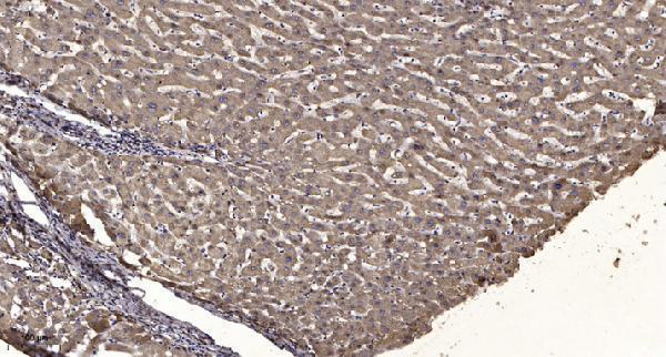
| Sample | Paraffin-embedded human liver cancer tissue; fixative not specified (datasheet A05423) |
| Fixation | Image fixative and duration unreported (datasheet A05423); verify before use. |
| Sectioning | 4–5 µm sections on charged slides (standard) |
| Deparaffinisation | Xylene, graded ethanol series to water (standard) |
| Antigen retrieval | Heat retrieval: Tris-EDTA pH 9.0 (datasheet A05423); 20 min, 95–100 °C (standard) |
| Peroxidase block | 3% H2O2, 10 min, room temperature (standard) |
| Blocking | 10% normal serum of the secondary host, 30 min, room temperature (standard) |
| Primary antibody | Rabbit anti-JMY, 1:50 recommended; image 1:200 (datasheet A05423) |
| Primary incubation | Overnight at 4 °C (standard) |
| Detection | HRP-polymer secondary, DAB chromogen 5–10 min (standard) |
| Counterstain | Hematoxylin, blue, dehydrate and mount (standard) |
| Expected result | JMY-positive staining in glandular cells of appendix (HPA tissue IHC: High). HPA tissue profile: Cytoplasmic expression in most tissues. No signal in the no-primary control. |
JMY is mainly nuclear in most cell types, but can also occupy the cytoplasm, cytoskeleton and cytoplasmic vesicles (UniProt Q8N9B5). In tissue IHC, expect predominantly cytoplasmic staining in the cell types reported as positive by HPA, such as appendix glandular cells and bronchial respiratory epithelial cells (HPA: High; HPA: cytoplasmic expression in most tissues). Interpret this pattern cautiously: HPA rates its tissue IHC evidence Uncertain, and JMY has no transmembrane segment (HPA: Uncertain; UniProt Q8N9B5 topology).
| Cytoplasmic chromogen in appendix glandular cells or bronchial respiratory epithelial cells, with clear cell boundaries. | This fits the reported tissue pattern (HPA: High in both cell types; HPA: cytoplasmic expression in most tissues). Compare intensity with the rest of the section and a same-run control; HPA rates the IHC evidence Uncertain (HPA: Uncertain). |
| Nuclear signal accompanies cytoplasmic staining, or the balance between compartments varies by cell. | Nuclear JMY is biologically plausible: UniProt places it in the nucleus in most cell types and describes redistribution with cell state (UniProt Q8N9B5). Nuclear staining alone cannot establish a biological shift in a paraffin section; HPA's nucleoplasmic ICC-IF assignment is Uncertain (HPA: subcellular, Uncertain). |
| Signal appears only on extracellular surfaces or as a continuous plasma-membrane rim. | That pattern does not match the listed nuclear, cytoplasmic and vesicular locations or the absence of a transmembrane segment (UniProt Q8N9B5). Check morphology, chromogen deposits and controls before calling it JMY (general IHC practice). |
| Strong signal appears in adipocytes or ovarian follicle cells, or staining spreads evenly across unrelated cell types. | HPA reports JMY as Not detected in those two cell types, so cross-reactivity or endogenous detection activity is possible (HPA: Not detected; general IHC practice). Because tissue IHC reliability is Uncertain, a mismatch prompts verification rather than an automatic rejection (HPA: Uncertain). |
| A diffuse haze obscures cell borders, or an expected positive compartment has no discernible signal. | Diffuse haze limits localization calls; absent staining in a reported positive tissue may reflect an assay failure or variable tissue staining (general IHC practice; HPA: Uncertain). Review controls and section quality before interpreting either result as JMY biology. |
| Tissue and cell selection | Use the reported High cell populations as candidate positive controls and adipocytes or ovarian follicle cells as comparison populations (HPA: High; HPA: Not detected). HPA labels the tissue IHC profile Uncertain, so these are observed patterns rather than independently verified controls (HPA: Uncertain). |
| Compartment and cell state | UniProt lists nuclear, cytoskeletal and vesicular locations, including autophagosome membrane during starvation-induced autophagy (UniProt Q8N9B5). Thus cytoplasmic staining need not be uniform; infer a state-dependent change only with suitable experimental controls (general IHC practice). |
| Antibody evidence and isoforms | The listed tissue IHC antibody HPA043308 has Uncertain validation, and the other listed antibody has no IHC validation entry (HPA: antibodies). UniProt records two JMY isoforms; without an epitope map here, isoform recognition cannot be predicted (UniProt Q8N9B5). |
| Topology and processing | JMY has no transmembrane segment, signal peptide or propeptide; UniProt lists a single chain spanning residues 1–988 (UniProt Q8N9B5). Treat a surface-only or secreted-looking pattern as a reason to inspect assay specificity, not as expected topology. |
| Retrieval and detection | No JMY-specific antigen-retrieval or fixation-sensitivity result is supplied (UniProt Q8N9B5; HPA: tissue IHC). If signal is weak, adjust retrieval and antibody dilution using section controls; check endogenous detection activity with the appropriate control (general IHC practice). |
| IF/ICC Q&A: Should it reproduce tissue IHC? | No direct match is established: HPA reports mainly nucleoplasmic ICC-IF localization, marked Uncertain, while tissue IHC is mainly cytoplasmic and also Uncertain (HPA: subcellular; HPA: tissue IHC). Assess each application's controls separately (general IHC practice). |
| Situation | Likely cause | Next action |
|---|---|---|
| No signal in appendix glandular cells or bronchial respiratory epithelium. | These are reported High populations, but HPA's tissue IHC reliability is Uncertain; a failed staining run is also possible (HPA: High; HPA: Uncertain; general IHC practice). | Check the same-run positive control, section integrity and detection reagents, then optimize retrieval and dilution within the IHC-validated antibody's instructions (general IHC practice). |
| Only a plasma-membrane rim or extracellular deposit stains. | The location conflicts with UniProt's nuclear and cytoplasmic assignments and lack of a transmembrane segment (UniProt Q8N9B5 topology). | Inspect chromogen distribution and tissue edges; compare a no-primary control and an independent specificity control before assigning the signal to JMY (general IHC practice). |
| Adipocytes or ovarian follicle cells stain strongly. | HPA reports JMY as Not detected in these populations; nonspecific antibody binding or endogenous detection activity may account for signal (HPA: Not detected; general IHC practice). | Compare no-primary and detection controls, then verify cell identity and staining in a reported High population on the same run (general IHC practice; HPA: High). |
| Brown haze obscures cytoplasm and nuclei. | Background can arise from incomplete blocking, excess antibody or endogenous detection activity (general IHC practice). | Review blocking and wash steps, titrate the primary antibody and inspect the no-primary control before scoring localization (general IHC practice). |
| Nuclear staining predominates over cytoplasmic staining. | Nuclear JMY is plausible, whereas HPA describes tissue IHC as mainly cytoplasmic and ICC-IF nucleoplasmic localization as Uncertain (UniProt Q8N9B5; HPA: tissue IHC; HPA: subcellular). | Score nuclei and cytoplasm separately, verify cell morphology and compare controlled sections; do not infer DNA damage from localization alone (general IHC practice). |
| Staining varies sharply between sections or cell populations. | UniProt describes context-dependent localization, and HPA reports low consistency between antibody staining and RNA data (UniProt Q8N9B5; HPA: Uncertain). | Run sections together, compare the same cell type and compartment, and record intensity and distribution separately before drawing a biological conclusion (general IHC practice). |


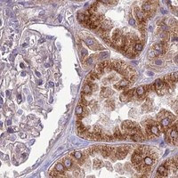


Comprehensive Human Protein Atlas IHC scoring per tissue (reliability: Uncertain — Low consistency between antibody staining and RNA expression data. Pending external verification.). Rows are taken directly from the HPA tissue chart — click any row's HPA link to view the source.
| Tissue | Cell type | Level | Evidence | Source |
|---|---|---|---|---|
| Appendix | Glandular cells | High | Protein (IHC) | HPA → |
| Bronchus | Respiratory epithelial cells | High | Protein (IHC) | HPA → |
| Cervix | Squamous epithelial cells | High | Protein (IHC) | HPA → |
| Colon | Endothelial cells | High | Protein (IHC) | HPA → |
| Duodenum | Glandular cells | High | Protein (IHC) | HPA → |
Troubleshooting for JMY chromogenic IHC in paraffin sections, with one entry for the separate IF/ICC workflow (datasheet A05423).
A05423 has an IHC image of paraffin-embedded human liver cancer (A05423 image caption). Human and Mouse reactivity is listed; no IF figure is provided (catalog reactivity; catalog IF images).
A05423 is listed for IHC and has an image of paraffin-embedded human liver cancer (catalog applications; A05423 image caption). It lists Human and Mouse reactivity, but the image documents human tissue only (catalog reactivity; A05423 image caption).
Which to pick: For tissue IHC, choose A05423 for paraffin sections; its image caption does not report the fixative (A05423 image caption). For IF/ICC, no listed SKU has an IF application or figure (catalog applications; catalog IF images). For cross-species work, A05423 lists Human and Mouse reactivity, though its IHC image shows human tissue only; its host is Rabbit and clonality is unreported (catalog reactivity/host/clone; A05423 image caption).