This website uses cookies to ensure you get the best experience on our website.


- Table of Contents
Plan chromogenic JPH1 IHC-P using skeletal muscle myocytes as a positive tissue reference (HPA tissue IHC). Compare their cytoplasmic staining with the catalog antibody’s 2.5 μg/mL IHC starting concentration (datasheet: mouse skeletal muscle IHC).

Expected localisation, validated protocols, controls and antibodies — the at-a-glance facts below, then the full design guide.
| Expected localisation | Cytoplasmic staining in skeletal myocytes (HPA tissue IHC) | |
| Staining pattern | Strong cytoplasmic staining in skeletal muscle myocytes (HPA tissue IHC) | |
| Antigen retrieval | Citrate pH 6.0 HIER, 95–98 °C, 20 min (rule: cytoplasmic / membrane antigen) | |
| Positive control | Skeletal muscle+1 more · see all | |
| Negative control | Adipose tissue+4 more · see all |
| Fixation | Matched tissue-IHC evidence does not establish the fixation claim. Validate the specimen-specific method before use. (selected-SKU IHC image A08565) | |
| Caveat | Presumed off-target binding can confound staining (HPA tissue IHC) | |
| Regulation | Abundant in skeletal muscle; very low in heart (UniProt) | |
| Isoform / epitope | No isoforms annotated; N-terminal region is cytoplasmic (UniProt) |
The catalog antibody’s paraffin-section protocol is followed by one published JPH1 chromogenic IHC protocol using mouse muscle cryosections (PMC10889371).
| Sample | Tissue sections; selected-image fixative not specified (standard IHC workflow) |
| Fixation | Image fixative and duration unreported (datasheet A08565); verify before use. |
| Sectioning | 4–5 µm sections on charged slides (standard) |
| Deparaffinisation | Xylene, graded ethanol series to water (standard) |
| Antigen retrieval | Heat-induced epitope retrieval in citrate buffer, pH 6.0, 20 min at 95–98 °C (standard rule: cytoplasmic / membrane antigen) |
| Peroxidase block | 3% H2O2, 10 min, room temperature (standard) |
| Blocking | 10% normal serum of the secondary host, 30 min, room temperature (standard) |
| Primary antibody | Rabbit anti-JPH1, 2.5 μg/mL (datasheet A08565) |
| Primary incubation | Overnight at 4 °C (standard) |
| Detection | HRP-polymer secondary, DAB chromogen 5–10 min (standard) |
| Counterstain | Hematoxylin, blue, dehydrate and mount (standard) |
| Expected result | JPH1-positive staining in myocytes of skeletal muscle (HPA tissue IHC: High). HPA tissue profile: Selective cytoplasmic expression in muscle, most abundant in skeletal muscle. No signal in the no-primary control. |
JPH1 links the plasma membrane to the endoplasmic or sarcoplasmic reticulum, with its C-terminal transmembrane segment anchored in the latter membrane (UniProt Q9HDC5 topology). In paraffin-section IHC, expect predominantly cytoplasmic staining in skeletal myocytes; spermatogonia also score high (HPA: tissue IHC). HPA rates the tissue profile Enhanced, while noting medium agreement with RNA and presumed off-target staining that was disregarded (HPA: tissue IHC reliability).
| Strong cytoplasmic staining in skeletal myocytes, with little staining in adjacent negative cells (HPA: High in skeletal myocytes). | This fits the most abundant tissue pattern reported by HPA (HPA: tissue IHC profile). JPH1 is associated with junctions between the cell surface and sarcoplasmic reticulum (UniProt Q9HDC5 function and topology). Judge the signal by its cell distribution and compartment; a sharply outlined plasma-membrane rim is not required by the HPA IHC profile (HPA: selective cytoplasmic expression). |
| Predominant nuclear staining in a paraffin section instead of the expected cytoplasmic pattern (HPA: tissue IHC profile). | Treat this as a discordant IHC result and check controls and staining conditions before assigning it to JPH1 (HPA: selective cytoplasmic IHC expression; general IHC practice). HPA separately reports enhanced nucleoplasmic localization in ICC-IF; that observation concerns a different assay and does not establish a nuclear pattern for tissue IHC (HPA: subcellular ICC-IF). |
| Strong signal in a cell population listed as not detected, such as adipocytes in adipose tissue (HPA: tissue IHC). | Consider cross-reactivity or endogenous detection activity, especially if the signal resembles staining in the positive control (general IHC practice). Do not apply that interpretation to spermatogonia: HPA reports high staining there (HPA: High in spermatogonia). Compare the suspect section with a negative reagent control and the expected distribution before calling it JPH1 (general IHC practice). |
| Broad, diffuse colour across cells and extracellular areas, obscuring cell boundaries (general IHC practice). | This limits compartment and cell-type interpretation; nonspecific reagent binding, incomplete blocking, or excess chromogen development are general possibilities (general IHC practice). Assess the negative reagent control and whether the skeletal-myocyte pattern remains discernible (general IHC practice; HPA: High in skeletal myocytes). Diffuse colour alone does not establish the HPA cytoplasmic pattern (HPA: tissue IHC profile). |
| No detectable signal in skeletal myocytes on a slide intended as a positive control (HPA: High in skeletal myocytes). | The run cannot support a negative interpretation of test tissue until the positive control works (general IHC practice). Check that primary antibody, detection reagents and chromogen were applied and that the chosen retrieval and antibody conditions follow the validated IHC method (general IHC practice). HPA's Enhanced rating describes evidence for its reported pattern, not a guarantee for every staining run (HPA: tissue IHC reliability). |
| Cellular location and topology (UniProt Q9HDC5 topology). | JPH1 has a cytoplasmic region at residues 1–639 and a transmembrane segment at 640–660 anchored in endoplasmic or sarcoplasmic reticulum membrane (UniProt Q9HDC5 topology). Interpret section staining at the cytoplasmic and membrane-junction level (UniProt Q9HDC5 topology; HPA: cytoplasmic tissue IHC), without assuming that light microscopy will resolve individual triads (general microscopy practice). |
| Tissue and cell context (HPA: tissue IHC). | HPA rates skeletal myocytes and spermatogonia High, cardiomyocytes and smooth muscle cells Low, and several listed cell types Not detected (HPA: tissue IHC). A faint heart or smooth-muscle result has a different expectation from a blank skeletal-muscle control (HPA: tissue IHC; general IHC practice). Use the named cell population when comparing sections (general IHC practice). |
| Evidence strength (HPA: tissue IHC reliability and antibody validation). | HPA labels the tissue profile Enhanced but reports medium agreement between antibody staining and RNA, with presumed off-target staining disregarded (HPA: tissue IHC reliability). HPA lists HPA008996 and HPA009413 as IHC Enhanced, and CAB013461 as IHC Supported (HPA: antibody validation). These labels support comparison with the reported pattern; they do not identify every unexpected stained cell as JPH1 (HPA: tissue IHC reliability). |
| IF/ICC Q: Should nucleoplasmic fluorescence define the expected paraffin-section IHC result? (HPA: subcellular ICC-IF). | A: No. HPA reports enhanced nucleoplasmic localization in ICC-IF images from A-431, U-251MG and U2OS, while its tissue IHC profile describes selective cytoplasmic expression, most abundant in skeletal muscle (HPA: subcellular ICC-IF; HPA: tissue IHC). Interpret each assay against its own reported pattern (general assay practice). |
| Situation | Likely cause | Next action |
|---|---|---|
| Skeletal-muscle positive control is blank (HPA: High in skeletal myocytes). | A failed reagent step or unsuitable retrieval or primary-antibody conditions can remove visible IHC signal (general IHC practice). | Confirm the antibody and detection sequence, then check retrieval and dilution against the IHC-validated antibody's method (general IHC practice). Repeat with a skeletal-muscle positive control before interpreting negative study sections (HPA: High in skeletal myocytes; general IHC practice). |
| Colour is diffuse across the section rather than concentrated in expected cells (HPA: tissue IHC profile). | Nonspecific reagent binding or excessive chromogen development can obscure localization (general IHC practice). | Inspect the negative reagent control, review blocking and washes, and shorten chromogen development if overdevelopment is evident (general IHC practice). Confirm that any remaining positive signal maps to skeletal myocytes or another reported positive cell type (HPA: tissue IHC). |
| Strong nuclear IHC signal dominates the skeletal-muscle section (HPA: cytoplasmic tissue IHC profile). | The compartment conflicts with HPA tissue IHC, even though HPA reports nucleoplasmic localization in ICC-IF (HPA: tissue IHC; HPA: subcellular ICC-IF). | Review control slides and primary-antibody conditions, then score the nuclear result as unresolved for tissue IHC unless independently supported (general IHC practice; HPA: tissue IHC profile). Do not use the ICC-IF localization alone to validate nuclear chromogenic staining (HPA: subcellular ICC-IF). |
| An HPA-listed negative cell type stains strongly (HPA: tissue IHC). | Off-target antibody staining or endogenous detection activity may account for an unexpected chromogenic signal (HPA: reliability note on presumed off-target staining; general IHC practice). | Compare a negative reagent control and a skeletal-muscle positive control, then check whether the unexpected colour tracks the cell type or the detection system (general IHC practice; HPA: High in skeletal myocytes). Keep the finding provisional (general IHC practice). |
| Heart or smooth-muscle staining is faint while skeletal muscle stains strongly (HPA: tissue IHC). | This relative intensity matches HPA's Low levels in cardiomyocytes and smooth muscle cells versus High in skeletal myocytes (HPA: tissue IHC). | Score each named cell population against its reported level and the same run's positive control (HPA: tissue IHC; general IHC practice). Investigate a technical failure if the skeletal-muscle control also loses signal (HPA: High in skeletal myocytes; general IHC practice). |
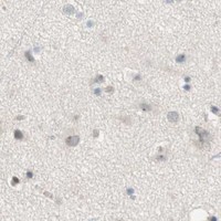


Comprehensive Human Protein Atlas IHC scoring per tissue (reliability: Enhanced — Medium consistency between antibody staining and RNA expression data. Presumed off target binding observed and disregarded.). Rows are taken directly from the HPA tissue chart — click any row's HPA link to view the source.
| Tissue | Cell type | Level | Evidence | Source |
|---|---|---|---|---|
| Skeletal muscle | Myocytes | High | Protein (IHC) | HPA → |
| Testis | Spermatogonia cells | High | Protein (IHC) | HPA → |
| Tissue | Cell type | Level | Evidence | Source |
|---|---|---|---|---|
| Adipose tissue | Adipocytes | Not detected | Protein (IHC) | HPA → |
| Adrenal gland | Glandular cells | Not detected | Protein (IHC) | HPA → |
| Appendix | Glandular cells | Not detected | Protein (IHC) | HPA → |
| Bone marrow | Hematopoietic cells | Not detected | Protein (IHC) | HPA → |
| Breast | Adipocytes | Not detected | Protein (IHC) | HPA → |
Troubleshoot JPH1 staining in paraffin sections by checking retrieval, compartment, and muscle cell identity against the available tissue evidence.
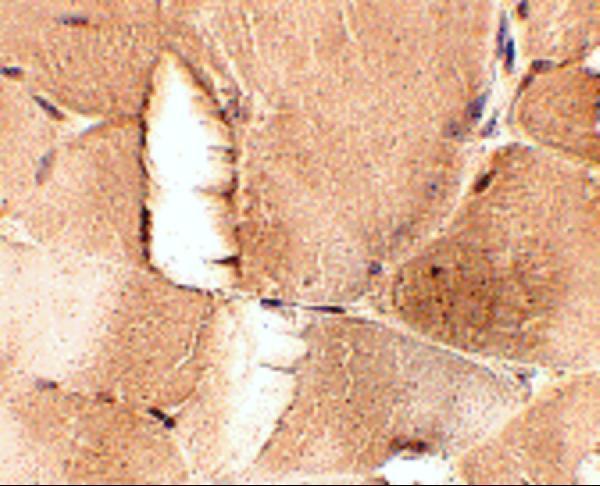
A08565 has real IHC data from mouse skeletal muscle (A08565 image caption). The catalog lists Human, Mouse, and Rat reactivity, but provides no IF image (catalog: reactivity and images).
A08565 renders with an IHC image of mouse skeletal muscle at 2.5 μg/mL (A08565 image caption). Its listed applications include IHC-P, and its listed reactivity covers Human, Mouse, and Rat; the supplied IHC image documents mouse tissue (catalog: applications and reactivity; A08565 image caption).
Which to pick: Choose A08565 for paraffin-section tissue IHC: IHC-P is listed, and its own image shows mouse skeletal muscle at 2.5 μg/mL (catalog: applications; A08565 image caption). The image caption does not report a fixative, and the catalog does not specify clonality (A08565 image caption; catalog: clone). No SKU has listed IF/ICC validation; for Human, Mouse, or Rat IHC, A08565 is the catalog choice by listed reactivity, with direct IHC image evidence limited to mouse skeletal muscle (catalog: applications and reactivity; A08565 image caption).