This website uses cookies to ensure you get the best experience on our website.


- Table of Contents
Plan chromogenic JUND IHC in paraffin sections around the nuclear tissue pattern (HPA tissue IHC), using adrenal glandular cells as a high-staining reference and adipocytes as a negative reference (HPA tissue IHC). This guide covers fixation, controls, nuclear scoring, and the catalog antibody’s 2–5 μg/ml IHC range (datasheet A05609-1).

Expected localisation, validated protocols, controls and antibodies — the at-a-glance facts below, then the full design guide.
| Expected localisation | Nuclear staining in tissue (HPA tissue IHC) | |
| Staining pattern | General nuclear staining across tissue cells (HPA tissue IHC) | |
| Antigen retrieval | EDTA pH 8.0 HIER, heat-mediated (datasheet A05609-1) | |
| Positive control | Adrenal gland+4 more · see all | |
| Negative control | Adipose tissue+2 more · see all |
| Fixation | Matched tissue-IHC evidence does not establish the fixation claim. Validate the specimen-specific method before use. (selected-SKU IHC image A05609-1) | |
| Caveat | Staining and RNA show medium consistency; verify locally (HPA tissue IHC) | |
| Regulation | Expression regulation not annotated (UniProt) | |
| Isoform / epitope | No isoforms annotated; one 1–347 chain (UniProt) |
Start with the catalog antibody’s paraffin-section protocol (datasheet A05609-1). The published JUND methods below describe mouse nucleus accumbens staining (PMC6082848) and hamster brain chromogenic staining (PMC3894818).
| Sample | Paraffin-embedded human renal cell carcinoma tissue; fixative not specified (datasheet A05609-1) |
| Fixation | Image fixative and duration unreported (datasheet A05609-1); verify before use. |
| Sectioning | 4–5 µm sections on charged slides (standard) |
| Deparaffinisation | Xylene, graded ethanol series to water (standard) |
| Antigen retrieval | Heat retrieval: EDTA pH 8.0 (datasheet A05609-1); 20 min, 95–100 °C (standard) |
| Peroxidase block | 3% H2O2, 10 min, room temperature (standard) |
| Blocking | 10% goat serum (datasheet A05609-1) |
| Primary antibody | Rabbit anti-JUND, 2-5μg/ml (datasheet A05609-1) |
| Primary incubation | Overnight at 4 °C (datasheet A05609-1) |
| Detection | HRP-conjugated secondary, DAB chromogen (datasheet A05609-1) |
| Counterstain | Hematoxylin, blue, dehydrate and mount (standard) |
| Expected result | JUND-positive staining in glandular cells of adrenal gland (HPA tissue IHC: High). HPA tissue profile: General nuclear expression. No signal in the no-primary control. |
JUND is a nuclear transcription factor with no transmembrane segment (UniProt P17535: nucleus, topology). In paraffin-section IHC, expect predominantly nuclear staining in glandular cells of several organs and esophageal squamous epithelium (HPA: general nuclear expression; High in listed cells). The HPA tissue profile is Supported, with medium agreement between staining and RNA data and external verification pending (HPA: tissue IHC reliability).
| Distinct nuclear chromogen in adrenal or duodenal glandular cells, with nuclei easier to identify against the counterstain. | This matches the expected compartment and a High-staining cell population (UniProt P17535: nucleus; HPA: High in adrenal and duodenal glandular cells). Judge the signal in identified cells rather than by whole-section color. |
| Predominantly cytoplasmic, membranous, or extracellular color, especially when nuclei remain clear. | That distribution conflicts with the reported nuclear pattern (UniProt P17535: nucleus, no transmembrane segment; HPA: general nuclear expression). Treat it as possible nonspecific staining or detection artifact; it does not establish a new JUND location. |
| Strong color in adipocytes or bone-marrow hematopoietic cells while the expected nuclear pattern is absent. | HPA reports JUND as Not detected in these cell populations (HPA: adipocytes; bone-marrow hematopoietic cells). Check cross-reactivity or endogenous detection activity. Their reference status does not make every stained cell an automatic false positive. |
| Diffuse color across tissue, luminal material, or blank areas, obscuring nuclear boundaries. | This is difficult to score as JUND because the reference pattern is nuclear (HPA: general nuclear expression). Uneven background may arise from blocking, washing, or chromogen detection conditions (standard IHC practice); compare the no-primary control. |
| No nuclear signal in an otherwise intact positive reference section, such as adrenal gland or esophagus. | Those reference cells are reported High (HPA: adrenal glandular cells; esophageal squamous epithelial cells). A blank result calls for a reagent and workflow check before interpreting test tissue as negative; HPA reliability remains Supported, pending external verification (HPA: tissue IHC reliability). |
| Reference cell population | Choose a named High population to assess whether nuclear detection works: adrenal, cervical, duodenal, endometrial, epididymal, fallopian-tube, or gallbladder glandular cells, or esophageal squamous cells (HPA: tissue IHC). Low populations, including gastric glandular cells and cardiomyocytes, are less decisive positive references (HPA: Low in those cells). |
| Strength of tissue evidence | The broad nuclear tissue pattern is Supported, with medium staining-to-RNA consistency and external verification pending (HPA: tissue IHC reliability). Low tissue RNA specificity does not predict uniform staining in every cell (HPA: RNA specificity; tissue IHC levels). |
| Antibody validation | HPA063029 and CAB005268 are Supported for IHC; HPA068905 has no IHC status in this payload (HPA: antibody validation). Use the catalog antibody's IHC-P instructions for its working conditions; do not transfer validation or a dilution between antibodies. |
| Detection background | Endogenous enzyme activity, inadequate blocking, or residual detection reagent can produce color unrelated to primary-antibody binding (standard chromogenic IHC practice). A no-primary control helps separate detection background from a nuclear staining pattern; this is general workflow guidance, not a reported JUND-specific effect. |
| IF/ICC Q: Where should JUND appear? | A: Predominantly in the nucleoplasm (HPA: enhanced nucleoplasm ICC-IF; UniProt P17535: nucleus). HPA lists MCF-7, SH-SY5Y, U2OS, and SK-MEL-30 as imaged cell lines (HPA: subcellular ICC-IF); this localization evidence does not supply an IF protocol for the paraffin IHC workflow. |
| Situation | Likely cause | Next action |
|---|---|---|
| The High-reference section has no nuclear chromogen. | Primary-antibody or detection conditions may have failed (standard IHC practice); HPA's High designation is an observed reference pattern, not a guarantee for each run (HPA: tissue IHC). | Check the catalog antibody's IHC-P conditions, reagent activity, and a same-run positive control; review the run before calling test tissue negative (standard IHC practice). |
| Signal is mostly cytoplasmic or outlines cell membranes. | The compartment disagrees with the reported nuclear localization (UniProt P17535: nucleus, no transmembrane segment; HPA: general nuclear expression). Nonspecific binding or detection background is possible. | Compare with a no-primary control and inspect whether identified nuclei stain in an HPA High cell population (HPA: tissue IHC; standard IHC practice). |
| Brown color persists in a no-primary control. | Detection-related background can persist without the primary antibody, including endogenous enzyme activity (standard chromogenic IHC practice). | Review the relevant enzyme block, detection reagents, washes, and control section before attributing color to JUND (standard IHC practice). |
| Background is diffuse, but some nuclei appear positive. | Excess detection signal or insufficient washing can obscure cell boundaries (standard IHC practice). HPA's reference pattern requires a readable nuclear distribution (HPA: general nuclear expression). | Assess the no-primary control, then adjust detection or washing under the catalog IHC-P workflow and rescore identifiable nuclei (standard IHC practice). |
| An HPA Not-detected population stains strongly. | Adipocytes, bone-marrow hematopoietic cells, and hippocampal glia are reported Not detected (HPA: tissue IHC); unexpected staining could reflect cross-reactivity or background, but cannot be diagnosed from color alone. | Compare compartment, nearby positive cells, and no-primary control; investigate specificity before assigning the unexpected signal to JUND (HPA: tissue IHC; standard IHC practice). |
| A Low-reference population looks blank. | Weak staining can be hard to distinguish from background; stomach glandular cells, cholangiocytes, cardiomyocytes, myocytes, and splenic red-pulp cells are listed Low (HPA: tissue IHC). | First verify nuclear staining in a High-reference population, then score the Low population conservatively with the same detection conditions (HPA: tissue IHC; standard IHC practice). |


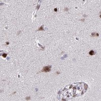
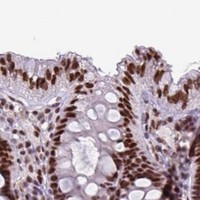
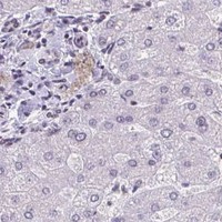
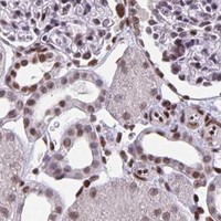


Comprehensive Human Protein Atlas IHC scoring per tissue (reliability: Supported — Medium consistency between antibody staining and RNA expression data. Pending external verification.). Rows are taken directly from the HPA tissue chart — click any row's HPA link to view the source.
| Tissue | Cell type | Level | Evidence | Source |
|---|---|---|---|---|
| Adrenal gland | Glandular cells | High | Protein (IHC) | HPA → |
| Cervix | Glandular cells | High | Protein (IHC) | HPA → |
| Duodenum | Glandular cells | High | Protein (IHC) | HPA → |
| Endometrium | Glandular cells | High | Protein (IHC) | HPA → |
| Epididymis | Glandular cells | High | Protein (IHC) | HPA → |
Troubleshoot JUND staining in paraffin sections by checking nuclear localisation, the validated retrieval condition and tissue controls before interpreting chromogenic signal.
JUND IHC images show staining in human renal cell carcinoma and appendix adenocarcinoma paraffin sections (A05609-1 image captions). Human, mouse, and rat reactivity is listed (A05609-1 catalog).
A05609-1, the SKU with a rendered IHC card, is shown staining human renal cell carcinoma paraffin tissue after EDTA pH 8 retrieval and DAB detection (A05609-1 image caption). A second caption shows staining in human appendix adenocarcinoma paraffin tissue (A05609-1 image caption).
Which to pick: Choose the polyclonal A05609-1 for tissue IHC: its images document paraffin sections, but the fixative is unreported (A05609-1 image captions; catalog). For IF/ICC, M05609 is listed for human, mouse, and rat, while M05609-1 is listed for human only; neither has an IF image in the payload (catalog). For cross-species tissue IHC, A05609-1 lists human, mouse, and rat reactivity, although its IHC images show human tissue only (A05609-1 catalog; image captions).