JUP / Junction plakoglobin · IHC design guide

Design Immunohistochemistry for JUP

Plan chromogenic JUP IHC in paraffin sections using its observed cytoplasmic and membranous staining in glandular and epithelial cells (HPA tissue IHC). The guide covers fixation, retrieval, detection, controls and interpretation, with the reported low consistency between antibody staining and RNA expression in mind (HPA tissue IHC).

Evidence assembled Oct 2026 · For research use; verify linked source records and product datasheet before use
Immunohistochemistry protocol sheet for JUP (IHC for JUP): expected localisation Glandular and epithelial cytoplasm and membranes (HPA tissue IHC), antibody PB9185, validated IHC image, and IHC protocol steps
Printable JUP IHC protocol sheet — expected localisation Glandular and epithelial cytoplasm and membranes (HPA tissue IHC), antibody PB9185, controls and protocol steps. Open the full JUP IHC guide →

JUP Immunohistochemistry Experimental Design Guide

Expected localisation, validated protocols, controls and antibodies — the at-a-glance facts below, then the full design guide.

Must know before staining
Expected localisation Glandular and epithelial cytoplasm and membranes (HPA tissue IHC)
Staining pattern Cytoplasmic and membranous staining in glandular and epithelial cells (HPA tissue IHC)
Antigen retrieval Citrate pH 6 HIER, heat-mediated (datasheet PB9185)
Positive control ⓘ Appendix+4 more · see all
Negative control ⓘ Adipose tissue+4 more · see all
Important caveats
Reasons your staining may differ from the expected pattern.
Fixation Keep fixation consistent (standard IHC practice; not target-specific); Matched tissue-IHC evidence does not establish the fixation claim. Validate the specimen-specific method before use. (selected-SKU IHC image PB9185)
Caveat Staining and RNA expression show low consistency (HPA tissue IHC)
Regulation RNA enhanced in esophagus and skin (HPA tissue RNA)
Isoform / epitope No annotated isoforms; one 1–745 chain and no transmembrane segment (UniProt)
Section 1

Recommended JUP IHC & IF Protocols

Compare the catalog antibody’s IHC-P protocol (datasheet: PB9185) with published JUP staining in prostate cancer tissue (PMC8253102) and renal cell carcinoma tissue (PMC13050451).

Recommended immunohistochemistry (IHC-P) protocol parameters
SampleParaffin-embedded human intestinal cancer tissue; fixative not specified (datasheet PB9185)
FixationImage fixative and duration unreported (datasheet PB9185); verify before use.
Sectioning4–5 µm sections on charged slides (standard)
DeparaffinisationXylene, graded ethanol series to water (standard)
Antigen retrievalHeat retrieval: Citrate pH 6, 20 min (datasheet PB9185)
Peroxidase block3% H2O2, 10 min, room temperature (standard)
Blocking10% goat serum (datasheet PB9185)
Primary antibodyRabbit anti-JUP, 0.5-1μg/ml (datasheet PB9185)
Primary incubationOvernight at 4 °C (datasheet PB9185)
DetectionStreptavidin-biotin complex (SABC), DAB chromogen (datasheet PB9185)
CounterstainHematoxylin, blue, dehydrate and mount (standard)
Expected resultJUP-positive staining in glandular cells of appendix (HPA tissue IHC: High). HPA tissue profile: Cytoplasmic and membranous expression in glandular and epithelial cells. No signal in the no-primary control.
💡Decision noteStart with heat-mediated citrate retrieval at pH 6 (datasheet: PB9185); the prostate study also used pH 6 heat retrieval (PMC8253102).
Section 2

What Is the Expected JUP Staining Pattern?

JUP should appear mainly at cell borders, with cytoplasmic staining in glandular and epithelial cells (HPA tissue IHC; UniProt P14923 localization). Its junctional pattern fits its adherens-junction and desmosomal roles; JUP has no transmembrane segment (UniProt P14923). HPA rates the tissue IHC pattern Supported, while noting low consistency between staining and RNA data (HPA tissue IHC).

What am I looking at on my slide?
Crisp cell-border staining, with some cytoplasmic signal, in appendix or colon glands.This fits the reported membranous and cytoplasmic profile; glandular cells in both tissues are scored High (HPA tissue IHC). Judge the pattern within the glands rather than treating every cell in the section as an expected positive (HPA tissue IHC).
Signal appears mainly nuclear, with little staining at epithelial cell borders.This differs from the dominant tissue IHC pattern (HPA tissue IHC). Check morphology and detection controls before assigning it to JUP. Nuclear localization is annotated, so nuclear signal alone cannot establish an artifact (UniProt P14923 localization).
Strong staining appears in adipocytes or lymph-node germinal-center cells.Those cell types are scored Not detected (HPA tissue IHC). Investigate antibody cross-reactivity or endogenous detection activity with appropriate controls (standard IHC practice); an unexpected positive does not establish JUP expression.
A broad, even haze covers cells and surrounding tissue.This obscures the cell-border pattern expected from JUP localization (HPA tissue IHC; UniProt P14923 localization). Review background controls and detection conditions before scoring weak cytoplasmic color as specific staining (standard IHC practice).
No signal appears in a well-preserved appendix or colon glandular-cell control.Both are scored High, making a blank control unexpected (HPA tissue IHC). First verify that the positive control and detection run worked; a single negative slide cannot distinguish technical failure from sample-specific biology (standard IHC practice).
💡Expected JUP appearanceCall a result positive when glandular or epithelial cells show clear membranous, especially cell-border, staining with possible cytoplasmic signal; appendix and colon glandular cells are scored High (HPA tissue IHC). Diffuse haze or isolated staining in an HPA-negative cell type is suspect (HPA tissue IHC; standard IHC practice).
How each factor affects the staining
Tissue and cell-type selectionUse a reported High population, such as appendix or colon glandular cells, to judge whether the run can reveal the expected pattern (HPA tissue IHC). Adipocytes and lymph-node germinal-center cells offer reported Not detected comparisons (HPA tissue IHC).
Compartment used for scoringGive cell-border staining weight because JUP is associated with adherens junctions and desmosomes (UniProt P14923 localization). Cytoplasmic signal can also fit the record; UniProt describes soluble and membrane-associated cytoplasmic forms (UniProt P14923 localization).
Strength of tissue-pattern evidenceHPA labels tissue IHC Supported and reports low consistency with RNA expression; external characterization supports the staining (HPA tissue IHC). Interpret an unexpected tissue result with controls rather than using RNA abundance alone to accept or reject the stain.
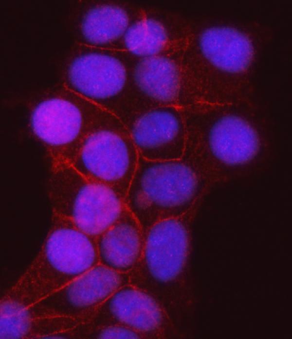
IF/ICC Q&A: Where should JUP appear?Mainly at the plasma membrane and cell junctions, with additional vesicular localization (HPA subcellular ICC-IF). HPA lists A-431, U2OS and MCF-7 images (HPA subcellular ICC-IF). This localization evidence does not supply an IF/ICC protocol.
Why is my staining missing, weak or wrong?
SituationLikely causeNext action
No staining in the appendix or colon glandular-cell positive control.A failed staining or detection run is possible; these cell populations are scored High (HPA tissue IHC).Check section integrity, primary-antibody application, retrieval and detection controls, then repeat the positive control alongside the test section (standard IHC practice). Do not infer a JUP-specific retrieval or fixation effect from this result.
The positive control stains, but a test population remains blank.Expression may differ by cell type; HPA reports both High and Not detected populations (HPA tissue IHC).Confirm the identity of the scored cells and compare with the matching HPA cell-type entry. Keep the working positive control in the next run (HPA tissue IHC; standard IHC practice).
Signal is diffuse and masks cell borders.Excess background can hide the expected membranous pattern (HPA tissue IHC; standard IHC practice).Inspect the no-primary control; review blocking, primary-antibody dilution, washes and chromogen development using the antibody instructions (standard IHC practice). Score only interpretable cells.
Staining is prominent in adipocytes or germinal-center cells.Those populations are scored Not detected; cross-reactivity or endogenous detection activity is possible (HPA tissue IHC; standard IHC practice).Compare a no-primary control and an appropriate detection control, then assess whether staining follows cell borders in a reported High population (HPA tissue IHC; standard IHC practice).
Only nuclei stain in an epithelial positive-control area.The dominant HPA tissue pattern is membranous and cytoplasmic, although UniProt also annotates nuclear localization (HPA tissue IHC; UniProt P14923 localization).Check whether cell borders stain in the same run and whether control slides show nuclear background. Describe isolated nuclear signal as unresolved until specificity is supported (standard IHC practice).
Two antibodies give different IHC patterns.HPA lists HPA032047 and CAB002139 as IHC Supported; that status does not guarantee identical results on every section (HPA antibodies; standard IHC practice).Compare staining in the same reported High and Not detected cell populations, with matched controls. Prioritize a reproducible cell-border pattern and document discordance (HPA tissue IHC; standard IHC practice).

Sample controls for JUP IHC & IF

🧪Run colon first: its glandular cells must show JUP staining (HPA: High in colon glandular cells). Use adipose tissue adipocytes as the biological negative (HPA: Not detected in adipocytes); on the colon slide, assess nonglandular cells for background without assuming they are JUP-negative, since the supplied HPA row scores only glandular cells (HPA: colon glandular cells High).
Positive control tissue: Appendix (Glandular cells, HPA High)
Negative control tissue: Adipose tissue (HPA Not detected)
ICC-IF cell lines (HPA subcellular resource): HPA ICC-IF images show JUP in A-431, U2OS, MCF-7, with annotated localisation: Plasma membrane (supported), Cell Junctions (enhanced) (HPA subcellular).
Technical controls: Include a no-primary, secondary-only slide and a concentration-matched isotype control with the primary antibody’s host species and clonality; a JUP knockout sample provides a biological specificity control (standard IHC practice). For the caption’s biotin-based DAB detection, block endogenous peroxidase and assess endogenous biotin background (PB9185 tissue-IHC caption: biotinylated secondary, SABC and DAB; standard IHC practice).
⚠️Feasibility: A target-specific fixation window or fixation effect is unreported, and the selected PB9185 tissue-IHC caption does not state a fixative (PB9185 tissue-IHC caption). The caption uses citrate retrieval at pH 6 for 20 minutes, but does not establish whether that step is required for every specimen (PB9185 tissue-IHC caption). The supplied evidence does not establish that frozen sections or IF are easier; for this colon DAB setup, check glandular background from endogenous peroxidase and biotin (HPA: High in colon glandular cells; PB9185 tissue-IHC caption: biotin-based DAB detection; standard IHC practice).

HPA tissue IHC evidence for JUP

Comprehensive Human Protein Atlas IHC scoring per tissue (reliability: Supported — Low consistency between antibody staining and RNA expression data. External characterization data supports antibody staining.). Rows are taken directly from the HPA tissue chart — click any row's HPA link to view the source.

Positive expression · recommended positive controls

TissueCell typeLevelEvidenceSource
Appendix Glandular cells High Protein (IHC) HPA →
Breast Glandular cells High Protein (IHC) HPA →
Bronchus Respiratory epithelial cells High Protein (IHC) HPA →
Cervix Squamous epithelial cells High Protein (IHC) HPA →
Colon Glandular cells High Protein (IHC) HPA →

Undetected expression · recommended negative controls

TissueCell typeLevelEvidenceSource
Adipose tissue Adipocytes Not detected Protein (IHC) HPA →
Caudate Glial cells Not detected Protein (IHC) HPA →
Lymph node Germinal center cells Not detected Protein (IHC) HPA →
Skeletal muscle Myocytes Not detected Protein (IHC) HPA →
Smooth muscle Smooth muscle cells Not detected Protein (IHC) HPA →
Section 3

Advanced JUP IHC Tips

Troubleshoot JUP staining in paraffin sections by checking retrieval, junctional localisation, controls and compartment-specific scoring.

What retrieval should I try when JUP staining is weak?
Start with heat-mediated retrieval in citrate buffer at pH 6 for 20 minutes (datasheet PB9185; PB9185 tissue-IHC caption). The documented paraffin-section workflow then used 10% goat serum and 1 μg/ml antibody overnight at 4°C (PB9185 tissue-IHC caption). If staining remains weak, compare a carefully controlled alternative retrieval condition on adjacent sections, keeping antibody and detection conditions constant (standard IHC practice). Include a previously positive epithelial section in each run so loss of signal can be separated from variation in the test specimen (HPA tissue IHC; standard IHC practice).
Could fixation explain weak or variable JUP staining?
Target-specific sensitivity to fixation is unknown: the PB9185 paraffin-section caption does not state the fixative, and the HPA tissue profile does not establish a fixation effect (PB9185 tissue-IHC caption; HPA tissue IHC). Record the actual fixative, time in fixative and tissue-processing history for each case, then compare sections processed alike (standard IHC practice). Run the documented citrate pH 6, 20-minute retrieval on a known positive control alongside the variable specimens (datasheet PB9185; HPA tissue IHC). If the control stains but a differently processed specimen does not, investigate processing and antigen preservation before assigning a biological absence of JUP (standard IHC practice).
Where should convincing JUP staining appear in tissue sections?
Look first for staining at contacts between neighboring cells: JUP is associated with adherens junctions and desmosomes (UniProt P14923), and HPA reports chiefly membranous and cytoplasmic staining in glandular and epithelial cells (HPA tissue IHC). A cytoplasmic component can be plausible because JUP has soluble and membrane-associated forms (UniProt P14923). Nuclear localisation is also annotated, but isolated diffuse nuclear DAB signal deserves comparison with junctional staining and controls before interpretation (UniProt P14923; standard IHC practice). JUP has no transmembrane segment, so do not require a uniform outline of the entire plasma membrane as the sole acceptable pattern (UniProt P14923).
How can epitope choice change the apparent JUP pattern?
The supplied record annotates 0 isoforms and one JUP chain spanning residues 1–745; it does not map the PB9185 epitope (UniProt P14923; PB9185 tissue-IHC caption). Therefore, a change in staining cannot be assigned to a particular isoform or epitope without additional antibody information (UniProt P14923; standard IHC practice). Documented modifications include phosphorylation at residues 99, 125, 182 and 665, but their effects on this antibody's tissue staining are unknown (UniProt P14923). If results disagree across antibodies, compare their stated immunogens and localisation patterns on adjacent sections before attributing the difference to biology (standard IHC practice).
How should I follow up an IHC pattern with multiplex IF?
Use IF as a separate validation experiment: the supplied PB9185 example documents chromogenic staining of a paraffin section, with no IF protocol or fixative specified (PB9185 tissue-IHC caption). Pair JUP with a marker that identifies the epithelial or glandular cells being evaluated, and check whether signal approaches cell-cell contacts (HPA tissue IHC; UniProt P14923). Choose a spectrally separated, preferably far-red fluorophore after checking tissue autofluorescence and single-channel controls (standard IF practice). Because JUP has no transmembrane segment and its antibody epitope is unspecified, compare mild permeabilisation with no permeabilisation rather than assuming an extracellular epitope (UniProt P14923; standard IF practice).
How do I reduce diffuse brown staining without losing junctional signal?
First compare a no-primary control with a known positive section to identify detection-related background (standard IHC practice; HPA tissue IHC). The documented workflow used 10% goat serum, a biotinylated secondary antibody, a streptavidin-biotin complex and DAB (PB9185 tissue-IHC caption). Apply a peroxidase block and assess whether endogenous biotin contributes to signal when using that detection system (standard IHC practice). If background persists, titrate the primary around the documented 1 μg/ml condition while holding retrieval, wash conditions and DAB development consistent (PB9185 tissue-IHC caption; standard IHC practice). Prefer staining that resolves at cell contacts over uniform precipitate across unrelated compartments (UniProt P14923; standard IHC practice).
How should I score JUP when membrane and cytoplasm both stain? ⚠ ANSWER MARKED FOR VERIFICATION
Score junctional or membranous and cytoplasmic staining separately, since both compartments are supported for JUP in tissue (HPA tissue IHC; UniProt P14923). For each compartment, record the percentage of positive target cells and an intensity grade of 0–3, then calculate an H-score of 0–300 if a combined measure is needed (standard IHC practice). Normalise cell counts to the number of evaluable cells of the same type, or report positive junctional length per tissue area in mm² when architecture is the endpoint (standard IHC practice). Keep section thickness, retrieval, illumination and DAB development consistent across compared samples, and exclude folds and necrotic regions before scoring (standard IHC practice).
When is a JUP-positive result convincing rather than artefactual?
A convincing result follows cell contacts in an appropriate cell population and is reproducible in a positive control; JUP is a junctional protein, and HPA reports membranous and cytoplasmic staining in glandular and epithelial cells (UniProt P14923; HPA tissue IHC). HPA lists high staining in colon glandular cells but no detection in adipocytes, making those cell types useful contextual comparators rather than absolute controls for every specimen (HPA tissue IHC). Treat isolated edge staining, necrotic deposits and signal reproduced by a no-primary control as possible artefacts (standard IHC practice). Check endogenous peroxidase signal when interpreting DAB, and avoid equating a diffuse nuclear-only pattern with the expected junctional pattern without independent support (standard IHC practice; UniProt P14923).
Boster reagents

Best JUP / Junction plakoglobin IHC Antibodies

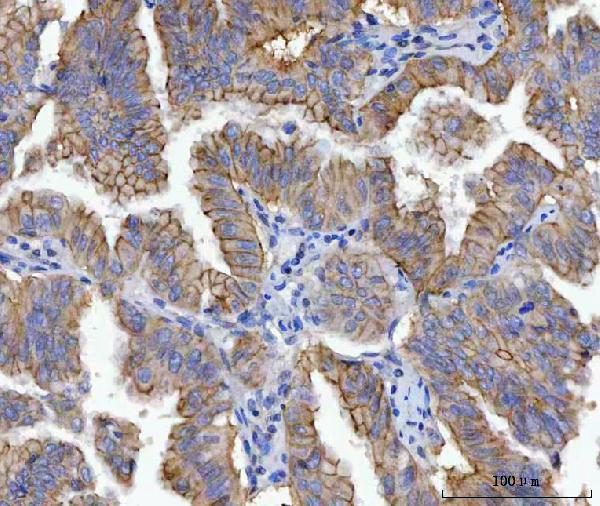
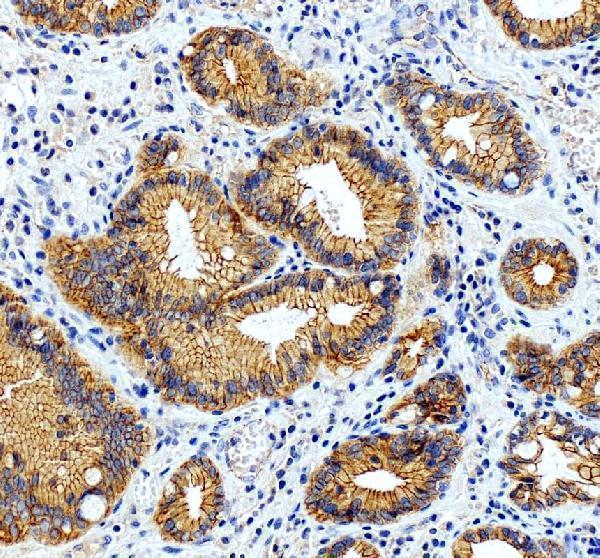
The catalog shows JUP staining in human paraffin tissue and human-cell IF (IHC and IF captions: PB9185, M01901-3, PA1117-1); PB9185 also shows mouse and rat intestine sections (PB9185 IHC captions).

Real IHC data IHC analysis of gamma Catenin using anti-gamma Catenin antibody (PB9185). gamma Catenin was detected in paraffin-embedded section of human intestinal cancer tissue. Heat mediated antigen retrieval was performed in citrate buffer (pH6, epitope retrieval solution) for 20 mins. The tissue section was blocked with 10% goat serum. The tissue section was then incubated with 1μg/ml rabbit anti-gamma Catenin Antibody (PB9185) overnight at 4°C. Biotinylated goat anti-rabbit IgG was used as secondary antibody and incubated for 30 minutes at 37°C. The tissue section was developed using Strepavidin-Biotin-Complex (SABC)(Catalog # SA1022) with DAB as the chromogen.
Anti-gamma Catenin/JUP Antibody ®
Cat # PB9185
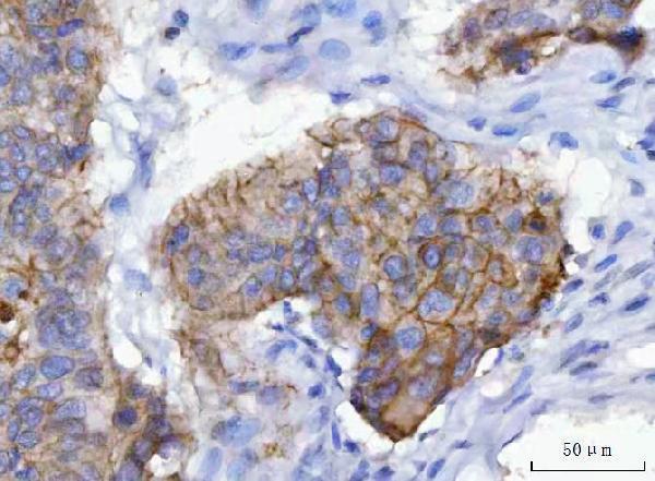
Real IHC data IHC analysis of gamma Catenin using anti-gamma Catenin antibody (M01901-3). gamma Catenin was detected in a paraffin-embedded section of human laryngeal squamous cell carcinoma tissue. Heat mediated antigen retrieval was performed in EDTA buffer (pH 8.0, epitope retrieval solution). The tissue section was blocked with 10% goat serum. The tissue section was then incubated with 2 μg/ml mouse anti-gamma Catenin Antibody (M01901-3) overnight at 4°C. Peroxidase Conjugated Goat Anti-mouse IgG was used as secondary antibody and incubated for 30 minutes at 37°C. The tissue section was developed using HRP Conjugated Mouse IgG Super Vision Assay Kit (Catalog # SV0001) with DAB as the chromogen.
Anti-gamma Catenin Antibody ® (monoclonal, 4C12D7)
Cat # M01901-3
Real IHC data IHC analysis of Gamma Catenin/JUP using anti-Gamma Catenin/JUP antibody (PA1117-1). Gamma Catenin/JUP was detected in a paraffin-embedded section of human stomach cancer tissue. Heat mediated antigen retrieval was performed in EDTA buffer (pH 8.0, epitope retrieval solution). The tissue section was blocked with 10% goat serum. The tissue section was then incubated with 2 μg/ml rabbit anti-Gamma Catenin/JUP Antibody (PA1117-1) overnight at 4°C. Peroxidase Conjugated Goat Anti-rabbit IgG was used as secondary antibody and incubated for 30 minutes at 37°C. The tissue section was developed using HRP Conjugated Rabbit IgG Super Vision Assay Kit (Catalog # SV0002) with DAB as the chromogen.
Anti-gamma Catenin/JUP Antibody ®
Cat # PA1117-1

PB9185 has IHC images from human intestinal cancer and mouse and rat intestine sections, plus IF in A431 cells (PB9185 image captions). M01901-3 has IHC images from human laryngeal squamous cell carcinoma and lung adenocarcinoma, plus IF in MCF-7 cells; PA1117-1 has an IHC image from human stomach cancer and IF in HeLa cells (M01901-3 and PA1117-1 image captions).

Which to pick: For paraffin-section IHC, choose PB9185 for its human, mouse, and rat tissue examples and listed IHC application, or mouse monoclonal M01901-3 for its human tissue examples and listed IHC application (PB9185 and M01901-3 catalog entries and IHC captions). For IF/ICC, all three list those applications and have human-cell IF images; PA1117-1 also has a paraffin-section image, although its application list omits IHC (catalog entries and IF/IHC captions: PB9185, M01901-3, PA1117-1). The paraffin-section captions do not report a fixative (IHC captions: PB9185, M01901-3, PA1117-1).

Each figure is that product's own IHC / IF validation image from its datasheet.

References

  1. UniProt Consortium. UniProt entry P14923 (PLAK_HUMAN, Junction plakoglobin).
  2. Human Protein Atlas. JUP tissue IHC expression (reliability: Supported).
  3. Human Protein Atlas. JUP subcellular location (ICC-IF): Mainly localized to the plasma membrane and cell junctions. In addition localized to vesicles..
  4. Human Protein Atlas. JUP antibody validation summary (2 antibodies).
  5. Opposing prognostic relevance of junction plakoglobin in distinct prostate cancer patient subsets. Molecular oncology 2021 — PMC8253102.
  6. SOX30 is a key regulator of desmosomal gene suppressing tumor growth and metastasis in lung adenocarcinoma. Journal of experimental & clinical cancer research : CR 2018 — PMC5984358.
  7. Suppression of ASNS expression by VHL-mediated ubiquitination hinders the progression of renal cell carcinoma through enhancing JUP expression and inhibiting PI3K-AKT and MAPK pathways. International journal of biological sciences 2026 — PMC13050451.
  8. Naxos Disease and Related Cardio-Cutaneous Syndromes. JACC. Advances 2025 — PMC11773020.
  9. PubMed PMID:2726765 — UniProt-cited evidence.
  10. PubMed PMID:11016852 — UniProt-cited evidence.
  11. PubMed PMID:16625196 — UniProt-cited evidence.