This website uses cookies to ensure you get the best experience on our website.


- Table of Contents
Plan chromogenic KDM1A IHC in paraffin sections around the expected nuclear staining (HPA tissue IHC). Use high staining bone marrow hematopoietic cells as a positive reference and interpret prostate glandular-cell staining cautiously (HPA tissue IHC).

Expected localisation, validated protocols, controls and antibodies — the at-a-glance facts below, then the full design guide.
| Expected localisation | Nuclear tissue staining (HPA tissue IHC) | |
| Staining pattern | Widespread nuclear staining across tissue cell types (HPA tissue IHC) | |
| Antigen retrieval | Tris-EDTA pH 9.0 HIER, 95–98 °C, 20 min (rule: nuclear antigen) | |
| Positive control | Adrenal gland+4 more · see all | |
| Negative control | Prostate |
| Fixation | Keep fixation consistent across sections (standard IHC practice; not target-specific) | |
| Caveat | Prostate glandular cells were not detected (HPA tissue IHC) | |
| Regulation | Ubiquitously expressed (UniProt) | |
| Isoform / epitope | Four isoforms; check antibody epitope coverage (UniProt) |
The catalog antibody protocol is followed by published KDM1A IHC protocols for papillary thyroid cancer and medulloblastoma tissue (PMC6653290; PMC3893444).
| Sample | Tissue sections; selected-image fixative not specified (standard IHC workflow) |
| Fixation | Image fixative and duration unreported (datasheet M00532-3); verify before use. |
| Sectioning | 4–5 µm sections on charged slides (standard) |
| Deparaffinisation | Xylene, graded ethanol series to water (standard) |
| Antigen retrieval | Heat-induced epitope retrieval in Tris-EDTA buffer, pH 9.0, 20 min at 95–98 °C (standard rule: nuclear antigen) |
| Peroxidase block | 3% H2O2, 10 min, room temperature (standard) |
| Blocking | 10% normal serum of the secondary host, 30 min, room temperature (standard) |
| Primary antibody | Rabbit anti-KDM1A, 1:200-1:1000 (datasheet M00532-3) |
| Primary incubation | Overnight at 4 °C (standard) |
| Detection | HRP-polymer secondary, DAB chromogen 5–10 min (standard) |
| Counterstain | Hematoxylin, blue, dehydrate and mount (standard) |
| Expected result | KDM1A-positive staining in glandular cells of adrenal gland (HPA tissue IHC: High). HPA tissue profile: Ubiquitous nuclear expression. No signal in the no-primary control. |
KDM1A should appear predominantly in nuclei, consistent with its chromatin association and lack of a transmembrane segment (UniProt O60341: subcellular location and topology). HPA describes ubiquitous nuclear expression, with high staining in several specified cell populations (HPA: tissue IHC). Treat tissue comparisons as guides rather than absolute controls because the tissue IHC assessment is Supported with medium consistency between staining and RNA data (HPA: reliability).
| Distinct nuclear chromogen in hematopoietic cells of bone marrow or cells in the cerebellar granular layer (HPA: High in both). | This fits the expected compartment and two reported high-staining populations (UniProt O60341: nucleus; HPA: tissue IHC). Judge intensity against nearby background and tissue morphology; a dark nucleus alone cannot establish antibody specificity (general IHC practice). |
| Predominantly cytoplasmic or membranous staining, with little nuclear signal (UniProt O60341: nucleus; HPA: ubiquitous nuclear expression). | This conflicts with the principal IHC pattern and warrants an artefact check (UniProt O60341: chromatin association; HPA: tissue IHC). Review morphology, counterstain and detection controls before assigning the signal to KDM1A (general IHC practice). |
| Strong staining of prostate glandular cells, reported as Not detected by HPA (HPA: prostate glandular cells). | An unexpected positive population can reflect cross-reactivity or endogenous chromogenic activity; it is not proof of either (general IHC practice). Compare nuclear localization, background and controls, allowing for HPA's medium consistency rating (HPA: reliability). |
| Diffuse color across nuclei, cytoplasm and extracellular areas, without clear cellular boundaries (general IHC practice). | Broad background makes nuclear scoring unreliable (general IHC practice). Assess the no-primary control, blocking and detection conditions before interpreting intensity; HPA's nuclear profile does not validate diffuse extracellular signal (HPA: tissue IHC). |
| No nuclear signal in a high-staining reference population, such as adrenal glandular cells (HPA: High in adrenal glandular cells). | Check tissue preservation, retrieval, antibody and detection performance before calling the specimen negative (general IHC practice). A failed reference weakens interpretation, while an HPA High designation does not guarantee every section will stain (HPA: tissue IHC). |
| Compartment and topology (UniProt O60341: nucleus, chromosome, chromatin association; no transmembrane segment). | Prioritize nuclear staining when scoring IHC. The record gives no basis to expect a membrane pattern from KDM1A itself (UniProt O60341: subcellular location and topology). |
| Tissue distribution and contrast (UniProt O60341: ubiquitous expression; HPA: low tissue specificity). | HPA reports High staining in selected cells of eight listed tissues, Low in several others, and Not detected in prostate glandular cells (HPA: tissue IHC). Interpret the specified cell population rather than an entire organ as uniformly positive. |
| Strength of tissue evidence (HPA: Supported; medium staining–RNA consistency). | Use HPA patterns as comparison points, with room for discordance between staining and RNA measurements (HPA: reliability description). Neither the rating nor a single slide independently proves that an unexpected signal is specific. |
| Antibody validation (HPA: CAB005884 IHC Supported; HPA053660 IHC status unreported). | The listed IHC support applies to CAB005884; the supplied record does not assign an IHC status to HPA053660 (HPA: antibody validation). Do not transfer validation between antibodies or infer that an unreported status means failure. |
| Isoforms and epitope (UniProt O60341: four isoforms; SWIRM domain at residues 174–273). | The supplied sources give no epitope coordinates for the catalog antibody, so isoform coverage and domain-specific staining cannot be predicted (UniProt O60341: isoforms and domain; supplied HPA antibody record). |
| Processing and modifications (UniProt O60341: no signal peptide or propeptide; 13 listed modified residues). | The annotated chain spans residues 1–852, with no annotated shedding step in this record (UniProt O60341: processing). Listed modifications do not establish an IHC intensity change or target-specific fixation sensitivity (UniProt O60341: modified residues). |
| IF/ICC Q&A: Where should signal appear? (HPA: subcellular ICC-IF). | Mainly in the nucleoplasm; HPA lists additional cytosolic localization as uncertain (HPA: supported nucleoplasm; uncertain cytosol). This IF/ICC observation does not turn cytoplasmic chromogen into an expected dominant IHC pattern (HPA: tissue IHC). |
| Situation | Likely cause | Next action |
|---|---|---|
| High-staining reference tissue lacks nuclear chromogen (HPA: High in adrenal glandular cells, among others). | The reference may have failed during staining or detection; HPA's High designation is an observed tissue pattern, not a guarantee for every section (general IHC practice; HPA: tissue IHC). | Check tissue morphology, the run's positive control, retrieval conditions and detection reagents before interpreting sample negatives (general IHC practice). Do not attribute the failure to KDM1A-specific fixation sensitivity; none is supplied. |
| Predominantly cytoplasmic color in an IHC section (HPA: ubiquitous nuclear tissue expression). | Background or nonspecific detection is possible (general IHC practice). HPA's additional cytosolic IF/ICC location is uncertain and comes from a different application (HPA: subcellular ICC-IF). | Compare the nuclear-to-cytoplasmic pattern with controls and morphology, then review antibody and detection conditions (general IHC practice). Score a dominant cytoplasmic IHC pattern cautiously (HPA: tissue IHC). |
| Prostate glandular cells stain strongly (HPA: Not detected in prostate glandular cells). | Possible causes include cross-reactivity or endogenous chromogenic activity; the unexpected pattern alone cannot identify its cause (general IHC practice; HPA: prostate tissue IHC). | Inspect nuclear localization and compare a no-primary control with a known high-staining cell population in the run (general IHC practice; HPA: tissue IHC). Interpret disagreement in light of HPA's medium consistency rating (HPA: reliability). |
| Diffuse chromogen obscures nuclei throughout the section (general IHC practice). | Nonspecific binding, inadequate washing or endogenous detection activity may elevate background in chromogenic IHC (general IHC practice). The supplied sources do not identify a KDM1A-specific background mechanism. | Review the no-primary control, blocking, washes and detection controls; restore readable nuclear boundaries before scoring (general IHC practice). Compare the resolved pattern with HPA's ubiquitous nuclear profile (HPA: tissue IHC). |
| One cell population stains weakly while another is strong (HPA: variable tissue IHC levels). | Expression varies by annotated population: HPA reports High in kidney glomerular cells and Low in small-intestine glandular cells, for example (HPA: tissue IHC). | Identify the cells on the counterstained section and compare each with its own HPA entry (general IHC practice; HPA: tissue IHC). Avoid treating a Low population as a failed positive control. |
| Two antibodies yield different nuclear patterns or intensities (HPA: antibody validation entries). | Their IHC evidence differs: CAB005884 is Supported, while HPA053660 has no reported IHC status in the supplied record (HPA: antibody validation). Epitope coordinates are not supplied, so the discrepancy cannot be assigned to isoform recognition. | Check each antibody's documented IHC validation and matched run controls, then compare compartment and cell population rather than intensity alone (HPA: antibody validation and tissue IHC; general IHC practice). |




Comprehensive Human Protein Atlas IHC scoring per tissue (reliability: Supported — Medium consistency between antibody staining and RNA expression data.). Rows are taken directly from the HPA tissue chart — click any row's HPA link to view the source.
| Tissue | Cell type | Level | Evidence | Source |
|---|---|---|---|---|
| Adrenal gland | Glandular cells | High | Protein (IHC) | HPA → |
| Bone marrow | Hematopoietic cells | High | Protein (IHC) | HPA → |
| Cerebellum | Cells in granular layer | High | Protein (IHC) | HPA → |
| Endometrium | Cells in endometrial stroma | High | Protein (IHC) | HPA → |
| Gallbladder | Glandular cells | High | Protein (IHC) | HPA → |
| Tissue | Cell type | Level | Evidence | Source |
|---|---|---|---|---|
| Prostate | Glandular cells | Not detected | Protein (IHC) | HPA → |
Troubleshoot KDM1A staining by checking nuclear localization, section processing, and cell-specific controls before interpreting chromogenic signal.
Both anti-KDM1A antibodies have IHC tissue images (catalog IHC captions) and list IF as an application (catalog applications); the catalog provides no IF images (catalog image inventory).
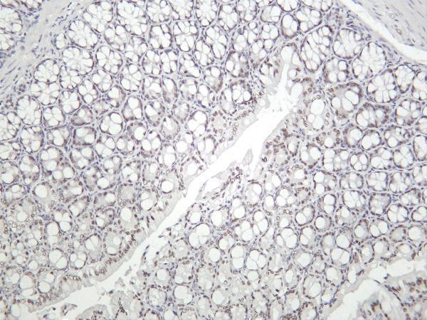
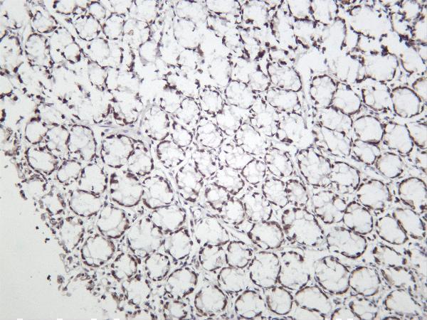
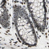
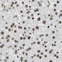
M00532-3 has IHC images of mouse and rat colon and human prostate and prostate carcinoma (catalog IHC captions); its listed reactivity is Human, Mouse, and Rat (catalog reactivity). M00532-1 has an IHC image of paraffin-embedded human colon (catalog IHC caption), lists IHC, IF, and ICC applications (catalog applications), and lists Human, Mouse, and Rat reactivity (catalog reactivity).
Which to pick: For tissue IHC across the pictured species, choose M00532-3: its own captions show mouse and rat colon and human prostate tissue, but do not report section processing or fixative (catalog IHC captions). For paraffin-section IHC in human colon or for ICC/IF, choose M00532-1: its own caption specifies paraffin-embedded human colon without reporting the fixative, and ICC/IF is listed as an application (catalog IHC caption; catalog applications). Both are rabbit monoclonals (catalog titles), list Human, Mouse, and Rat reactivity (catalog reactivity), and list IF as an application without an IF image in the payload (catalog applications; catalog image inventory).