KIF2A / Kinesin-like protein KIF2A · IHC design guide

Design Immunohistochemistry for KIF2A

Plan KIF2A staining in paraffin sections using the catalog antibody at 2–5 μg/ml (datasheet A03979-1). Compare cytoplasmic and nuclear staining with the tissue IHC profile, while assessing spindle staining in mitotic cells separately (HPA tissue IHC; UniProt).

Evidence assembled Oct 2026 · For research use; verify linked source records and product datasheet before use
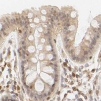
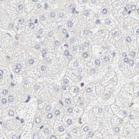
Immunohistochemistry protocol sheet for KIF2A (IHC for KIF2A): expected localisation Cytoplasmic and nuclear staining in most tissues (HPA tissue IHC), antibody A03979-1, validated IHC image, and IHC protocol steps
Printable KIF2A IHC protocol sheet — expected localisation Cytoplasmic and nuclear staining in most tissues (HPA tissue IHC), antibody A03979-1, controls and protocol steps. Open the full KIF2A IHC guide →

KIF2A Immunohistochemistry Experimental Design Guide

Expected localisation, validated protocols, controls and antibodies — the at-a-glance facts below, then the full design guide.

Must know before staining
Expected localisation Cytoplasmic and nuclear staining in most tissues (HPA tissue IHC)
Staining pattern Most tissues show cytoplasmic and nuclear staining (HPA tissue IHC)
Antigen retrieval EDTA pH 8.0 HIER, heat-mediated (datasheet A03979-1)
Positive control ⓘ Adrenal gland+4 more · see all
Negative control ⓘ Heart muscle+4 more · see all
Important caveats
Reasons your staining may differ from the expected pattern.
Fixation Keep fixation consistent across paraffin sections. (standard IHC practice; not target-specific)
Caveat Spindle enrichment varies with mitotic stage (UniProt)
Regulation PLK1 activity supports spindle targeting (UniProt)
Isoform / epitope 5 isoforms; check whether the epitope is shared (UniProt)
Section 1

Recommended KIF2A IHC & IF Protocols

The catalog antibody’s IHC-P protocol (datasheet A03979-1) is paired with two published KIF2A chromogenic IHC methods (PMC5537331; PMC9102491).

Recommended immunohistochemistry (IHC-P) protocol parameters
SampleParaffin-embedded human lung cancer tissue; fixative not specified (datasheet A03979-1)
FixationImage fixative and duration unreported (datasheet A03979-1); verify before use.
Sectioning4–5 µm sections on charged slides (standard)
DeparaffinisationXylene, graded ethanol series to water (standard)
Antigen retrievalHeat retrieval: EDTA pH 8.0 (datasheet A03979-1); 20 min, 95–100 °C (standard)
Peroxidase block3% H2O2, 10 min, room temperature (standard)
Blocking10% goat serum (datasheet A03979-1)
Primary antibodyRabbit anti-KIF2A, 2-5 μg/ml (datasheet A03979-1)
Primary incubationOvernight at 4 °C (datasheet A03979-1)
DetectionStreptavidin-biotin complex (SABC), DAB chromogen (datasheet A03979-1)
CounterstainHematoxylin, blue, dehydrate and mount (standard)
Expected resultKIF2A-positive staining in glandular cells of adrenal gland (HPA tissue IHC: High). HPA tissue profile: Cytoplasmic and nuclear expression in most tissues. No signal in the no-primary control.
💡Decision noteStart with heat retrieval in EDTA pH 8.0 for the catalog antibody (datasheet A03979-1); the published excerpts do not specify retrieval conditions (PMC5537331; PMC9102491).
Section 2

What Is the Expected KIF2A Staining Pattern?

KIF2A staining is expected in the cytoplasm and nuclei of many tissues (HPA tissue IHC: cytoplasmic and nuclear expression in most tissues). Cytoplasmic staining is consistent with KIF2A at centrosomes and mitotic spindles (UniProt O00139: subcellular location). Suitable high-staining cells include cerebellar Purkinje cells, colon glandular cells and bone marrow hematopoietic cells (HPA tissue IHC: High). HPA rates its tissue IHC evidence Approved, with medium agreement between staining and RNA data (HPA tissue IHC: reliability). KIF2A has no transmembrane segment (UniProt O00139: topology).

What am I looking at on my slide?
Cytoplasmic and nuclear staining in the expected cells, with occasional concentrated signal at a mitotic spindle or pole.This fits the broad tissue pattern (HPA tissue IHC: cytoplasmic and nuclear expression). Spindle and pole localisation is biologically plausible from prophase through metaphase (UniProt O00139: subcellular location). Assess those structures only where mitotic cells are identifiable; a diffuse chromogenic deposit alone cannot establish spindle localisation (general IHC interpretation).
Staining is confined to cell borders or another unexpected compartment, without a matching cytoplasmic or nuclear pattern.A border-only pattern is discordant with the reported tissue pattern (HPA tissue IHC: cytoplasmic and nuclear expression) and the absence of a transmembrane segment (UniProt O00139: topology). Treat it as a possible artefact and compare the positive control, morphology and detection-only control (general IHC practice). Nuclear staining by itself is not inherently misplaced (HPA tissue IHC: nuclear expression).
Strong staining appears in cardiomyocytes or kidney glomerular cells.These specific populations were reported as not detected (HPA tissue IHC: heart cardiomyocytes; kidney glomerular cells). Check whether the signal follows tissue pigment, endogenous detection activity or an unrelated antibody reaction (general chromogenic IHC practice). An HPA negative observation is a comparison point, not proof that every specimen must be negative (HPA tissue IHC: Approved; medium consistency).
The whole section develops diffuse colour, including spaces outside cells.A widespread deposit that ignores cell boundaries is inconsistent with the reported cell-associated pattern (HPA tissue IHC: cytoplasmic and nuclear expression). Review detection-only staining, endogenous enzyme blocking, antibody concentration and washes (general chromogenic IHC practice). Do not score diffuse background as KIF2A-positive cells (general IHC interpretation).
No staining is visible in an intended positive cell population.Confirm that the sampled section contains the chosen cells: Purkinje cells, colon glandular cells and bone marrow hematopoietic cells have High reported staining (HPA tissue IHC). If they are present, review the IHC-validated antibody, retrieval and detection controls (general IHC practice). Absence in one section does not establish absence of KIF2A across that tissue (HPA tissue IHC: medium consistency).
💡Expected KIF2A appearanceCall positive when identifiable high-staining cells show cell-associated cytoplasmic and/or nuclear chromogen, with spindle or pole concentration only in suitable mitotic cells (HPA tissue IHC: High cell populations; cytoplasmic and nuclear profile; UniProt O00139: spindle localisation); widespread extracellular colour or isolated border staining is suspect (general IHC interpretation; UniProt O00139: no transmembrane segment).
How each factor affects the staining
Which cells make useful tissue comparisons?Purkinje cells, colon glandular cells and bone marrow hematopoietic cells are reported High; cardiomyocytes and kidney glomerular cells are not detected (HPA tissue IHC). Compare the named cell populations, since a tissue contains multiple cell types (general IHC interpretation).
How strong is the tissue-pattern evidence?HPA labels the IHC profile Approved and reports medium consistency between antibody staining and RNA expression (HPA tissue IHC: reliability). HPA004716 has Approved IHC validation; HPA004715 has Enhanced ICC validation, which does not establish IHC validation for that antibody (HPA antibodies: validation status).
Do isoforms or modifications predict this antibody's staining?Five isoforms and nine modified residues are listed (UniProt O00139: isoforms; modified residues). The supplied sources give no antibody epitope or isoform coverage, so they cannot predict an isoform-specific tissue pattern or retrieval response (UniProt O00139: record scope; HPA antibodies: validation status).
Does topology suggest surface staining?KIF2A has no transmembrane segment and no signal peptide; its annotated chain spans residues 1–706 (UniProt O00139: topology; processing). A surface-only result therefore warrants review, while these annotations alone do not specify an antibody epitope or its accessibility in paraffin sections (UniProt O00139: record scope).
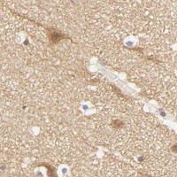
What should an IF/ICC image show?HPA reports enhanced nucleoplasm, centrosome and cytosol localisation; mitotic spindle localisation is uncertain in its ICC-IF summary (HPA subcellular: location and evidence labels). These cellular images can guide compartment checks, but they do not replace tissue IHC interpretation or provide an IF/ICC protocol (HPA subcellular: ICC-IF; HPA tissue IHC: tissue profile).
Why is my staining missing, weak or wrong?
SituationLikely causeNext action
The positive-control section has no visible signal.The expected cells may be absent from the section, or retrieval, primary incubation or chromogenic detection may have failed (general IHC practice).First locate the intended cell population; colon glandular and cerebellar Purkinje cells are reported High (HPA tissue IHC). Then check the IHC-validated antibody and each detection step with appropriate process controls (general IHC practice).
A test tissue stains weakly while the positive control works.The sampled population may have low reported staining: stomach glandular cells and adipocytes are listed Low (HPA tissue IHC).Score the named cells against their expected level and inspect section morphology (HPA tissue IHC: Low populations; general IHC practice). Avoid interpreting a weak signal as an assay failure solely because another tissue stains strongly (HPA tissue IHC: variable levels).
Unexpected cell populations show strong chromogen.Cross-reactivity or endogenous detection activity is possible (general chromogenic IHC practice); HPA reports some named populations as not detected (HPA tissue IHC).Run a primary-omission control and assess endogenous enzyme blocking; compare the same cell population with HPA rather than the tissue name alone (general IHC practice; HPA tissue IHC: cell-specific levels).
Brown colour covers the section without cellular structure.Background may arise from inadequate blocking, excess detection reagents or insufficient washing (general chromogenic IHC practice).Check the detection-only control, wash steps and reagent concentration, then score only resolved cellular signal (general IHC practice). The reported pattern is cytoplasmic and nuclear within cells (HPA tissue IHC).
Signal appears only along membranes or tissue edges.Edge-associated deposition or other nonspecific staining may mimic positivity (general IHC practice); KIF2A has no transmembrane segment (UniProt O00139: topology).Inspect internal cells away from section edges and compare morphology and controls (general IHC practice). Seek cytoplasmic or nuclear staining in expected populations (HPA tissue IHC: tissue profile).
Nuclear IHC staining seems inconsistent with spindle-related KIF2A biology.The sources describe different observations: tissue IHC includes nuclear staining, while UniProt annotates cytoplasmic spindle localisation (HPA tissue IHC: profile; UniProt O00139: subcellular location).Retain nuclear staining as potentially expected when it is cell-associated and controls behave appropriately (HPA tissue IHC: nuclear expression; general IHC practice). Use cell morphology before attributing a discrete focus to a mitotic pole (UniProt O00139: spindle-pole localisation).

Sample controls for KIF2A IHC & IF

🧪Run adrenal gland first and require staining in glandular cells (HPA: High in adrenal gland glandular cells); use heart muscle cardiomyocytes as the negative tissue (HPA: Not detected in heart muscle cardiomyocytes). On the adrenal slide, assess any unstained neighboring cells for background, but do not designate a specific internal negative cell type because none is identified in the supplied adrenal row (HPA: High in adrenal gland glandular cells).
Positive control tissue: Adrenal gland (Glandular cells, HPA High)
Negative control tissue: Heart muscle (HPA Not detected)
ICC-IF cell lines (HPA subcellular resource): HPA ICC-IF images show KIF2A in A-549, Rh30, U2OS, ASC52telo, RPTEC/TERT1, hTERT-RPE1 (serum starved), Sperm, HeLa BAC 6513, HeLa , with annotated localisation: Nucleoplasm (enhanced), Nucleoli (uncertain), Centrosome (enhanced), Basal body (uncertain), Annulus (approved) (HPA subcellular).
Technical controls: Include a no-primary, secondary-only section, a concentration-matched rabbit isotype control, and a KIF2A knockout specimen or immunizing-peptide competition when available (caption: rabbit primary antibody; standard IHC controls). For the SABC/DAB assay, block endogenous peroxidase and assess endogenous biotin with the no-primary section (caption: biotinylated secondary, SABC and DAB).
⚠️Feasibility: A target-specific fixation window or fixation effect is unreported, and the exact A03979-1 paraffin-section caption does not state a fixative (caption: paraffin section; fixative unreported). The reported IHC procedure uses heat retrieval in EDTA at pH 8.0 and 2 μg/ml primary antibody overnight at 4°C; whether retrieval is required under other conditions is unreported (caption: A03979-1 IHC). Frozen sections have no supplied matched protocol, and the HPA ICC-IF images do not establish that IF is easier than IHC-P; check the chromogenic assay for endogenous peroxidase and biotin signal (HPA: ICC-IF images; caption: SABC/DAB detection).

HPA tissue IHC evidence for KIF2A

Comprehensive Human Protein Atlas IHC scoring per tissue (reliability: Approved — Medium consistency between antibody staining and RNA expression data.). Rows are taken directly from the HPA tissue chart — click any row's HPA link to view the source.

Positive expression · recommended positive controls

TissueCell typeLevelEvidenceSource
Adrenal gland Glandular cells High Protein (IHC) HPA →
Appendix Glandular cells High Protein (IHC) HPA →
Bone marrow Hematopoietic cells High Protein (IHC) HPA →
Bronchus Respiratory epithelial cells High Protein (IHC) HPA →
Caudate Glial cells High Protein (IHC) HPA →

Undetected expression · recommended negative controls

TissueCell typeLevelEvidenceSource
Heart muscle Cardiomyocytes Not detected Protein (IHC) HPA →
Kidney Cells in glomeruli Not detected Protein (IHC) HPA →
Liver Cholangiocytes Not detected Protein (IHC) HPA →
Oral mucosa Squamous epithelial cells Not detected Protein (IHC) HPA →
Ovary Follicle cells Not detected Protein (IHC) HPA →
Section 3

Advanced KIF2A IHC Tips

Troubleshoot KIF2A staining in paraffin sections by checking retrieval, compartment patterns, controls and scoring against the documented IHC evidence.

What retrieval should I try first when KIF2A staining is weak?
Start with heat-mediated retrieval in EDTA at pH 8.0 (datasheet A03979-1). The documented paraffin-section image used that retrieval before overnight incubation with 2 μg/ml catalog antibody at 4°C (datasheet A03979-1). If staining remains weak, compare retrieval duration or a different buffer on matched sections while keeping antibody concentration and detection constant (standard IHC practice). Include a known staining region, such as bronchial respiratory epithelium, to distinguish failed retrieval from low expression in the test region (HPA: High in bronchus respiratory epithelial cells). Check tissue preservation and section adhesion before increasing retrieval intensity, since excessive heating can impair morphology (standard IHC practice).
Could fixation explain faint or uneven KIF2A staining?
Target-specific fixation sensitivity is unknown: the selected paraffin-section caption does not state a fixative (datasheet A03979-1). Record the fixative, fixation duration and processing history for each section before attributing weak staining to KIF2A biology (standard IHC practice). Compare similarly processed sections with a documented positive cell population, such as cerebellar Purkinje cells, using the same retrieval and detection run (HPA: High in cerebellar Purkinje cells; standard IHC practice). Assess whether weak signal tracks folds, thick areas or damaged morphology, which can indicate a preparation problem (standard IHC practice). Neither tissue staining patterns nor KIF2A topology establish a fixation-dependent loss of this epitope (HPA: tissue IHC; UniProt O00139 topology).
How should I assess cytoplasmic, nuclear and mitotic KIF2A staining?
Score compartments separately because tissue IHC reports cytoplasmic and nuclear expression in most tissues (HPA: tissue IHC profile). Cytoplasmic staining can be assessed alongside centrosomal or spindle-associated staining in dividing cells; UniProt places KIF2A at centrosomes, spindle poles and spindle microtubules from prophase to metaphase (UniProt O00139 subcellular). Do not require a visible spindle pattern in every section, since mitotic structures depend on the sampled cell state and section plane (UniProt O00139 subcellular; standard IHC practice). Compare nuclear signal with adjacent cytoplasmic signal and a no-primary control before scoring it (HPA: tissue IHC profile; standard IHC practice). Record distinct patterns rather than combining them into one intensity grade (standard IHC practice).
Could an isoform or modified epitope explain discordant IHC staining?
KIF2A has 5 listed isoforms, a kinesin motor domain at residues 223–553, and multiple modified residues (UniProt O00139 isoforms, domains and modified residues). Those features make antibody epitope mapping relevant, but the supplied catalog caption does not identify this antibody’s epitope (datasheet A03979-1). Check the antibody’s immunogen or mapped epitope before claiming isoform selectivity or modification sensitivity (standard IHC practice). If sections disagree, compare matched processing and retrieval conditions, then assess whether the same cellular compartments stain with an independently validated antibody (standard IHC practice). Do not assign an isoform from chromogenic staining intensity alone (UniProt O00139 isoforms; standard IHC practice).
How can IF help assess a questionable KIF2A IHC pattern?
Use IF as a separate corroborating assay for an uncertain IHC pattern, since the catalog evidence here documents chromogenic staining of a paraffin section (datasheet A03979-1). Multiplex KIF2A with a validated marker for the expected cell type; bronchial respiratory epithelial cells are one documented high-staining population (HPA: High in bronchus respiratory epithelial cells; standard IF practice). Select spectrally separated fluorophores and consider a far-red channel if the tissue has strong shorter-wavelength autofluorescence (standard IF practice). KIF2A lacks a transmembrane segment and has intracellular locations, so permeabilisation must allow access to the intracellular epitope; its precise epitope is unreported here (UniProt O00139 topology and subcellular; datasheet A03979-1). Include single-label and no-primary controls when comparing IF with IHC (standard IF practice).
How do I reduce diffuse or granular DAB background?
First inspect a no-primary section to separate detection-system background from signal dependent on the KIF2A antibody (standard IHC practice). The documented chromogenic image used 10% goat-serum block, a biotinylated secondary, streptavidin–biotin detection and DAB (datasheet A03979-1). Block endogenous peroxidase before DAB development, and assess whether endogenous biotin contributes signal when using a biotin-based system (standard IHC practice). If background persists, compare shorter primary exposure or a lower antibody concentration against a matched positive section, changing one variable at a time (standard IHC practice). Interpret diffuse deposit cautiously where folds, damaged tissue or section edges also stain (standard IHC practice).
How should I quantify KIF2A staining across sections? ⚠ ANSWER MARKED FOR VERIFICATION
Define the cell population and compartment before scoring, since tissue IHC reports both cytoplasmic and nuclear KIF2A staining (HPA: tissue IHC profile). For a cell-based readout, record percent positive cells and intensity or calculate an H-score from intensity bins; use positive-cell density per mm² when cell abundance itself matters (standard IHC practice). Normalise counts to the number of eligible cells or analysed viable tissue area, and apply the same threshold to all matched sections (standard IHC practice). Exclude folds, necrosis and section edges using a prespecified rule (standard IHC practice). Report nuclear, cytoplasmic and mitotic patterns separately so changes in cell-cycle composition do not masquerade as uniform expression changes (UniProt O00139 function; standard IHC practice).
What makes a KIF2A-positive IHC result convincing?
A convincing result combines reproducible cellular staining with appropriate controls and morphology (standard IHC practice). Cytoplasmic or nuclear staining can fit the tissue IHC profile, while centrosomal and spindle-associated patterns fit reported KIF2A locations in dividing cells (HPA: tissue IHC profile; UniProt O00139 subcellular). Compare the sampled cell type with documented patterns: bronchial respiratory epithelial cells are High, whereas heart muscle cardiomyocytes are Not detected (HPA: tissue IHC). Treat isolated staining at cut edges, in necrotic areas or on a no-primary section as possible artefact, including endogenous enzyme signal (standard IHC practice). HPA labels its tissue staining Approved with medium RNA–protein consistency, so resolve discordant cases with independent evidence (HPA: tissue IHC reliability; standard IHC practice).
Boster reagents

Best KIF2A / Kinesin-like protein KIF2A IHC Antibodies

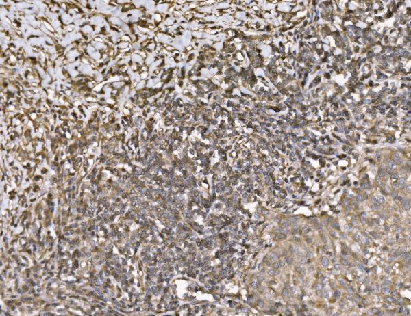
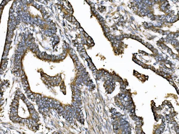
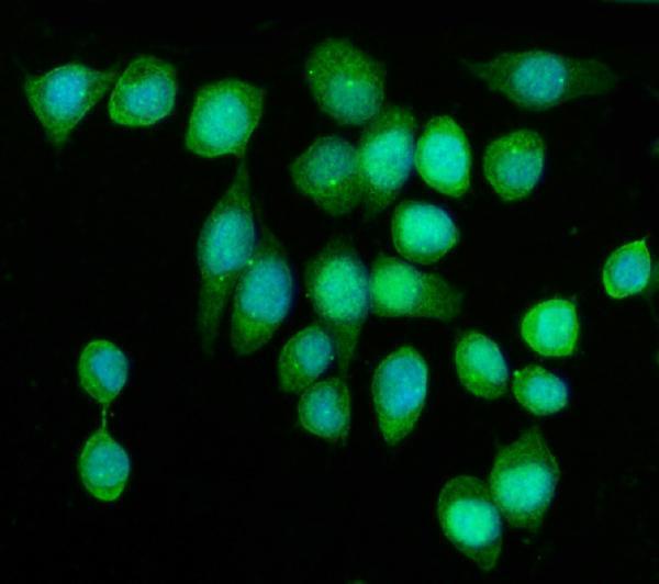
A03979-1 has IHC images from human paraffin sections and an IF image from SiHa cells (catalog image captions). Listed reactivity covers human, mouse, rat, and monkey (catalog reactivity).

Real IHC data IHC analysis of KIF2A using anti-KIF2A antibody (A03979-1). KIF2A was detected in a paraffin-embedded section of human lung cancer tissue. Heat mediated antigen retrieval was performed in EDTA buffer (pH 8.0, epitope retrieval solution). The tissue section was blocked with 10% goat serum. The tissue section was then incubated with 2 μg/ml rabbit anti-KIF2A Antibody (A03979-1) overnight at 4°C. Biotinylated goat anti-rabbit IgG was used as secondary antibody and incubated for 30 minutes at 37°C. The tissue section was developed using Strepavidin-Biotin-Complex (SABC) (Catalog # SA1022) with DAB as the chromogen.
Anti-KIF2A Antibody ®
Cat # A03979-1

A03979-1 is shown in IHC on paraffin sections of human lung cancer, pancreatic cancer, and placenta (catalog IHC captions). The same SKU is shown in IF on SiHa cells and lists both IF and ICC applications (catalog IF caption; catalog applications).

Which to pick: For tissue IHC, choose A03979-1: its caption documents a human paraffin section with EDTA retrieval and DAB detection; the fixative is unreported (A03979-1 IHC caption). For IF/ICC, A03979-1 lists both applications and shows IF in SiHa cells (catalog applications; A03979-1 IF caption). For cross-species work, A03979-1 lists human, mouse, rat, and monkey reactivity, while its provided IHC images document human tissue (catalog reactivity; catalog IHC captions).

Each figure is that product's own IHC / IF validation image from its datasheet.

References

  1. UniProt Consortium. UniProt entry O00139 (KIF2A_HUMAN, Kinesin-like protein KIF2A).
  2. Human Protein Atlas. KIF2A tissue IHC expression (reliability: Approved).
  3. Human Protein Atlas. KIF2A subcellular location (ICC-IF): Mainly localized to the nucleoplasm, nucleoli, centrosome, basal body and annulus. In addition localized to the cytosol, mitotic spindle, mid piece and principal piece..
  4. Human Protein Atlas. KIF2A antibody validation summary (2 antibodies).
  5. High KIF2A expression predicts unfavorable prognosis in diffuse large B cell lymphoma. Annals of hematology 2017 — PMC5537331.
  6. Kinesin family member 2A links with advanced tumor stage, reduced chemosensitivity and worse prognosis in gastric cancer. Journal of clinical laboratory analysis 2022 — PMC9102491.
  7. Kinesin Family Member 2A Serves as a Potential Biomarker Reflecting More Frequent Lymph Node Metastasis and Tumor Recurrence Risk in Basal-Like Breast Cancer Patients. Frontiers in surgery 2022 — PMC9243457.
  8. Aberrant kinesin family member 2A signifies tumor size and invasion, and may help predict prognosis of patients with papillary thyroid carcinoma. Oncology letters 2022 — PMC9219030.
  9. PubMed PMID:9177777 — UniProt-cited evidence.
  10. PubMed PMID:14702039 — UniProt-cited evidence.
  11. PubMed PMID:17974005 — UniProt-cited evidence.