This website uses cookies to ensure you get the best experience on our website.


- Table of Contents
Plan paraffin-section LONP1 IHC around granular cytoplasmic staining in glandular cells (HPA tissue IHC) and its mitochondrial matrix location (UniProt). Use adrenal glandular cells as a high-staining reference and thyroid glandular cells as a not-detected reference (HPA tissue IHC); the IHC-validated antibody is listed at 0.5–1 μg/mL (datasheet).

Expected localisation, validated protocols, controls and antibodies — the at-a-glance facts below, then the full design guide.
| Expected localisation | Granular cytoplasm (HPA tissue IHC); mitochondrial matrix (UniProt) | |
| Staining pattern | Granular cytoplasmic staining in glandular cells (HPA tissue IHC) | |
| Antigen retrieval | Citrate pH 6 HIER, heat-mediated (datasheet A03808-2) | |
| Positive control | Adrenal gland+4 more · see all | |
| Negative control | Thyroid gland |
| Fixation | Keep fixation conditions consistent across sections. (standard IHC practice; not target-specific) | |
| Caveat | Thyroid glandular cells are not detected, despite positive glands elsewhere (HPA tissue IHC) | |
| Regulation | Adrenal gland RNA is tissue enhanced (HPA tissue RNA) | |
| Isoform / epitope | 3 isoforms; mature chain 68–959; epitope impact unknown (UniProt) |
The catalog antibody’s IHC-P protocol uses citrate pH 6 retrieval (datasheet A03808-2). One published LONP1 tumor-section protocol follows (PMC11840119).
| Sample | Paraffin-embedded human mammary cancer tissues; fixative not specified (datasheet A03808-2) |
| Fixation | Image fixative and duration unreported (datasheet A03808-2); verify before use. |
| Sectioning | 4–5 µm sections on charged slides (standard) |
| Deparaffinisation | Xylene, graded ethanol series to water (standard) |
| Antigen retrieval | Heat retrieval: Citrate pH 6, 20 min (datasheet A03808-2) |
| Peroxidase block | 3% H2O2, 10 min, room temperature (standard) |
| Blocking | 10% goat serum (datasheet A03808-2) |
| Primary antibody | Rabbit anti-LONP1, 0.5-1μg/ml (datasheet A03808-2) |
| Primary incubation | Overnight at 4 °C (datasheet A03808-2) |
| Detection | Streptavidin-biotin complex (SABC), DAB chromogen (datasheet A03808-2) |
| Counterstain | Hematoxylin, blue, dehydrate and mount (standard) |
| Expected result | LONP1-positive staining in glandular cells of adrenal gland (HPA tissue IHC: High). HPA tissue profile: Granular cytoplasmic expression. No signal in the no-primary control. |
LONP1 is a mitochondrial matrix protein with no transmembrane segment (UniProt P36776). In paraffin-section IHC, expect granular cytoplasmic staining, especially in adrenal, duodenal, appendiceal and colonic glandular cells and bronchial respiratory epithelium (HPA: granular cytoplasmic profile; High in listed cells). HPA rates its tissue staining Supported, citing consistency with RNA expression (HPA: Supported).
| Granular cytoplasmic staining in adrenal or duodenal glandular cells. | This fits the reported pattern and provides a useful positive reference (HPA: High in these cells; granular cytoplasmic profile). Score the relevant cells and their cytoplasmic distribution; staining of a tissue section alone does not establish that every cell is positive (general IHC practice). |
| Predominantly nuclear or cell-surface staining, without a convincing granular cytoplasmic component. | Question the signal's specificity: LONP1 resides in the mitochondrial matrix and has no transmembrane segment (UniProt P36776). HPA reports uncertain nucleoplasmic and cytosolic signals in ICC-IF; those observations do not establish a predominantly nuclear or surface IHC pattern (HPA: subcellular ICC-IF). |
| Strong signal in a cell population where the HPA tissue profile reports little or no staining. | Compare the precise cell type before calling it positive. Thyroid glandular cells are Not detected, while heart cardiomyocytes and liver cholangiocytes are Low in the supplied IHC profile (HPA: tissue IHC). Unexpected strong staining warrants checks for cross-reactivity or endogenous detection activity (general IHC practice). |
| Diffuse chromogen across tissue, including spaces or structures without identifiable cells. | A widespread deposit is difficult to reconcile with the reported granular cytoplasmic pattern (HPA: tissue IHC). Check background in the relevant controls and reassess blocking, washing and detection reagents before interpreting cell-specific LONP1 expression (general IHC practice). |
| No detectable signal in adrenal or duodenal glandular cells. | These are reported High in the HPA IHC profile, so a blank result calls for a technical review before a biological negative call (HPA: High in these cells). Inspect section quality, antigen retrieval, antibody use and chromogen development as general IHC checks; target-specific fixation sensitivity is unreported here. |
| Cell and tissue context | HPA reports High staining in adrenal, appendix, colon and duodenal glandular cells and bronchial respiratory epithelium; thyroid glandular cells are Not detected (HPA: tissue IHC). These are cell-specific observations, so compare like cells when choosing a reference section (general IHC practice). |
| Subcellular interpretation | Matrix localisation and the absence of a transmembrane segment support a cytoplasmic, mitochondrial interpretation (UniProt P36776). HPA describes the tissue-IHC profile as granular cytoplasmic; routine chromogenic IHC does not by itself prove mitochondrial colocalisation (HPA: tissue IHC; general IHC practice). |
| Antibody validation | HPA002192 carries an IHC Supported rating; HPA002034 has no supplied IHC rating (HPA: antibody validation). Do not transfer the antibodies' ICC Enhanced ratings to paraffin IHC (HPA: antibody validation). Interpret an unexpected pattern alongside appropriate assay controls (general IHC practice). |
| Processing and isoforms | UniProt lists 3 isoforms and a mature chain spanning residues 68–959 (UniProt P36776). Epitope-dependent differences are possible in principle, but no epitope position or isoform-specific IHC result is supplied; do not assign a particular staining pattern to either feature. |
| IF/ICC Q&A | What location should IF/ICC show? Mainly mitochondria; HPA also lists nucleoplasm and cytosol as uncertain additional locations (HPA: subcellular ICC-IF). This localisation answer is not an IF/ICC protocol or a substitute for interpreting the paraffin-section IHC pattern. |
| Situation | Likely cause | Next action |
|---|---|---|
| Positive reference tissue is blank. | A technical step may have failed; adrenal and duodenal glandular cells are reported High (HPA: tissue IHC). The supplied sources do not identify a LONP1-specific fixation effect. | Review section integrity, retrieval, antibody application and detection development with assay controls (general IHC practice). Avoid interpreting the blank section as biological absence until those checks pass. |
| Signal is mostly nuclear. | This conflicts with mitochondrial matrix localisation and the granular cytoplasmic tissue-IHC profile (UniProt P36776; HPA: tissue IHC). HPA's nucleoplasmic ICC-IF location is uncertain (HPA: subcellular ICC-IF). | Recheck morphology and background controls; compare with a known High cell population in the same staining workflow (HPA: tissue IHC; general IHC practice). |
| Thyroid glandular cells stain strongly. | HPA reports this cell population as Not detected, making strong signal unexpected for the supplied tissue-IHC reference (HPA: thyroid glandular cells). Cross-reactivity or endogenous detection activity is possible (general IHC practice). | Verify cell identity and inspect reagent-control staining; reassess blocking and detection if controls also develop chromogen (general IHC practice). |
| Weak staining in cardiomyocytes is scored as a failed assay. | HPA reports Low staining in cardiomyocytes, although UniProt lists heart among tissues expressing LONP1 (HPA: heart muscle; UniProt P36776 tissue specificity). These statements address different levels of evidence. | Use a reported High cell population to judge technical performance, then record cardiomyocyte intensity separately (HPA: tissue IHC; general IHC practice). |
| Granules are obscured by diffuse background. | Excess background can mask the reported granular cytoplasmic pattern (HPA: tissue IHC; general IHC practice). The supplied sources do not establish a LONP1-specific retrieval or fixation cause. | Inspect control sections and review blocking, washing and chromogen development as general IHC workflow checks before scoring localisation (general IHC practice). |
| Two antibodies appear to give different IHC patterns. | The supplied HPA ratings differ: HPA002192 is IHC Supported, while HPA002034 has no supplied IHC rating; both are ICC Enhanced (HPA: antibody validation). Their ICC status cannot resolve an IHC discrepancy. | Compare cell-level localisation against the HPA granular cytoplasmic profile and assess each IHC assay with its own controls (HPA: tissue IHC; general IHC practice). |


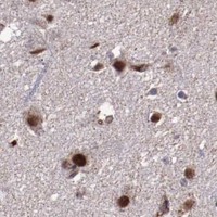
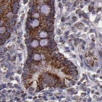
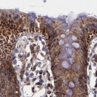
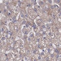


Comprehensive Human Protein Atlas IHC scoring per tissue (reliability: Supported — High consistency between antibody staining and RNA expression data.). Rows are taken directly from the HPA tissue chart — click any row's HPA link to view the source.
| Tissue | Cell type | Level | Evidence | Source |
|---|---|---|---|---|
| Adrenal gland | Glandular cells | High | Protein (IHC) | HPA → |
| Appendix | Glandular cells | High | Protein (IHC) | HPA → |
| Bronchus | Respiratory epithelial cells | High | Protein (IHC) | HPA → |
| Colon | Glandular cells | High | Protein (IHC) | HPA → |
| Duodenum | Glandular cells | High | Protein (IHC) | HPA → |
| Tissue | Cell type | Level | Evidence | Source |
|---|---|---|---|---|
| Thyroid gland | Glandular cells | Not detected | Protein (IHC) | HPA → |
Troubleshoot LONP1 staining in paraffin sections by checking retrieval, compartment, cell type and controls before comparing chromogenic signal across specimens.
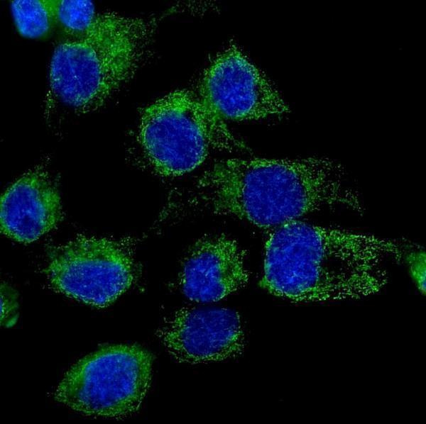
The catalog antibody has IHC data from human paraffin sections and IF/ICC data from A431 cells (image captions); listed reactivity includes human, mouse and rat (catalog: reactivity).
A03808-2 will render with IHC images from human mammary, rectal and lung cancer paraffin sections (A03808-2: IHC image captions). Its IF/ICC image shows staining in A431 cells (A03808-2: IF image caption).
Which to pick: Choose A03808-2 for paraffin-section IHC: its images show human tissue staining after citrate retrieval, while the fixative is unreported (A03808-2: IHC image captions). The same rabbit-host antibody is listed for IF/ICC and has an A431 cell image; its clonality is unreported (A03808-2: catalog applications, host and clone; IF image caption). For mouse or rat samples, A03808-2 lists reactivity with both species, but the supplied IHC images show human tissue only (A03808-2: catalog reactivity; IHC image captions).