This website uses cookies to ensure you get the best experience on our website.


- Table of Contents
Plan LPIN1 IHC-P around cytoplasmic staining in most tissues, with high signal in kidney tubules, lung macrophages and Leydig cells (HPA tissue IHC). This guide covers fixation consistency, controls, chromogenic detection and cell-specific scoring.

Expected localisation, validated protocols, controls and antibodies — the at-a-glance facts below, then the full design guide.
| Expected localisation | Cytoplasmic staining in most tissues (HPA tissue IHC) | |
| Staining pattern | Cytoplasmic signal; high in lung macrophages and kidney tubules (HPA tissue IHC) | |
| Antigen retrieval | Tris-EDTA pH 9.0 HIER, 95–98 °C, 20 min (rule: nuclear antigen) | |
| Positive control | Kidney+4 more · see all | |
| Negative control | Cerebellum+4 more · see all |
| Fixation | Matched tissue-IHC evidence does not establish the fixation claim. Validate the specimen-specific method before use. (selected-SKU IHC image A02467) | |
| Caveat | Staining has medium consistency with RNA expression (HPA tissue IHC) | |
| Regulation | Muscle enriched; adipose abundant (UniProt) | |
| Isoform / epitope | 7 isoforms; no extracellular region; check epitope coverage (UniProt) |
The catalog antibody IHC-P protocol is accompanied by published LPIN1 IHC conditions for colonic tissue (PMC12395820) and breast cancer tissue (PMC8124251).
| Sample | Tissue sections; selected-image fixative not specified (standard IHC workflow) |
| Fixation | Image fixative and duration unreported (datasheet A02467); verify before use. |
| Sectioning | 4–5 µm sections on charged slides (standard) |
| Deparaffinisation | Xylene, graded ethanol series to water (standard) |
| Antigen retrieval | Heat-induced epitope retrieval in Tris-EDTA buffer, pH 9.0, 20 min at 95–98 °C (standard rule: nuclear antigen) |
| Peroxidase block | 3% H2O2, 10 min, room temperature (standard) |
| Blocking | 10% normal serum of the secondary host, 30 min, room temperature (standard) |
| Primary antibody | Rabbit anti-LPIN1, 1:100–1:500 starting range (standard) |
| Primary incubation | Overnight at 4 °C (standard) |
| Detection | HRP-polymer secondary, DAB chromogen 5–10 min (standard) |
| Counterstain | Hematoxylin, blue, dehydrate and mount (standard) |
| Expected result | LPIN1-positive staining in cells in tubules of kidney (HPA tissue IHC: High). HPA tissue profile: Cytoplasmic expression in most tissues. No signal in the no-primary control. |
LPIN1 should usually appear cytoplasmic in tissue sections (HPA tissue IHC: cytoplasmic in most tissues), with strong staining in kidney tubular cells, lung macrophages and testis Leydig cells (HPA tissue IHC: High). HPA rates the tissue pattern Approved, with medium consistency between staining and RNA data (HPA tissue IHC). Cytosolic and membrane-associated localisation is plausible for a protein without a transmembrane segment (UniProt Q14693: localisation and topology).
| Distinct cytoplasmic staining in kidney tubules, lung macrophages or testis Leydig cells. | This matches the reported High cell-specific tissue pattern (HPA tissue IHC). Compare cells within the section; staining of the entire tissue is not the expected readout (HPA tissue IHC: cell-level observations). |
| Predominantly nuclear interior staining, with little cytoplasmic signal. | Treat a nuclear-only pattern as suspect: tissue IHC is mainly cytoplasmic (HPA tissue IHC), although UniProt lists the nuclear membrane (UniProt Q14693: localisation). Check whether any apparent nuclear signal follows the rim before calling it discordant. |
| Strong staining in cells reported as undetected, such as pancreatic exocrine glandular cells. | This conflicts with that cell-level reference (HPA tissue IHC: Not detected). Cross-reactivity or endogenous detection activity is possible (standard IHC practice); confirm cell identity and inspect controls before assigning LPIN1. |
| Uniform colour across stroma, empty spaces and several unrelated cell types. | This is more consistent with background than the reported cell-specific pattern (HPA tissue IHC). Review the no-primary control, blocking and detection steps (standard chromogenic IHC practice). |
| No staining in kidney tubular cells or lung macrophages. | A blank known-positive cell population suggests an assay problem, though HPA reports only medium staining–RNA consistency (HPA tissue IHC). Check section quality, controls and the antibody's documented IHC use before interpreting the absence biologically (standard IHC practice). |
| Cell and tissue context | HPA reports High staining in kidney tubules, lung macrophages and Leydig cells, but Medium in adipocytes (HPA tissue IHC). UniProt describes skeletal muscle specificity and abundant adipose expression (UniProt Q14693); RNA or tissue-level descriptions do not replace cell-level IHC observations. |
| Compartment and movement | LPIN1 is listed in cytosol, ER membrane and nuclear membrane, and can move from cytosol to ER after KAT5 acetylation (UniProt Q14693: localisation). Interpret variable cytoplasmic distribution cautiously; these annotations do not predict a particular paraffin-section pattern. |
| Antibody validation by application | HPA038021 is IHC Approved, while HPA024591 is ICC Approved (HPA antibodies). These are distinct application records; the ICC result does not establish an IHC pattern for HPA024591. HPA's tissue profile has medium staining–RNA consistency (HPA tissue IHC). |
| Isoforms and antibody target | UniProt lists 7 LPIN1 isoforms (UniProt Q14693). The supplied records give no epitope or isoform coverage for the IHC antibody, so a difference between sections cannot be assigned to a particular isoform from staining alone. |
| Protein topology and processing | LPIN1 has no transmembrane segment, signal peptide or propeptide in the supplied record (UniProt Q14693: topology and processing). Membrane-associated localisation therefore does not establish integral membrane staining or extracellular deposition; evaluate the observed cellular compartment instead (UniProt Q14693: localisation). |
| Situation | Likely cause | Next action |
|---|---|---|
| Known-positive tissue is blank. | The assay may have failed; kidney tubular cells and lung macrophages are reported High (HPA tissue IHC). | Check the tissue and run controls, then review the IHC antibody's documented conditions and detection reagents (standard IHC practice). No LPIN1-specific retrieval or fixation effect is supplied. |
| Colour is diffuse across the section. | Non-specific binding, residual endogenous detection activity or incomplete washing can produce background (standard chromogenic IHC practice). | Inspect the no-primary control and review blocking, washes and detection chemistry (standard IHC practice). Score only cell-associated staining against the HPA cell-level reference (HPA tissue IHC). |
| Nuclear-interior staining dominates. | A nuclear-only result differs from the usual cytoplasmic tissue profile (HPA tissue IHC); the nuclear membrane annotation alone does not explain filled nuclei (UniProt Q14693: localisation). | Compare cytoplasm and nuclear rim in known-positive cells, then check controls and antibody specificity before accepting the pattern (standard IHC practice). |
| An expected negative cell type stains strongly. | Cross-reactivity or endogenous chromogen activity may mimic signal (standard IHC practice); pancreatic exocrine glandular cells are reported Not detected (HPA tissue IHC). | Verify the cell type and compare positive tissue with no-primary and detection controls (standard IHC practice). Treat the HPA negative as a reference observation, not proof of universal absence. |
| Only a low-expression tissue looks blank. | Glial cells in hippocampus and caudate are reported Low, whereas several other cell types are Not detected (HPA tissue IHC). A faint or absent result there is less informative than a blank High reference. | Assess kidney tubular cells or lung macrophages in the same run as positive references (HPA tissue IHC); use controls to judge assay performance (standard IHC practice). |
| IF/ICC shows peripheral signal: is that an IHC positive pattern? | ICC-IF reports approved cytosolic localisation plus additional plasma membrane localisation (HPA subcellular); tissue IHC reports a generally cytoplasmic profile (HPA tissue IHC). | Interpret the IF/ICC observation in its own application context and assess paraffin IHC against the tissue cell pattern (HPA subcellular; HPA tissue IHC). The two application records do not establish identical staining distributions. |
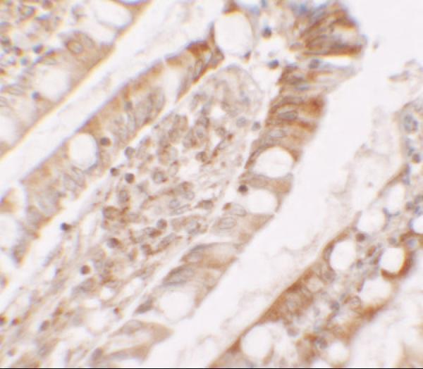
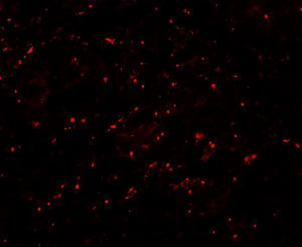
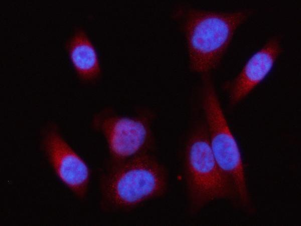
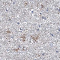
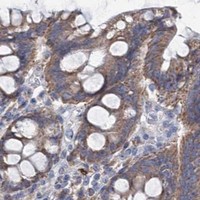
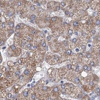
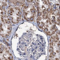


Comprehensive Human Protein Atlas IHC scoring per tissue (reliability: Approved — Medium consistency between antibody staining and RNA expression data.). Rows are taken directly from the HPA tissue chart — click any row's HPA link to view the source.
| Tissue | Cell type | Level | Evidence | Source |
|---|---|---|---|---|
| Kidney | Cells in tubules | High | Protein (IHC) | HPA → |
| Lung | Macrophages | High | Protein (IHC) | HPA → |
| Testis | Leydig cells | High | Protein (IHC) | HPA → |
| Adipose tissue | Adipocytes | Medium | Protein (IHC) | HPA → |
| Adrenal gland | Glandular cells | Medium | Protein (IHC) | HPA → |
| Tissue | Cell type | Level | Evidence | Source |
|---|---|---|---|---|
| Cerebellum | Cells in granular layer | Not detected | Protein (IHC) | HPA → |
| Cerebral cortex | Endothelial cells | Not detected | Protein (IHC) | HPA → |
| Endometrium | Cells in endometrial stroma | Not detected | Protein (IHC) | HPA → |
| Fallopian tube | Glandular cells | Not detected | Protein (IHC) | HPA → |
| Ovary | Ovarian stroma cells | Not detected | Protein (IHC) | HPA → |
Troubleshoot LPIN1 staining in paraffin sections by checking retrieval, tissue controls, subcellular pattern, and cell-specific scoring before interpreting signal.
A02467 has human small-intestine IHC and IF images (catalog: image captions); A02467-2 has human PC-3 ICC/IF data (catalog: IF image caption).
A02467 will render with a human small-intestine IHC image; its listed applications also include IF (catalog: IHC image caption; applications). A02467-2 will render with a human PC-3 ICC/IF image (catalog: IF image caption).
Which to pick: Choose A02467 for tissue IHC: it lists IHC-P and shows staining in human small intestine (catalog: applications; IHC image caption); the caption does not report the fixative (catalog: IHC image caption). Choose A02467-2 for human cell ICC/IF: its PC-3 image used 5 μg/mL (catalog: applications; IF image caption). For planned mouse or rat work, A02467 lists both species and IHC-P, while its reported IHC and IF images are from human tissue (catalog: reactivity; applications; image captions).