This website uses cookies to ensure you get the best experience on our website.


- Table of Contents
Plan chromogenic LRRC1 IHC on paraffin sections with colon glandular cells as a positive control and bone marrow hematopoietic cells as a negative comparator (HPA tissue IHC). Start catalog antibody A13956 at 10 μg/mL (datasheet: IHC-P) and score membranous glandular staining (HPA tissue IHC).

Expected localisation, validated protocols, controls and antibodies — the at-a-glance facts below, then the full design guide.
| Expected localisation | Membranous staining in glandular epithelium (HPA tissue IHC) | |
| Staining pattern | Most glandular epithelia show membranous staining (HPA tissue IHC) | |
| Antigen retrieval | Citrate pH 6.0 HIER, 95–98 °C, 20 min (rule: cytoplasmic / membrane antigen) | |
| Positive control | Cerebellum+4 more · see all | |
| Negative control | Adipose tissue+4 more · see all |
| Fixation | Matched tissue-IHC evidence does not establish the fixation claim. Validate the specimen-specific method before use. (selected-SKU IHC image A13956) | |
| Caveat | Staining has medium consistency with RNA expression (HPA tissue IHC) | |
| Regulation | Expression varies by tissue and cell type (HPA tissue IHC) | |
| Isoform / epitope | 2 isoforms; epitope differences are unspecified (UniProt) |
Compare the catalog antibody’s IHC-P protocol with two published LRRC1 liver-section methods (PMC12691226; PMC10932271).
| Sample | Tissue sections; selected-image fixative not specified (standard IHC workflow) |
| Fixation | Image fixative and duration unreported (datasheet A13956); verify before use. |
| Sectioning | 4–5 µm sections on charged slides (standard) |
| Deparaffinisation | Xylene, graded ethanol series to water (standard) |
| Antigen retrieval | Heat-induced epitope retrieval in citrate buffer, pH 6.0, 20 min at 95–98 °C (standard rule: cytoplasmic / membrane antigen) |
| Peroxidase block | 3% H2O2, 10 min, room temperature (standard) |
| Blocking | 10% normal serum of the secondary host, 30 min, room temperature (standard) |
| Primary antibody | Rabbit anti-LRRC1, 10 μg/mL (datasheet A13956) |
| Primary incubation | Overnight at 4 °C (standard) |
| Detection | HRP-polymer secondary, DAB chromogen 5–10 min (standard) |
| Counterstain | Hematoxylin, blue, dehydrate and mount (standard) |
| Expected result | LRRC1-positive staining in purkinje cells - nucleus of cerebellum (HPA tissue IHC: High). HPA tissue profile: Membranous expression in most glandular epithelia. No signal in the no-primary control. |
LRRC1 is reported in the cytoplasm and at membranes, including the basolateral side of epithelial cells (UniProt Q9BTT6 localisation). In tissue IHC, expect prominent membranous staining in glandular epithelia, including colon and prostate glandular cells (HPA tissue IHC: High). HPA rates its tissue IHC profile Enhanced, while noting medium agreement with RNA data and pending external verification (HPA tissue IHC reliability).
| Strong staining outlines glandular cells in colon or prostate; some cytoplasmic signal is present. | This fits the reported membranous pattern in glandular epithelia (HPA tissue IHC: High in colon and prostate glandular cells) and LRRC1's cytoplasmic and epithelial basolateral localisation (UniProt Q9BTT6 localisation). Judge the cell boundary and tissue context together; HPA's Enhanced rating still carries a pending external-verification caveat (HPA tissue IHC reliability). |
| Nuclei dominate the signal in glandular epithelium, with little convincing membrane staining. | That differs from the reported glandular pattern (HPA tissue IHC: membranous expression in most glandular epithelia). Check for counterstain confusion, detection artefact or antibody-dependent staining (general IHC practice). Do not treat every nuclear signal as impossible: Purkinje-cell nuclei are reported High in cerebellum (HPA tissue IHC: cerebellum). |
| A supposedly negative cell population stains as strongly as adjacent glandular cells. | First confirm the cell type: adipocytes, cardiomyocytes and smooth muscle cells are reported Not detected (HPA tissue IHC). In those cells, staining may reflect cross-reactivity or endogenous detection activity, especially if the expected epithelial pattern is absent (general IHC practice); morphology alone cannot identify which cause applies. |
| Brown colour spreads across stroma, extracellular spaces or nearly every cell. | A diffuse, tissue-wide deposit does not resemble the reported glandular membrane pattern (HPA tissue IHC profile). Consider excess detection signal, endogenous peroxidase activity or inadequate blocking, then compare with a negative reagent control (general chromogenic IHC practice). A uniform background cannot establish LRRC1 distribution. |
| A colon glandular-cell section shows no convincing staining. | Colon glandular cells are reported High (HPA tissue IHC: colon), so a blank result calls for a technical check before interpreting the sample as LRRC1-negative. Review tissue preservation, retrieval, antibody application and detection using a same-run positive control (general IHC practice). The supplied sources give no LRRC1-specific retrieval or fixation requirement. |
| Cellular location and topology (UniProt Q9BTT6 localisation and topology) | LRRC1 is cytoplasmic and membrane-associated, with epithelial basolateral localisation and no transmembrane segment (UniProt Q9BTT6). A membrane-associated IHC pattern therefore need not imply a membrane-spanning protein; assess staining against cell borders and cytoplasm. |
| Choice of reference tissue (HPA tissue IHC) | Colon, endometrium, gallbladder, prostate and rectum glandular cells are reported High, while adipocytes and smooth muscle cells are Not detected (HPA tissue IHC). Select a reported High cell population for the positive control and interpret adjacent compartments separately; an organ name alone is insufficient. |
| Antibody evidence (HPA antibody validation; HPA tissue IHC reliability) | HPA031603 has Enhanced IHC validation (HPA antibodies), and the overall tissue profile is Enhanced with medium RNA agreement and pending external verification (HPA tissue IHC reliability). Use this as support for the reported pattern, while treating unexpected staining as a result to verify. |
| IF/ICC? (HPA subcellular ICC-IF; HPA antibodies) | HPA reports supported cytosolic localisation in ICC-IF and lists A-431, RT-4 and U2OS images (HPA subcellular ICC-IF); HPA031604 is ICC Supported (HPA antibodies). Those cell-image observations answer the localisation question for IF/ICC but do not replace the tissue IHC pattern. |
| Isoforms and epitope information (UniProt Q9BTT6 isoforms) | Two isoforms are listed (UniProt Q9BTT6 isoforms). The supplied evidence does not locate the IHC antibody epitope or show isoform-specific staining, so an unusual pattern cannot be assigned to one isoform from these sources. |
| Situation | Likely cause | Next action |
|---|---|---|
| No signal in a reported High glandular control (HPA tissue IHC: colon). | The section, primary antibody or detection workflow may have failed (general IHC practice); the supplied sources do not establish LRRC1-specific fixation sensitivity. | Check that glandular cells are present, inspect the same-run control and verify antibody and detection steps (general IHC practice). If retrieval is changed, label it an empirical workflow adjustment rather than a documented LRRC1 requirement. |
| Signal appears mainly in nuclei of glandular cells. | This conflicts with the reported glandular membrane profile (HPA tissue IHC profile), though nuclear staining is reported in cerebellar Purkinje cells (HPA tissue IHC: cerebellum). | Recheck cell identity, focus and counterstain; compare an independent antibody or controlled repeat when available (general IHC practice). Record glandular and Purkinje-cell findings separately. |
| Stroma or negative-reference cells stain as strongly as glandular cells. | Broad staining may arise from nonspecific binding or endogenous detection activity (general chromogenic IHC practice); adipocytes and smooth muscle cells are reported Not detected (HPA tissue IHC). | Compare morphology and a negative reagent control, review blocking and endogenous-enzyme control, then reassess the glandular pattern (general chromogenic IHC practice). |
| The section has a uniform brown haze that obscures cell borders. | Background can result from excessive antibody or detection signal, insufficient washing or endogenous activity (general chromogenic IHC practice). It cannot be scored as the reported membranous pattern (HPA tissue IHC profile). | Check reagent controls and wash steps; adjust staining conditions empirically until cell boundaries can be assessed (general IHC practice). No LRRC1-specific dilution is supplied. |
| Heart tissue seems positive by expression information, but cardiomyocytes lack IHC staining. | UniProt reports strong expression in heart (UniProt Q9BTT6 tissue specificity), whereas HPA reports cardiomyocytes Not detected by tissue IHC (HPA tissue IHC: heart muscle). These observations have different scopes. | Identify the stained cell population and report the discordance without converting tissue-level expression into a cardiomyocyte IHC expectation (UniProt Q9BTT6 tissue specificity; HPA tissue IHC: heart muscle). |
| IF/ICC shows cytosol while tissue IHC emphasises glandular cell boundaries. | HPA reports supported cytosolic ICC-IF localisation (HPA subcellular ICC-IF) and membranous expression in most glandular epithelia by IHC (HPA tissue IHC profile). | Interpret each result in its assay and cell context; use the dedicated IF/ICC guide for IF workflow decisions. Do not infer that the tissue IHC or ICC-IF observation alone proves the other assay is wrong. |
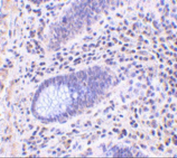
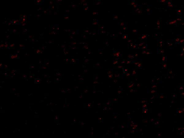


Comprehensive Human Protein Atlas IHC scoring per tissue (reliability: Enhanced — Medium consistency between antibody staining and RNA expression data. Pending external verification.). Rows are taken directly from the HPA tissue chart — click any row's HPA link to view the source.
| Tissue | Cell type | Level | Evidence | Source |
|---|---|---|---|---|
| Cerebellum | Purkinje cells - nucleus | High | Protein (IHC) | HPA → |
| Colon | Glandular cells | High | Protein (IHC) | HPA → |
| Endometrium | Glandular cells | High | Protein (IHC) | HPA → |
| Gallbladder | Glandular cells | High | Protein (IHC) | HPA → |
| Nasopharynx | Respiratory epithelial cells | High | Protein (IHC) | HPA → |
| Tissue | Cell type | Level | Evidence | Source |
|---|---|---|---|---|
| Adipose tissue | Adipocytes | Not detected | Protein (IHC) | HPA → |
| Bone marrow | Hematopoietic cells | Not detected | Protein (IHC) | HPA → |
| Heart muscle | Cardiomyocytes | Not detected | Protein (IHC) | HPA → |
| Hippocampus | Glial cells | Not detected | Protein (IHC) | HPA → |
| Lymph node | Germinal center cells | Not detected | Protein (IHC) | HPA → |
Troubleshoot LRRC1 staining by checking retrieval, tissue preservation, cellular compartment and cell type before comparing signal intensity across sections.
A13956 is a human-reactive anti-LRRC1 antibody with IHC-P and IF images from human colon tissue (catalog: applications/reactivity; IHC/IF image captions).
A13956 has IHC-P data from human colon tissue at 10 μg/mL (catalog: applications; IHC image caption). A13956 also has IF data from human colon tissue at 20 μg/mL (catalog: applications; IF image caption).
Which to pick: Choose A13956 for human paraffin-section IHC (catalog: IHC-P, Human); its IHC caption shows human colon tissue, but does not report the fixative (IHC image caption). Choose A13956 for tissue IF; ICC validation is not listed (catalog: applications; IF image caption). No cross-species option is listed: A13956 is human-reactive, rabbit-hosted, and has no clone specified (catalog: reactivity, host, clone).