This website uses cookies to ensure you get the best experience on our website.


- Table of Contents
Plan LRRC41 staining in paraffin sections using the reported cytoplasmic tissue pattern as a provisional reference (HPA tissue IHC). The guide covers the catalog antibody’s IHC dilution range of 1:100–1:300 (datasheet A30736), controls and staining assessment.

Expected localisation, validated protocols, controls and antibodies — the at-a-glance facts below, then the full design guide.
| Expected localisation | Cytoplasmic staining in most tissues (HPA tissue IHC) | |
| Staining pattern | Cytoplasmic signal in colon glands and lung macrophages (HPA tissue IHC) | |
| Antigen retrieval | Tris-EDTA pH 9.0 HIER, heat-mediated (datasheet A30736) | |
| Positive control | Breast+4 more · see all | |
| Negative control | Adipose tissue+4 more · see all |
| Fixation | Keep fixation consistent across sections (standard IHC practice; not target-specific) | |
| Caveat | Tissue staining awaits external verification (HPA tissue IHC) | |
| Regulation | Expression regulation unreported (UniProt) | |
| Isoform / epitope | 2 isoforms listed; epitope impact unknown (UniProt) |
The catalog antibody protocol and two published LRRC41 IHC protocols provide starting conditions for paraffin section staining (datasheet A30736; PMC10773795; PMC13555846).
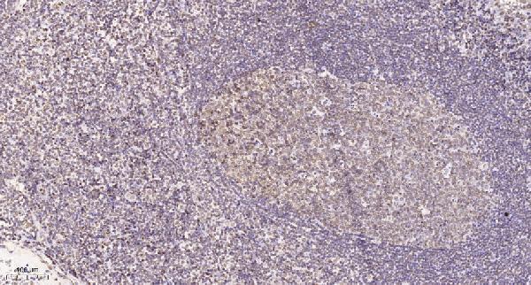
| Sample | Paraffin-embedded human tonsil tissue; fixative not specified (datasheet A30736) |
| Fixation | Image fixative and duration unreported (datasheet A30736); verify before use. |
| Sectioning | 4–5 µm sections on charged slides (standard) |
| Deparaffinisation | Xylene, graded ethanol series to water (standard) |
| Antigen retrieval | Heat retrieval: Tris-EDTA pH 9.0 (datasheet A30736); 20 min, 95–100 °C (standard) |
| Peroxidase block | 3% H2O2, 10 min, room temperature (standard) |
| Blocking | 10% normal serum of the secondary host, 30 min, room temperature (standard) |
| Primary antibody | Rabbit anti-LRRC41, 1:100 - 1:300 (datasheet A30736) |
| Primary incubation | Overnight at 4 °C (standard) |
| Detection | HRP-polymer secondary, DAB chromogen 5–10 min (standard) |
| Counterstain | Hematoxylin, blue, dehydrate and mount (standard) |
| Expected result | LRRC41-positive staining in myoepithelial cells of breast (HPA tissue IHC: Medium). HPA tissue profile: Cytoplasmic expression in most tissues. No signal in the no-primary control. |
For paraffin-section IHC, expect predominantly cytoplasmic LRRC41 staining in many tissues, including colon glandular cells and lung macrophages with medium reported intensity (HPA tissue IHC). LRRC41 has no annotated transmembrane segment (UniProt Q15345 topology). Treat this as a provisional pattern: the tissue IHC result is rated Uncertain, while separate ICC-IF data show approved nuclear-body and nucleoplasmic localization (HPA tissue IHC; HPA subcellular).
| Cytoplasmic signal in colon glandular cells or lung macrophages, with discernible cell boundaries. | This fits the reported medium staining and broad cytoplasmic tissue profile (HPA tissue IHC). Compare positive cells with nearby cells and controls; medium is an HPA category, not a calibrated intensity threshold. Because tissue IHC reliability is Uncertain, matching the pattern alone does not establish antibody specificity (HPA tissue IHC). |
| Predominantly membranous or extracellular signal, with little cytoplasmic staining. | This conflicts with the reported cytoplasmic tissue pattern (HPA tissue IHC) and the absence of an annotated transmembrane segment (UniProt Q15345 topology). Consider nonspecific deposition or detection artefact and repeat with controls. Nuclear puncta alone need separate assessment: approved ICC-IF images place LRRC41 in nuclear bodies (HPA subcellular). |
| Strong staining in adipocytes or adrenal glandular cells, especially when expected positive cells are weak. | Those cell populations are listed as Not detected, whereas colon glandular cells and lung macrophages are Medium (HPA tissue IHC). Review cell identification, cross-reactivity, and endogenous chromogen-generating activity using appropriate controls (general IHC practice). A Not detected HPA entry is an observation with Uncertain IHC reliability, not proof of biological absence (HPA tissue IHC). |
| Diffuse color across tissue, blank areas, or many cell types without clear cellular localization. | A uniform haze is hard to reconcile with a readable cytoplasmic cellular pattern (HPA tissue IHC). Background can arise from detection reagents, inadequate blocking or washing, or excess antibody (general IHC practice). Assess a no-primary control and inspect unstained spaces before assigning the color to LRRC41. |
| No visible signal in colon glandular cells or lung macrophages. | Both are reported Medium and can serve as reference cell populations (HPA tissue IHC). Verify that the reference cells are present, then check retrieval, antibody conditions, detection reagents, and counterstain on a control section (general IHC practice). An absent signal does not alone establish absent LRRC41, particularly with Uncertain tissue IHC reliability (HPA tissue IHC). |
| Assay-specific localization | Tissue IHC reports cytoplasmic staining, whereas approved ICC-IF reports mainly nuclear bodies with additional nucleoplasmic signal (HPA tissue IHC; HPA subcellular). Interpret each in its assay context; this record does not establish why the patterns differ. |
| Antibody validation | HPA051941 is rated Uncertain for IHC and Approved for ICC (HPA antibodies). The ICC rating does not validate a paraffin-section chromogenic pattern; use tissue morphology and controls when interpreting IHC (general IHC practice). |
| Isoforms and antigen features | UniProt lists isoforms 2 and 3, several phosphorylated residues, no signal peptide, and no annotated transmembrane segment (UniProt Q15345). Their effects on this antibody's epitope or IHC intensity are unreported; do not infer tissue-specific loss or membrane localization from them. |
| Situation | Likely cause | Next action |
|---|---|---|
| Expected positive cells are blank. | The section may lack the identified reference cells, or the IHC workflow may have failed (general IHC practice); HPA reports Medium staining in colon glandular cells and lung macrophages (HPA tissue IHC). | Confirm the cells by morphology. Check the positive control, retrieval conditions, primary-antibody step, and chromogenic detection in sequence (general IHC practice). Avoid assigning target-specific retrieval sensitivity from these data. |
| Every cell and tissue space appears brown. | Diffuse color may reflect reagent background, excess primary antibody, or incomplete washing (general IHC practice), rather than the described cytoplasmic tissue pattern (HPA tissue IHC). | Review a no-primary control, blocking and wash steps, then titrate the primary antibody within its documented instructions (general IHC practice). Score only localized cellular signal. |
| Adipocytes stain as strongly as the intended positive population. | Adipocytes are listed as Not detected (HPA tissue IHC); staining could reflect mistaken cell identification, cross-reactivity, or endogenous detection activity (general IHC practice). | Verify morphology and compare a no-primary control. Review the detection chemistry and reassess specificity before counting those cells as LRRC41 positive (general IHC practice; HPA tissue IHC: Uncertain). |
| Signal outlines cell membranes or pools outside cells. | This differs from cytoplasmic tissue staining (HPA tissue IHC); UniProt annotates no transmembrane segment or signal peptide (UniProt Q15345). | Inspect section edges and blank spaces for deposited chromogen, repeat with a no-primary control, and reassess the staining call (general IHC practice). Do not interpret the outline as established LRRC41 localization. |
| Only nuclear puncta are visible. | Nuclear bodies are an approved ICC-IF location, but HPA describes tissue IHC as cytoplasmic and rates it Uncertain (HPA subcellular; HPA tissue IHC). The supplied evidence does not resolve the assay difference. | Record the compartment and cell type, compare the no-primary and positive controls, and report the IHC result as discordant rather than automatically calling nuclear puncta false positive (general IHC practice; HPA subcellular). |
| IF/ICC images seem inconsistent with the IHC slide. | Approved ICC-IF localization is mainly nuclear bodies with additional nucleoplasm; the tissue IHC profile is cytoplasmic and Uncertain (HPA subcellular; HPA tissue IHC). | Interpret the images under their respective assay evidence and use the separate IF/ICC guide for that assay. Do not transfer an ICC-IF localization call into an IHC specificity verdict (HPA antibodies; HPA subcellular). |


Comprehensive Human Protein Atlas IHC scoring per tissue (reliability: Uncertain — Pending external verification. Antibody staining in cells/structures not annotated, view images.). Rows are taken directly from the HPA tissue chart — click any row's HPA link to view the source.
| Tissue | Cell type | Level | Evidence | Source |
|---|---|---|---|---|
| Breast | Myoepithelial cells | Medium | Protein (IHC) | HPA → |
| Colon | Glandular cells | Medium | Protein (IHC) | HPA → |
| Duodenum | Glandular cells | Medium | Protein (IHC) | HPA → |
| Heart muscle | Cardiomyocytes | Medium | Protein (IHC) | HPA → |
| Lung | Macrophages | Medium | Protein (IHC) | HPA → |
| Tissue | Cell type | Level | Evidence | Source |
|---|---|---|---|---|
| Adipose tissue | Adipocytes | Not detected | Protein (IHC) | HPA → |
| Adrenal gland | Glandular cells | Not detected | Protein (IHC) | HPA → |
| Appendix | Glandular cells | Not detected | Protein (IHC) | HPA → |
| Caudate | Glial cells | Not detected | Protein (IHC) | HPA → |
| Cerebellum | Cells in granular layer | Not detected | Protein (IHC) | HPA → |
Troubleshoot LRRC41 staining by checking retrieval, compartment, cell identity and controls before interpreting chromogenic IHC signal (HPA tissue IHC; HPA subcellular).
A30736 has a real IHC image from paraffin-embedded human tonsil (A30736 IHC caption); both antibodies list IF and Human, Mouse, and Rat reactivity (catalog application and reactivity lists).
A30736 will render with an IHC figure of paraffin-embedded human tonsil and lists IHC, IF, and ICC applications (A30736 IHC caption; catalog application list). A13640 will render as an IHC- and IF-listed antibody with Human, Mouse, and Rat reactivity, but no IHC or IF image is supplied (A13640 catalog application, reactivity, and image lists).
Which to pick: For tissue IHC, pick A30736 because its own caption documents paraffin-embedded human tonsil, Tris-EDTA pH 9.0 retrieval, and a 1:200 primary dilution overnight at 4°C; the fixative is unreported (A30736 IHC caption). For IF/ICC, pick A30736 when ICC is needed because ICC and IF are both listed; A13640 lists IF without ICC (catalog application lists). Both list Human, Mouse, and Rat reactivity, while the supplied tissue image documents only human tonsil (catalog reactivity lists; A30736 IHC caption).