This website uses cookies to ensure you get the best experience on our website.


- Table of Contents
MAEA IHC shows general nuclear and cytoplasmic tissue staining, with high signal in colon glandular cells and bronchial respiratory epithelial cells (HPA tissue IHC). This guide pairs those expectations with the catalog antibody’s 1:100–1:300 IHC dilution range (datasheet A08766-1).

Expected localisation, validated protocols, controls and antibodies — the at-a-glance facts below, then the full design guide.
| Expected localisation | Nuclear and cytoplasmic tissue staining (HPA tissue IHC) | |
| Staining pattern | High glandular and epithelial staining; nuclear and cytoplasmic (HPA tissue IHC) | |
| Antigen retrieval | Tris-EDTA pH 9.0 HIER, heat-mediated (datasheet A08766-1) | |
| Positive control | Breast+4 more · see all | |
| Negative control | None in HPA (detected in all 45 tissues); use no-primary + isotype controls |
| Fixation | Keep fixation consistent across paraffin sections (standard IHC practice; not target-specific) | |
| Caveat | Staining and RNA show medium consistency (HPA tissue IHC) | |
| Regulation | Low tissue specificity (HPA tissue RNA) | |
| Isoform / epitope | 5 isoforms; epitope coverage is unspecified (UniProt) |
Compare the catalog antibody’s IHC-P protocol (datasheet: A08766-1) with two published MAEA IHC workflows (PMC12450695; PMC10506284).
| Sample | Paraffin-embedded human tonsil tissue; fixative not specified (datasheet A08766-1) |
| Fixation | Image fixative and duration unreported (datasheet A08766-1); verify before use. |
| Sectioning | 4–5 µm sections on charged slides (standard) |
| Deparaffinisation | Xylene, graded ethanol series to water (standard) |
| Antigen retrieval | Heat retrieval: Tris-EDTA pH 9.0 (datasheet A08766-1); 20 min, 95–100 °C (standard) |
| Peroxidase block | 3% H2O2, 10 min, room temperature (standard) |
| Blocking | 10% normal serum of the secondary host, 30 min, room temperature (standard) |
| Primary antibody | Rabbit anti-MAEA, 1:100 - 1:300 (datasheet A08766-1) |
| Primary incubation | Overnight at 4 °C (standard) |
| Detection | HRP-polymer secondary, DAB chromogen 5–10 min (standard) |
| Counterstain | Hematoxylin, blue, dehydrate and mount (standard) |
| Expected result | MAEA-positive staining in glandular cells of breast (HPA tissue IHC: High). HPA tissue profile: General nuclear and cytoplasmic expression. No signal in the no-primary control. |
MAEA staining should be assessed in nuclei and cytoplasm across many cell types; high staining is reported in selected epithelial and neuronal cells (HPA: general nuclear and cytoplasmic expression; Low tissue specificity). Macrophages may also show cell-surface staining that varies with maturation (UniProt Q7L5Y9: macrophage localisation). The tissue IHC profile is Approved, with medium consistency between staining and RNA data (HPA: reliability). MAEA has no transmembrane segment (UniProt Q7L5Y9: topology).
| Distinct nuclear and cytoplasmic staining in colon glandular cells or bronchial respiratory epithelial cells. | This fits the reported compartment pattern and high staining in those cells (HPA: general nuclear and cytoplasmic expression; High in colon and bronchus). Judge intensity against cells in the same section; HPA's Approved rating has medium consistency with RNA data (HPA: reliability). |
| Signal confined to an unexpected compartment, with no convincing nuclear or cytoplasmic staining. | Check for an artefact before treating that distribution as MAEA: the tissue profile is nuclear and cytoplasmic (HPA: tissue IHC). Cell-surface staining can be plausible in macrophages, particularly mature ones (UniProt Q7L5Y9: macrophage localisation). |
| Strong staining in a cell population outside the expected high-staining populations. | Compare its identity with the tissue profile and controls; cross-reactivity or endogenous detection activity is possible (general IHC practice). An unlisted cell type is not automatically negative because MAEA is ubiquitous (UniProt Q7L5Y9: tissue specificity). |
| Diffuse colour over tissue and empty areas, obscuring cell boundaries. | Treat this as background until controls and a cleaner run establish a cellular pattern (general IHC practice). It cannot be scored reliably against HPA's nuclear and cytoplasmic profile (HPA: tissue IHC). |
| No signal in colon glandular cells or cerebellar Purkinje cells. | Investigate the run: both are reported as High, though the HPA profile has medium consistency with RNA data (HPA: tissue IHC; reliability). A single negative section does not establish MAEA absence (general IHC practice). |
| Tissue and cell selection | Colon glandular cells and cerebellar Purkinje cells are reported High; liver hepatocytes and soft-tissue fibroblasts are Low (HPA: tissue IHC). Use the latter as lower-signal comparators, not proven negatives; HPA lists no negative population (HPA: tissue IHC). |
| Macrophage maturation and compartment | Immature macrophages are predominantly nuclear, whereas mature macrophages are predominantly cell-surface positive (UniProt Q7L5Y9: subcellular location). Interpret a macrophage membrane-like outline in that context; MAEA has no transmembrane segment (UniProt Q7L5Y9: topology). |
| Antibody evidence | HPA058185 has Approved IHC status; HPA036886 has no listed IHC status (HPA: antibodies). Those ratings are antibody-specific. The overall tissue profile is Approved with medium RNA–staining consistency (HPA: reliability). |
| Isoforms and processing | Five isoforms are listed, with no signal peptide, propeptide or processed chain reported (UniProt Q7L5Y9: isoforms; processing). These facts do not identify which isoforms an antibody detects or predict loss of staining after tissue preparation. |
| Target-specific fixation sensitivity | The supplied HPA staining profile and UniProt record provide no MAEA-specific fixation effect (HPA: tissue IHC; UniProt Q7L5Y9). Assess retrieval empirically as a general IHC workflow variable; do not infer its effect from tissue staining levels (general IHC practice). |
| Situation | Likely cause | Next action |
|---|---|---|
| Reported High tissue has no discernible nuclear or cytoplasmic signal. | A failed staining or detection step is possible (general IHC practice); colon glandular cells are reported High (HPA: tissue IHC). | Check the run's positive control and detection reagents; review retrieval and primary-antibody conditions as general IHC variables (general IHC practice). Do not assign an MAEA-specific fixation cause from these sources. |
| Colour spreads across the section or appears where no cells are present. | Nonspecific binding, incomplete washing or detection-system background can obscure localisation (general IHC practice). | Compare with a primary-omission control, then review blocking, washing and detection conditions (general IHC practice). Score MAEA only when cellular staining is resolved. |
| Signal appears mainly at macrophage surfaces. | A surface-associated pattern is reported for mature macrophages (UniProt Q7L5Y9: macrophage localisation). | Confirm macrophage identity and compare nuclear and surface distributions before calling the outline artefactual (general IHC practice; UniProt Q7L5Y9: macrophage localisation). |
| Unexpected cells stain strongly while reported High cells do not. | Cross-reactivity or endogenous detection activity is possible (general IHC practice); MAEA is also described as ubiquitous (UniProt Q7L5Y9: tissue specificity). | Check cell identity and primary-omission controls; compare with reported High populations on a separate section (general IHC practice; HPA: tissue IHC). Avoid declaring every unlisted cell negative. |
| Liver hepatocytes show weak staining. | Low staining is reported for hepatocytes (HPA: tissue IHC). Weak signal alone does not demonstrate assay failure. | Interpret alongside a reported High population and the run's controls before changing staining conditions (HPA: tissue IHC; general IHC practice). |
| IF/ICC shows mostly nucleoplasmic signal; is that consistent with MAEA? | Nucleoplasm is the enhanced main location in HPA ICC-IF images (HPA: subcellular); tissue IHC has a broader nuclear and cytoplasmic profile (HPA: tissue IHC). | Interpret IF/ICC on its own guide page; HPA lists ICC as Supported for HPA036886 and HPA058185 (HPA: antibodies). Do not use the IF pattern alone to reject a tissue IHC result. |


Comprehensive Human Protein Atlas IHC scoring per tissue (reliability: Approved — Medium consistency between antibody staining and RNA expression data.). Rows are taken directly from the HPA tissue chart — click any row's HPA link to view the source.
| Tissue | Cell type | Level | Evidence | Source |
|---|---|---|---|---|
| Breast | Glandular cells | High | Protein (IHC) | HPA → |
| Bronchus | Respiratory epithelial cells | High | Protein (IHC) | HPA → |
| Cerebellum | Purkinje cells | High | Protein (IHC) | HPA → |
| Cervix | Squamous epithelial cells | High | Protein (IHC) | HPA → |
| Colon | Glandular cells | High | Protein (IHC) | HPA → |
| Tissue | Cell type | Level | Evidence | Source |
|---|---|---|---|---|
| None in HPA: MAEA is detected in all 45 scored tissues. Use a no-primary (secondary-only) and an isotype control instead. | ||||
These questions focus on interpreting MAEA staining in paraffin sections using chromogenic IHC; the immunofluorescence entry addresses the separate IF workflow.
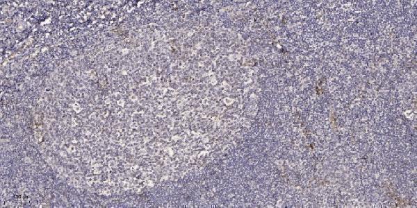
A08766-1 has an IHC figure from paraffin-embedded human tonsil (catalog image caption). IF/ICC are listed applications without an IF figure (catalog applications and image inventory); reactivity lists human, mouse and rat (catalog reactivity).
A08766-1 is shown by IHC on paraffin-embedded human tonsil (catalog image caption). A08766-1 also lists IF/ICC applications and human, mouse and rat reactivity, but has no IF image (catalog applications, reactivity and image inventory).
Which to pick: For tissue IHC, choose A08766-1: its figure documents paraffin-embedded human tonsil, Tris-EDTA retrieval at pH 9.0, and primary antibody at 1:200 overnight at 4 °C (catalog IHC image caption). For IF/ICC, A08766-1 lists those applications and an IF dilution of 1:50, but has no IF figure (catalog applications, IF dilution and image inventory). For cross-species work, A08766-1 is polyclonal and lists human, mouse and rat reactivity (catalog dilution data and reactivity); its IHC figure shows only human tonsil, and the fixative is unreported (catalog IHC image caption).