This website uses cookies to ensure you get the best experience on our website.


- Table of Contents
Plan chromogenic MAP1A IHC in paraffin sections using cerebellum or cerebral cortex as positive tissue (HPA tissue IHC). This guide covers cytoplasmic neuronal staining (HPA tissue IHC) and how MAP1A processing affects epitope selection (UniProt).

Expected localisation, validated protocols, controls and antibodies — the at-a-glance facts below, then the full design guide.
| Expected localisation | Cytoplasmic tissue staining (HPA tissue IHC) | |
| Staining pattern | Cytoplasm of neurons, Purkinje cells and molecular-layer subsets (HPA tissue IHC) | |
| Antigen retrieval | Tris-EDTA pH 9.0 HIER, heat-mediated (datasheet A03897) | |
| Positive control | Cerebellum+4 more · see all | |
| Negative control | Adipose tissue+4 more · see all |
| Fixation | Keep fixation consistent across sections. (standard IHC practice; not target-specific) | |
| Caveat | Ciliated cells also stain in bronchus and nasopharynx (HPA tissue IHC) | |
| Regulation | Brain-enriched expression (UniProt) | |
| Isoform / epitope | 2 isoforms; heavy-chain and LC2 processing makes epitope location relevant (UniProt) |
The catalog antibody IHC-P protocol (datasheet: A03897) is accompanied by one published prostate tissue microarray protocol (PMC4599305).
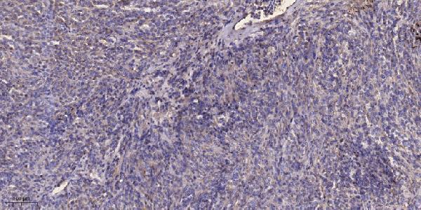
| Sample | Paraffin-embedded human Colon cancer tissue; fixative not specified (datasheet A03897) |
| Fixation | Image fixative and duration unreported (datasheet A03897); verify before use. |
| Sectioning | 4–5 µm sections on charged slides (standard) |
| Deparaffinisation | Xylene, graded ethanol series to water (standard) |
| Antigen retrieval | Heat retrieval: Tris-EDTA pH 9.0 (datasheet A03897); 20 min, 95–100 °C (standard) |
| Peroxidase block | 3% H2O2, 10 min, room temperature (standard) |
| Blocking | 10% normal serum of the secondary host, 30 min, room temperature (standard) |
| Primary antibody | Rabbit anti-MAP1A, 1:50 recommended; image 1:200 (datasheet A03897) |
| Primary incubation | Overnight at 4 °C (standard) |
| Detection | HRP-polymer secondary, DAB chromogen 5–10 min (standard) |
| Counterstain | Hematoxylin, blue, dehydrate and mount (standard) |
| Expected result | MAP1A-positive staining in gLUC cells - cytoplasm/membrane of cerebellum (HPA tissue IHC: High). HPA tissue profile: Cytoplasmic expression most abundant in neuronal cells, Purkinje cells and subset of cells in molecular layer of cerebellum. No signal in the no-primary control. |
MAP1A is a cytoplasmic cytoskeletal protein without a transmembrane segment (UniProt P78559 topology). In paraffin section IHC, expect staining chiefly in neuronal cells, including cerebellar Purkinje cells and a subset of cells in the molecular layer (HPA tissue IHC: Enhanced reliability). Cerebral cortex neuronal staining is high, while hippocampal neuronal staining is medium (HPA tissue IHC). Interpret intensity within the identified cell type and tissue.
| Neuronal cell bodies show cytoplasmic chromogen in cerebral cortex; cerebellar Purkinje cells and some molecular layer cells stain. | This fits the reported brain enriched pattern (HPA tissue IHC). Cerebral cortex neuronal staining is high; the cerebellar profile identifies Purkinje cells and a subset of molecular layer cells (HPA tissue IHC). Assess the stained cells against their local tissue context. |
| Predominantly nuclear signal appears in otherwise identifiable neuronal cells. | A nuclear dominant pattern does not fit the reported cytoplasmic cytoskeletal location (UniProt P78559 subcellular location; HPA tissue IHC). Treat it as suspect staining and compare it with an appropriate control and the expected cytoplasmic pattern (general IHC practice). |
| Strong staining appears broadly in adipocytes or bone marrow hematopoietic cells. | Those cell types are reported as not detected (HPA tissue IHC). Consider antibody cross reactivity or endogenous detection activity; an unexpected positive result alone cannot distinguish them (general IHC practice). Compare a no primary antibody control with a known positive section. |
| Chromogen is spread across cells and surrounding tissue, obscuring cell boundaries. | This is difficult to score as MAP1A because the reported tissue pattern identifies particular cells and cytoplasmic staining (HPA tissue IHC). Review blocking, antibody concentration, washes and detection controls as general IHC background checks (general IHC practice). |
| No neuronal cytoplasmic signal appears in a cerebral cortex section expected to be positive. | That conflicts with high neuronal staining reported in cerebral cortex (HPA tissue IHC). Check that the expected cells are present, then review the IHC assay and its controls; a single negative section cannot establish absent MAP1A expression (general IHC practice). |
| Tissue and cell context | Brain is tissue enriched, but reported staining is not exclusive to neurons: ciliated bronchial and nasopharyngeal cells and small intestine endocrine cells show medium staining (HPA tissue IHC). Do not call every extra neural positive an artefact. |
| Intracellular location | MAP1A is cytoplasmic and cytoskeletal, with no transmembrane segment (UniProt P78559 topology). HPA also annotates cerebellar GLUC cells as cytoplasm/membrane, so a membrane associated appearance there needs cell context before rejection (HPA tissue IHC). |
| Processing and antibody recognition | The precursor yields a MAP1A heavy chain and LC2 light chain; two isoforms are listed (UniProt P78559 processing, isoforms). The supplied evidence gives no antibody epitope, so it cannot establish which processed form or isoform produces a given stain. |
| IHC evidence | The tissue profile has Enhanced reliability for consistency between antibody staining and RNA expression (HPA tissue IHC). HPA039063 and HPA039064 have Enhanced IHC status (HPA antibodies); this supports the reported pattern, without validating every unexpected cell or compartment. |
| IF/ICC: what localisation should I expect? | Mainly cytosolic signal is approved; additional mid piece and principal piece localisation is reported (HPA subcellular ICC-IF). Those IF observations provide a localisation check, while the tissue and intensity calls on this page come from paraffin section IHC (HPA tissue IHC). |
| Situation | Likely cause | Next action |
|---|---|---|
| Cerebral cortex neurons have no visible stain. | High neuronal staining is expected there (HPA tissue IHC); an absent signal may reflect an IHC run or specimen issue (general IHC practice). | Confirm neuronal cells are present and review the positive control, antibody dilution, retrieval and detection steps used for this run (general IHC practice). |
| Signal is mainly nuclear. | Nuclear dominance conflicts with the reported cytoplasmic location (UniProt P78559 subcellular location; HPA tissue IHC). | Check the no primary antibody control and compare cytoplasmic signal in a known positive neuronal section before scoring (general IHC practice). |
| Adipocytes or bone marrow hematopoietic cells stain strongly. | Both are reported as not detected (HPA tissue IHC); cross reactivity or endogenous detection activity are possible (general IHC practice). | Compare a no primary antibody control and a positive control, then reassess specificity if the unexpected staining remains (general IHC practice). |
| Diffuse chromogen prevents identification of positive cells. | The result cannot be matched confidently to the reported cell restricted cytoplasmic pattern (HPA tissue IHC). | Review blocking, antibody concentration, washing and detection controls to reduce general IHC background (general IHC practice). |
| A cerebellar cell shows apparent membrane associated signal. | The HPA cerebellar GLUC annotation includes cytoplasm/membrane staining (HPA tissue IHC); appearance alone is inconclusive. | Identify the stained cell and assess accompanying cytoplasmic staining before deciding whether the pattern is unexpected (HPA tissue IHC; general IHC practice). |
| Hippocampal staining looks weaker than cerebral cortex staining. | HPA reports medium hippocampal neuronal staining and high cerebral cortex neuronal staining (HPA tissue IHC). | Score each tissue against its own reported level and cell type; use run controls before treating weaker hippocampal signal as assay failure (HPA tissue IHC; general IHC practice). |


![HPA ICC-IF BJ [Human fibroblast] — MAP1A (HPA066488)](https://images.proteinatlas.org/66488/1534_A8_1_blue_red_green.jpg)
![HPA ICC-IF BJ [Human fibroblast] — MAP1A (HPA066488)](https://images.proteinatlas.org/66488/1534_A8_2_blue_red_green.jpg)
Comprehensive Human Protein Atlas IHC scoring per tissue (reliability: Enhanced — High consistency between antibody staining and RNA expression data.). Rows are taken directly from the HPA tissue chart — click any row's HPA link to view the source.
| Tissue | Cell type | Level | Evidence | Source |
|---|---|---|---|---|
| Cerebellum | GLUC cells - cytoplasm/membrane | High | Protein (IHC) | HPA → |
| Cerebral cortex | Neuronal cells | High | Protein (IHC) | HPA → |
| Bronchus | Ciliated cells (cell body) | Medium | Protein (IHC) | HPA → |
| Hippocampus | Neuronal cells | Medium | Protein (IHC) | HPA → |
| Nasopharynx | Ciliated cells (cell body) | Medium | Protein (IHC) | HPA → |
| Tissue | Cell type | Level | Evidence | Source |
|---|---|---|---|---|
| Adipose tissue | Adipocytes | Not detected | Protein (IHC) | HPA → |
| Adrenal gland | Glandular cells | Not detected | Protein (IHC) | HPA → |
| Appendix | Endocrine cells | Not detected | Protein (IHC) | HPA → |
| Bone marrow | Hematopoietic cells | Not detected | Protein (IHC) | HPA → |
| Breast | Adipocytes | Not detected | Protein (IHC) | HPA → |
Troubleshoot MAP1A staining in paraffin sections by checking retrieval, cell type, cytoplasmic localisation and appropriate controls before scoring chromogenic signal.
A03897 is listed for MAP1A IHC and IF in human, mouse, and rat (catalog: applications and reactivity); its IHC figure shows a paraffin-embedded human colon cancer section (A03897 image caption).
A03897 has an IHC figure of paraffin-embedded human colon cancer (A03897 image caption). The catalog lists A03897 for IHC and IF in human, mouse, and rat, but supplies no IF figure (catalog: applications, reactivity, IF image alts).
Which to pick: For tissue IHC, choose A03897: its own figure documents a paraffin-embedded section with Tris-EDTA retrieval at pH 9.0 and primary antibody at 1:200 overnight at 4°C; the fixative is unreported (A03897 image caption). For IF, A03897 is listed, but no IF figure or ICC validation is supplied; its host is rabbit and clonality is unspecified (catalog: applications, IF image alts, host, clone). For cross-species work, A03897 lists human, mouse, and rat reactivity (catalog: reactivity).