MCAT / Malonyl-CoA-acyl carrier protein transacylase, mitochondrial · IHC design guide

Design Immunohistochemistry for MCAT

Plan MCAT paraffin-section IHC around the observed cytoplasmic tissue pattern (HPA tissue IHC) and annotated mitochondrial location (UniProt). Use liver hepatocytes as a high-staining reference (HPA tissue IHC) and start within the 2–5 μg/ml antibody range (datasheet A04806-1).

Evidence assembled Oct 2026 · For research use; verify linked source records and product datasheet before use
Immunohistochemistry protocol sheet for MCAT (IHC for MCAT): expected localisation General cytoplasmic staining (HPA tissue IHC), antibody A04806-1, validated IHC image, and IHC protocol steps
Printable MCAT IHC protocol sheet — expected localisation General cytoplasmic staining (HPA tissue IHC), antibody A04806-1, controls and protocol steps. Open the full MCAT IHC guide →

MCAT Immunohistochemistry Experimental Design Guide

Expected localisation, validated protocols, controls and antibodies — the at-a-glance facts below, then the full design guide.

Must know before staining
Expected localisation General cytoplasmic staining (HPA tissue IHC)
Staining pattern Cytoplasmic staining in hepatocytes and adipocytes (HPA tissue IHC)
Antigen retrieval EDTA pH 8.0 HIER, heat-mediated (datasheet A04806-1)
Positive control ⓘ Adipose tissue+4 more · see all
Negative control ⓘ Lymph node+4 more · see all
Important caveats
Reasons your staining may differ from the expected pattern.
Fixation Keep section fixation consistent (standard IHC practice; not target-specific); assess staining empirically.
Caveat Staining and RNA show medium consistency (HPA tissue IHC)
Regulation Expression regulation not annotated (UniProt)
Isoform / epitope 2 isoforms; epitope coverage is unspecified (UniProt)
Section 1

Recommended MCAT IHC & IF Protocols

The catalog antibody protocol uses EDTA pH 8.0 retrieval (datasheet A04806-1); the published human artery protocol provides a second IHC workflow (PMC6726794).

Recommended immunohistochemistry (IHC-P) protocol parameters
SampleParaffin-embedded human lung cancer tissue; fixative not specified (datasheet A04806-1)
FixationImage fixative and duration unreported (datasheet A04806-1); verify before use.
Sectioning4–5 µm sections on charged slides (standard)
DeparaffinisationXylene, graded ethanol series to water (standard)
Antigen retrievalHeat retrieval: EDTA pH 8.0 (datasheet A04806-1); 20 min, 95–100 °C (standard)
Peroxidase block3% H2O2, 10 min, room temperature (standard)
Blocking10% goat serum (datasheet A04806-1)
Primary antibodyRabbit anti-MCAT, 2-5 μg/ml (datasheet A04806-1)
Primary incubationOvernight at 4 °C (datasheet A04806-1)
DetectionHRP-conjugated secondary, DAB chromogen (datasheet A04806-1)
CounterstainHematoxylin, blue, dehydrate and mount (standard)
Expected resultMCAT-positive staining in adipocytes of adipose tissue (HPA tissue IHC: High). HPA tissue profile: General cytoplasmic expression. No signal in the no-primary control.
💡Decision noteStart with heat-mediated EDTA pH 8.0 retrieval (datasheet A04806-1). The published protocol specifies microwave retrieval but gives no buffer (PMC6726794).
Section 2

What Is the Expected MCAT Staining Pattern?

MCAT localizes to mitochondria and has no transmembrane segment (UniProt Q8IVS2). In paraffin IHC, expect cytoplasmic staining in adipocytes, cardiomyocytes and hepatocytes, each rated High by HPA (HPA tissue IHC). HPA describes the tissue profile as general cytoplasmic expression; its IHC reliability is Approved, with medium consistency between staining and RNA data and external verification pending (HPA tissue IHC).

What am I looking at on my slide?
Clear cytoplasmic chromogen in adipocytes, cardiomyocytes or hepatocytes (HPA tissue IHC: High).This agrees with the reported tissue pattern (HPA tissue IHC). A granular appearance can fit mitochondrial localization, but chromogenic IHC need not resolve individual mitochondria (UniProt Q8IVS2; general IHC practice).
Predominantly nuclear or crisp cell-surface staining, with little cytoplasmic signal.That distribution conflicts with mitochondrial localization and the reported cytoplasmic IHC profile (UniProt Q8IVS2; HPA tissue IHC). Check controls and staining conditions before interpreting it as MCAT.
Strong signal in lymph-node germinal-center cells or oral-mucosa squamous cells.HPA reports MCAT as Not detected in those cells (HPA tissue IHC). Cross-reactivity or endogenous chromogen activity are possibilities; a no-primary control tests the latter (general IHC practice).
Diffuse color across cells and surrounding tissue, with little cellular distinction.This is difficult to score against HPA’s cytoplasmic pattern (HPA tissue IHC). Excess detection background or insufficient washing can cause diffuse color (general IHC practice); compare the no-primary control.
No cellular signal in a slide containing hepatocytes or cardiomyocytes.That disagrees with their High HPA staining levels (HPA tissue IHC). Check whether the positive-control section and detection reagents worked before calling the sample MCAT-negative (general IHC practice).
💡Expected MCAT appearanceA convincing positive is distinct cytoplasmic staining in HPA High cells such as hepatocytes, with intensity judged against controls; dominant nuclear or cell-surface color is discordant (HPA tissue IHC; UniProt Q8IVS2).
How each factor affects the staining
Compartment and optical resolutionMCAT is mitochondrial without a transmembrane segment (UniProt Q8IVS2). HPA reports general cytoplasmic tissue IHC, so absence of visible mitochondrial puncta alone does not reject a chromogenic result (HPA tissue IHC; general IHC practice).
Processing and epitopeUniProt assigns the mature chain to residues 22–390 (UniProt Q8IVS2). The antibody epitope is unspecified here; any effect of processing on antibody binding therefore remains unknown.
IsoformsTwo isoforms are listed (UniProt Q8IVS2). Their antibody recognition is unspecified, so staining cannot be assigned to one isoform from this record.
Strength of IHC evidenceThe tissue antibody is IHC Approved, while external verification remains pending (HPA tissue IHC; HPA antibodies: HPA035471). Treat tissue levels as reported observations, not proof that every signal is specific.
IF/ICC Q&AWhere should MCAT appear by IF/ICC? In mitochondria: HPA calls this location supported and lists HEK293, PC-3 and KOLF2.1J images (HPA subcellular). That finding does not establish an IHC-P dilution or retrieval condition.
Chromogenic detection backgroundEndogenous enzyme activity can generate color in chromogenic IHC (general IHC practice). Its contribution to a particular MCAT slide requires a no-primary control; HPA tissue levels do not establish that contribution.
Why is my staining missing, weak or wrong?
SituationLikely causeNext action
Positive-control hepatocytes or cardiomyocytes are blank.Those cell types are High in HPA tissue IHC; a failed staining or detection step is possible (HPA tissue IHC; general IHC practice).Check control-section integrity, reagent sequence and chromogen development; review retrieval and antibody dilution as general IHC variables. MCAT-specific sensitivity to either is unreported here.
Nuclei dominate the stain.Nuclear dominance conflicts with the mitochondrial and cytoplasmic assignments (UniProt Q8IVS2; HPA tissue IHC).Compare with a no-primary control and inspect counterstain and detection background (general IHC practice); withhold a positive call if the discordant pattern persists.
Signal appears in HPA Not detected cells.A signal in germinal-center or oral-mucosa squamous cells conflicts with their HPA categories (HPA tissue IHC). Cross-reactivity or detection background may explain it.Check a no-primary control for detection background and compare neighboring cell types with the HPA pattern (general IHC practice; HPA tissue IHC).
Color is diffuse and obscures cell boundaries.Excess antibody or detection reagent, or inadequate washing, can raise background (general IHC practice). The resulting slide cannot be scored reliably against HPA’s cytoplasmic profile.Review wash and reagent conditions, then compare a no-primary control (general IHC practice).
A positive slide lacks distinct mitochondrial dots.Chromogenic IHC may show cytoplasmic color without resolving mitochondria (general IHC practice), matching HPA’s general cytoplasmic tissue description (HPA tissue IHC).Judge cell distribution and controls first. For a separate IF/ICC interpretation, HPA supports mitochondrial localization (HPA subcellular).
One tissue stains while another appears weak.HPA reports differing cell-level categories, including High hepatocytes and Low skeletal-muscle myocytes (HPA tissue IHC). Category differences alone do not identify a technical failure.Compare the specific cell types with their HPA categories and score controls on the same run before changing general IHC conditions (HPA tissue IHC; general IHC practice).

Sample controls for MCAT IHC & IF

🧪Run adipose tissue first and look for staining in adipocytes (HPA: High in adipocytes). Use oral mucosa squamous epithelial cells as the negative tissue (HPA: Not detected); treat non-adipocyte cells on the adipose slide as provisional internal negatives only, since the supplied HPA row gives no negative call for them.
Positive control tissue: Adipose tissue (Adipocytes, HPA High)
Negative control tissue: Lymph node (HPA Not detected)
ICC-IF cell lines (HPA subcellular resource): HPA ICC-IF images show MCAT in HEK293, PC-3, KOLF2.1J, with annotated localisation: Mitochondria (supported) (HPA subcellular).
Technical controls: Include a no-primary (secondary-only) slide, a concentration-matched rabbit IgG isotype control matched to the primary antibody’s clonality, and MCAT knockout material or a validated antigen-peptide block (caption: rabbit primary; standard IHC controls). Quench endogenous peroxidase before HRP/DAB detection and check adipocyte vacuole edges for nonspecific chromogen deposits (caption: HRP/DAB; standard IHC practice).
⚠️Feasibility: A target-specific fixation window or fixation effect is unreported, and the selected A04806-1 tissue-IHC caption does not state a fixative (A04806-1 caption). That caption uses heat retrieval in EDTA at pH 8.0 on a paraffin section, but does not establish that retrieval is required; the supplied evidence does not establish whether frozen sections or IF are easier (A04806-1 caption). Adipocyte vacuoles can make rim staining difficult to judge, so assess cellular staining against the negative tissue and controls (HPA: High in adipocytes; standard IHC practice).

HPA tissue IHC evidence for MCAT

Comprehensive Human Protein Atlas IHC scoring per tissue (reliability: Approved — Medium consistency between antibody staining and RNA expression data. Pending external verification.). Rows are taken directly from the HPA tissue chart — click any row's HPA link to view the source.

Positive expression · recommended positive controls

TissueCell typeLevelEvidenceSource
Adipose tissue Adipocytes High Protein (IHC) HPA →
Adrenal gland Glandular cells High Protein (IHC) HPA →
Bronchus Respiratory epithelial cells High Protein (IHC) HPA →
Epididymis Glandular cells High Protein (IHC) HPA →
Fallopian tube Glandular cells High Protein (IHC) HPA →

Undetected expression · recommended negative controls

TissueCell typeLevelEvidenceSource
Lymph node Germinal center cells Not detected Protein (IHC) HPA →
Oral mucosa Squamous epithelial cells Not detected Protein (IHC) HPA →
Ovary Ovarian stroma cells Not detected Protein (IHC) HPA →
Spleen Cells in red pulp Not detected Protein (IHC) HPA →
Vagina Squamous epithelial cells Not detected Protein (IHC) HPA →
Section 3

Advanced MCAT IHC Tips

Troubleshoot MCAT staining in paraffin sections using the catalog antibody’s tissue-IHC conditions and mitochondrial localisation as reference points.

What retrieval should I use if MCAT staining is weak?
Start with heat-mediated antigen retrieval in EDTA at pH 8.0 (datasheet A04806-1). The catalog antibody detected MCAT in a paraffin section after that retrieval, followed by 2 μg/ml primary antibody overnight at 4°C (datasheet A04806-1). If signal is weak, first check heating consistency, section adhesion and antibody incubation before changing retrieval conditions (standard IHC practice). Compare any fallback buffer on adjacent sections against EDTA pH 8.0, keeping detection and exposure identical (standard IHC practice). Assess improvement in cytoplasmic staining within appropriate cells, while checking whether stronger retrieval also raises background (HPA tissue IHC; standard IHC practice).
Could fixation explain inconsistent MCAT staining between specimens?
Target-specific MCAT sensitivity to fixation is unknown: the selected paraffin-section caption does not state a fixative (datasheet A04806-1). Record each specimen’s fixative, fixation duration and processing history, then compare sections processed as similarly as possible (standard IHC practice). Keep EDTA retrieval at pH 8.0 and the 2 μg/ml overnight primary incubation constant while assessing the fixation variable (datasheet A04806-1; standard IHC practice). Use morphology and internal staining patterns to flag sections with poor preservation or uneven processing (standard IHC practice). Neither the reported tissue patterns nor MCAT’s mitochondrial location establishes a fixation effect (HPA tissue IHC; UniProt Q8IVS2).
How should I judge a diffuse cytoplasmic MCAT stain?
MCAT is assigned to mitochondria, and supported cellular imaging places it there (UniProt Q8IVS2; HPA subcellular). Tissue IHC reports general cytoplasmic expression, so chromogenic staining need not resolve individual mitochondria to be plausible (HPA tissue IHC; standard IHC practice). Compare the pattern with cell morphology and adjacent negative-control sections, particularly where diffuse DAB obscures intracellular detail (standard IHC practice). MCAT has no annotated transmembrane segment, so a sharp plasma-membrane outline should prompt scrutiny rather than a localisation claim (UniProt Q8IVS2). If compartment assignment matters, evaluate a separately validated fluorescence colocalisation experiment rather than inferring mitochondrial overlap from DAB alone (standard IF practice).
Can this antibody distinguish MCAT isoforms or modified epitopes?
MCAT has 2 annotated isoforms and a processed chain spanning residues 22–390 (UniProt Q8IVS2). An N6-succinyllysine is annotated at residue 314, but the payload does not map this antibody’s epitope or establish modification-sensitive binding (UniProt Q8IVS2; datasheet A04806-1). Treat positive DAB staining as MCAT-associated signal, not as evidence for a particular isoform or modification state (standard IHC interpretation). If those distinctions are essential, obtain epitope mapping or isoform-specific validation and test appropriate controls before assigning them to tissue staining (standard antibody-validation practice). Keep retrieval and detection matched across comparison sections so technical variation does not mimic an epitope difference (standard IHC practice).
How could IF clarify an ambiguous chromogenic MCAT pattern?
Use IF as a separate localisation check when DAB cannot resolve the cytoplasmic pattern seen in tissue IHC (HPA tissue IHC; standard IF practice). Multiplex MCAT with a mitochondrial marker and, in an appropriate specimen, a hepatocyte marker because hepatocytes show high tissue staining (UniProt Q8IVS2; HPA tissue IHC). Choose fluorophores after checking specimen autofluorescence, favouring channels with clear signal separation and including single-stain controls (standard IF practice). MCAT has no annotated transmembrane segment, but the antibody epitope’s accessibility is unspecified; adjust permeabilisation according to whether the mapped epitope lies behind a mitochondrial membrane (UniProt Q8IVS2; standard IF practice). Do not transfer the paraffin-section retrieval or incubation conditions directly to IF (datasheet A04806-1; standard IF practice).
What should I check when MCAT DAB staining looks widespread?
The reported tissue pattern is generally cytoplasmic, with high staining in several cell types, so widespread signal alone does not prove nonspecificity (HPA tissue IHC). Compare a no-primary control and inspect whether deposit follows tissue edges, damaged areas or endogenous peroxidase activity (standard IHC practice). The selected tissue caption used 10% goat serum blocking, 2 μg/ml primary antibody and peroxidase-based DAB detection (datasheet A04806-1). Check peroxidase blocking and secondary-antibody controls before reducing primary concentration or changing retrieval (standard IHC practice). Reassess staining against the expected cytoplasmic pattern and documented low or undetected cell populations on the same slide when available (HPA tissue IHC; standard IHC practice).
How should I quantify MCAT staining across paraffin sections? ⚠ ANSWER MARKED FOR VERIFICATION
Define the cell population and tissue region before scoring, because MCAT staining varies among documented cell types (HPA tissue IHC; standard IHC practice). For chromogenic IHC, record an H-score from intensity and percentage of positive cells, or report percentage positive and DAB-positive area per mm² when those better fit the question (standard IHC practice). Normalise cell-based scores to the number of evaluable cells of the same type and area-based measurements to evaluable tissue area (standard IHC practice). Apply the same threshold, counterstain assessment and imaging settings to every section, excluding folds and necrotic regions by a preset rule (standard IHC practice). Report cell identity alongside the score rather than treating a whole-section average as uniform MCAT expression (HPA tissue IHC; standard IHC practice).
When is a positive MCAT stain credible rather than artefactual?
A credible result shows cytoplasmic staining in intact cells, consistent with tissue-IHC observations and MCAT’s mitochondrial assignment (HPA tissue IHC; UniProt Q8IVS2). High staining in hepatocytes or cardiomyocytes can support interpretation, whereas germinal-center cells were reported as undetected; these are comparison patterns, not specimen-specific guarantees (HPA tissue IHC). Investigate staining confined to a plasma-membrane outline, section edge or necrotic area, and signal persisting in a no-primary control (UniProt Q8IVS2; standard IHC practice). Check endogenous peroxidase when DAB appears despite omitted primary antibody (standard IHC practice). The HPA tissue-IHC profile has medium staining-to-RNA consistency and awaits external verification, so confirm consequential findings independently (HPA tissue IHC).
Boster reagents

Best MCAT / Malonyl-CoA-acyl carrier protein transacylase, mitochondrial IHC Antibodies

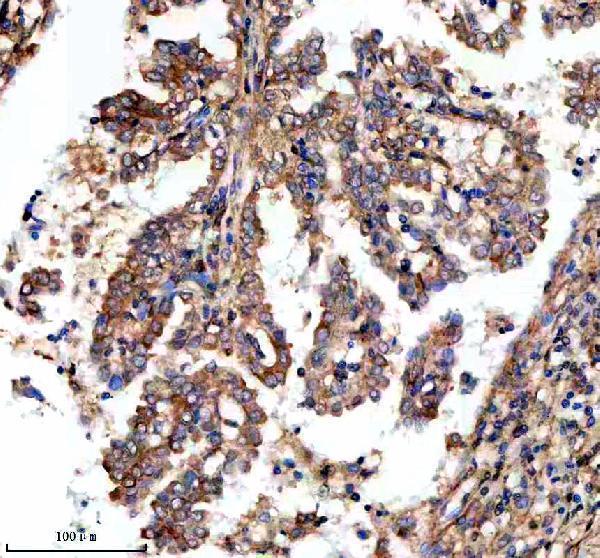
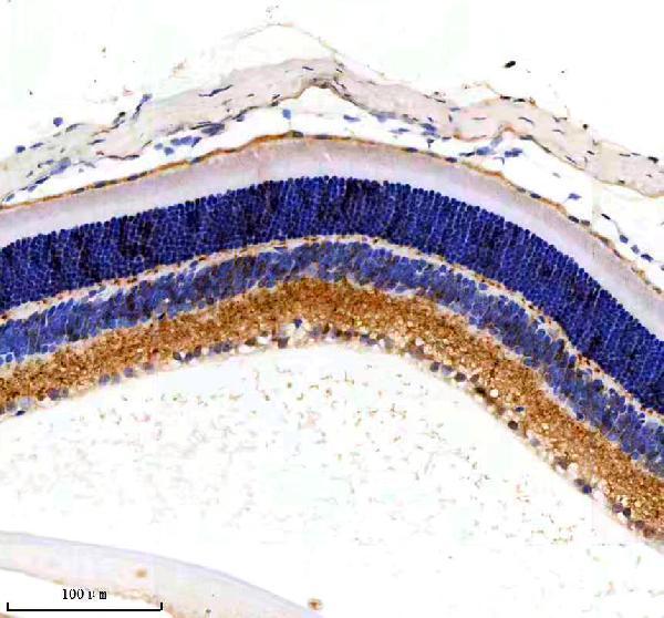
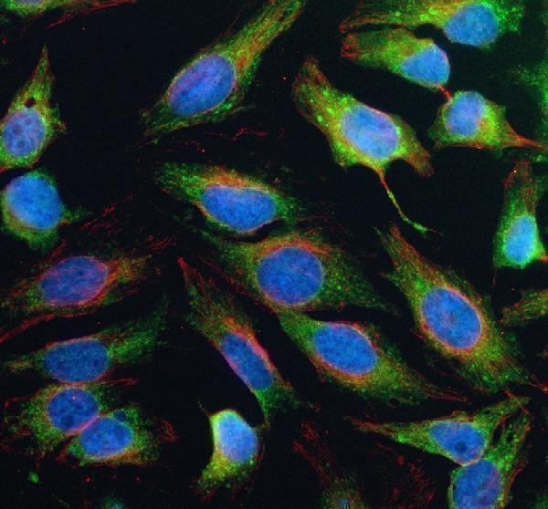
The anti-MCAT antibody A04806-1 has IHC images from paraffin-embedded human lung cancer and liver, mouse eye, and rat eye (catalog IHC captions), plus cellular IF in HeLa cells (catalog IF caption).

Real IHC data IHC analysis of MCAT using anti-MCAT antibody (A04806-1). MCAT was detected in a paraffin-embedded section of human lung cancer tissue. Heat mediated antigen retrieval was performed in EDTA buffer (pH 8.0, epitope retrieval solution). The tissue section was blocked with 10% goat serum. The tissue section was then incubated with 2 μg/ml rabbit anti-MCAT Antibody (A04806-1) overnight at 4°C. Peroxidase Conjugated Goat Anti-rabbit IgG was used as secondary antibody and incubated for 30 minutes at 37°C. The tissue section was developed using HRP Conjugated Rabbit IgG Super Vision Assay Kit (Catalog # SV0002) with DAB as the chromogen.
Anti-MCAT Antibody ®
Cat # A04806-1

A04806-1 has IHC examples in paraffin-embedded human lung cancer and liver, mouse eye, and rat eye (catalog IHC captions). Its IF example uses HeLa cells (catalog IF caption), and the catalog lists IHC and ICC/IF applications with human, mouse, and rat reactivity (catalog applications/reactivity).

Which to pick: Choose A04806-1 for chromogenic IHC on paraffin sections: its own caption documents EDTA retrieval at pH 8.0, 2 μg/ml primary antibody, and HRP/DAB detection; the fixative is unreported (A04806-1 IHC caption). For IF/ICC, A04806-1 has a HeLa cell example at 5 μg/ml (A04806-1 IF caption); clonality is unreported (catalog clone field). For cross-species IHC, A04806-1 has paraffin-section images from human, mouse, and rat samples (catalog IHC captions).

Each figure is that product's own IHC / IF validation image from its datasheet.

References

  1. UniProt Consortium. UniProt entry Q8IVS2 (FABD_HUMAN, Malonyl-CoA-acyl carrier protein transacylase, mitochondrial).
  2. Human Protein Atlas. MCAT tissue IHC expression (reliability: Approved).
  3. Human Protein Atlas. MCAT subcellular location (ICC-IF): Localized to the mitochondria..
  4. Human Protein Atlas. MCAT antibody validation summary (2 antibodies).
  5. Late-life restoration of mitochondrial function reverses cardiac dysfunction in old mice. eLife 2020 — PMC7377906.
  6. Hydrogen sulfide-mediated endothelial function and the interaction with eNOS and PDE5A activity in human internal mammary arteries. The Journal of international medical research 2019 — PMC6726794.
  7. Nuclear WRAP53 promotes neuronal survival and functional recovery after stroke. Science advances 2020 — PMC7541066.
  8. Mitochondrial targeted catalase suppresses invasive breast cancer in mice. BMC cancer 2011 — PMC3123323.
  9. PubMed PMID:12529303 — UniProt-cited evidence.
  10. PubMed PMID:14702039 — UniProt-cited evidence.
  11. PubMed PMID:10591208 — UniProt-cited evidence.