This website uses cookies to ensure you get the best experience on our website.


- Table of Contents
Plan MFSD2A chromogenic IHC on paraffin sections using the observed cytoplasmic tissue profile (HPA tissue IHC) and annotated membrane and ER localisation (UniProt). Use the reported high staining in appendix, breast, or salivary gland glandular cells as a reference, while accounting for the uncertain reliability of the tissue staining (HPA tissue IHC).

Expected localisation, validated protocols, controls and antibodies — the at-a-glance facts below, then the full design guide.
| Expected localisation | Cytoplasmic tissue staining (HPA tissue IHC); membrane and ER localisation (UniProt) | |
| Staining pattern | Cytoplasmic staining in most tissues (HPA tissue IHC) | |
| Antigen retrieval | EDTA pH 8.0 HIER, heat-mediated (datasheet A01967-2) | |
| Positive control | Appendix+4 more · see all | |
| Negative control | Heart muscle+4 more · see all |
| Fixation | Keep fixation consistent across sections (standard IHC practice; not target-specific) | |
| Caveat | Staining has low consistency with RNA expression (HPA tissue IHC) | |
| Regulation | Tissue-enhanced RNA expression (HPA tissue RNA) | |
| Isoform / epitope | 3 isoforms; check extracellular versus cytoplasmic epitope (UniProt) |
The catalog antibody’s IHC-P protocol (datasheet: A01967-2) is accompanied by published paraffin-section workflows for MFSD2A (PMC2846890; PMC6814593).
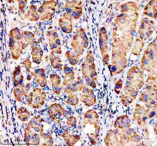
| Sample | Paraffin-embedded human liver cancer tissue; fixative not specified (datasheet A01967-2) |
| Fixation | Image fixative and duration unreported (datasheet A01967-2); verify before use. |
| Sectioning | 4–5 µm sections on charged slides (standard) |
| Deparaffinisation | Xylene, graded ethanol series to water (standard) |
| Antigen retrieval | Heat retrieval: EDTA pH 8.0 (datasheet A01967-2); 20 min, 95–100 °C (standard) |
| Peroxidase block | 3% H2O2, 10 min, room temperature (standard) |
| Blocking | 10% goat serum (datasheet A01967-2) |
| Primary antibody | Rabbit anti-MFSD2A, 2-5 μg/ml (datasheet A01967-2) |
| Primary incubation | Overnight at 4 °C (datasheet A01967-2) |
| Detection | HRP-conjugated secondary, DAB chromogen (datasheet A01967-2) |
| Counterstain | Hematoxylin, blue, dehydrate and mount (standard) |
| Expected result | MFSD2A-positive staining in glandular cells of appendix (HPA tissue IHC: High). HPA tissue profile: Cytoplasmic expression in most tissues. No signal in the no-primary control. |
MFSD2A is a 12-pass membrane protein assigned to the cell and endoplasmic reticulum membranes, with possible cytoplasmic puncta (UniProt Q8NA29 topology and subcellular location). In tissue IHC, HPA reports mainly cytoplasmic staining, including high staining in selected glandular cells and cerebellar Purkinje cells (HPA: tissue IHC). Treat this as a provisional pattern: HPA rates its tissue staining Uncertain because antibody staining and RNA expression have low consistency (HPA: reliability).
| Strong cytoplasmic staining in appendix, breast, or salivary gland glandular cells, or in cerebellar Purkinje cells (HPA: High in these cells). | This matches reported tissue staining. A membrane-associated component is biologically plausible for a transmembrane transporter, but the tissue IHC profile is described as cytoplasmic; do not require a crisp membrane outline to call the pattern concordant (UniProt Q8NA29 topology; HPA: tissue IHC). |
| Predominantly nuclear staining, with little cytoplasmic or membrane-associated signal (HPA: cytoplasmic tissue profile; UniProt Q8NA29 subcellular location). | Nuclear dominance conflicts with the supplied localisation evidence. Consider nonspecific staining or a detection artefact, and compare with a no-primary control before assigning the signal to MFSD2A (HPA: tissue IHC; UniProt Q8NA29 subcellular location; general IHC practice). |
| Strong staining in cardiomyocytes, skeletal myocytes, or prostate glandular cells (HPA: Not detected in these cells). | This disagrees with HPA's reported negatives and raises cross-reactivity or endogenous detection activity as possibilities. HPA's overall IHC reliability is Uncertain, so investigate the discrepancy rather than treating those cells as a definitive biological negative (HPA: tissue IHC and reliability; general IHC practice). |
| Diffuse chromogen across many cell types and tissue spaces, without clear cellular boundaries (general IHC practice). | A widespread haze cannot establish MFSD2A localisation. Check the no-primary control and detection background, then reassess whether any cell-associated staining remains; HPA's broad cytoplasmic profile alone cannot validate diffuse signal (general IHC practice; HPA: tissue IHC and reliability). |
| No staining in an appendix, breast, or salivary gland section where the sampled glandular cells are present (HPA: High in glandular cells). | The result differs from HPA's reported pattern, but those tissues are provisional comparison material because HPA rates IHC reliability Uncertain. Check tissue preservation, retrieval, antibody and detection performance, and a concurrent positive control before interpreting absence as biological (HPA: tissue IHC and reliability; general IHC practice). |
| Membrane topology (UniProt Q8NA29 topology) | MFSD2A has 12 transmembrane segments and both cytoplasmic and extracellular regions. Epitope position could affect antibody access, but the supplied record does not locate the IHC antibody's epitope or establish a retrieval requirement (UniProt Q8NA29 topology; general IHC practice). |
| Subcellular distribution (UniProt Q8NA29 subcellular location; HPA: subcellular ICC-IF) | UniProt places MFSD2A at cell and endoplasmic reticulum membranes and notes possible cytoplasmic puncta. HPA ICC-IF supports plasma-membrane localisation, calls cytosol uncertain, and lists cytoplasmic bodies as an additional approved location; these observations do not set an IHC scoring threshold (UniProt Q8NA29 subcellular location; HPA: subcellular ICC-IF). |
| Tissue evidence and antibody validation (HPA: tissue IHC and antibodies) | HPA reports high staining in selected cells but rates the overall tissue profile Uncertain; antibody HPA043107 is also marked IHC Uncertain. Use concordant cell and compartment patterns as supporting evidence, not proof of target specificity (HPA: tissue IHC, reliability, and antibodies). |
| Isoforms and glycosylation (UniProt Q8NA29 isoforms and glycosylation) | UniProt lists 3 isoforms and glycosylation sites at residues 230 and 240. The payload does not show which isoforms the IHC antibody recognises or whether these sites alter staining, so neither feature predicts a specific positive or negative section (UniProt Q8NA29 isoforms and glycosylation). |
| Situation | Likely cause | Next action |
|---|---|---|
| The expected glandular cells or Purkinje cells have no visible signal (HPA: High in those cells). | The assay may have failed, the sampled cells may be absent, or this section may differ from HPA's uncertain profile (HPA: tissue IHC and reliability; general IHC practice). | Confirm the relevant cells are present; review retrieval, antibody dilution and chromogenic detection against the catalog IHC-P instructions; include a concurrent positive control. Do not infer a target-specific fixation effect from this result alone (general IHC practice; HPA: reliability). |
| The entire section shows diffuse brown background (general IHC practice). | Nonspecific antibody binding or endogenous detection activity can obscure cell-associated staining (general IHC practice). | Inspect no-primary and detection controls, then review blocking, washes and antibody dilution. Score MFSD2A only after distinct cellular staining can be separated from background (general IHC practice). |
| Nuclei dominate the stain while cytoplasm is faint (HPA: cytoplasmic tissue profile). | The pattern conflicts with UniProt membrane localisation and HPA tissue staining; its cause cannot be assigned from morphology alone (UniProt Q8NA29 subcellular location; HPA: tissue IHC). | Compare with a no-primary control and an HPA-listed high-staining cell population on the same or a parallel section. Record persistent nuclear signal as discordant rather than counting it as MFSD2A-positive (general IHC practice; HPA: tissue IHC). |
| HPA-listed negative cells stain as strongly as the intended comparison cells (HPA: tissue IHC). | Cross-reactivity or endogenous detection activity is possible; HPA's uncertain reliability also limits how decisively its negative calls can be used (HPA: tissue IHC and reliability; general IHC practice). | Check no-primary and detection controls, compare staining by cell type and compartment, and seek independent target validation before interpreting the unexpected cells as expressing MFSD2A (general IHC practice; HPA: reliability). |
| Membrane edges and cytoplasmic puncta appear together (UniProt Q8NA29 subcellular location). | Both can fit the reported cell-membrane and possible vesicular distribution, although morphology alone cannot identify the puncta (UniProt Q8NA29 subcellular location). | Score each compartment separately and compare the stained cell types with HPA tissue IHC. Avoid calling every punctum specific because the HPA tissue profile remains Uncertain (HPA: tissue IHC and reliability; general IHC practice). |
| Can ICC-IF localisation settle an ambiguous chromogenic IHC result? (HPA: subcellular ICC-IF and tissue IHC) | HPA supports plasma-membrane localisation by ICC-IF, while its tissue IHC profile and the listed antibody's IHC validation are Uncertain; the assays provide different evidence (HPA: subcellular ICC-IF, tissue IHC, and antibodies). | Use the separate IF/ICC guide for that application. Here, compare IHC cell types, compartments and controls, and report any disagreement with the ICC-IF localisation explicitly (HPA: subcellular ICC-IF and tissue IHC; general IHC practice). |


Comprehensive Human Protein Atlas IHC scoring per tissue (reliability: Uncertain — Low consistency between antibody staining and RNA expression data. Pending external verification.). Rows are taken directly from the HPA tissue chart — click any row's HPA link to view the source.
| Tissue | Cell type | Level | Evidence | Source |
|---|---|---|---|---|
| Appendix | Glandular cells | High | Protein (IHC) | HPA → |
| Breast | Glandular cells | High | Protein (IHC) | HPA → |
| Cerebellum | Purkinje cells | High | Protein (IHC) | HPA → |
| Salivary gland | Glandular cells | High | Protein (IHC) | HPA → |
| Adipose tissue | Adipocytes | Medium | Protein (IHC) | HPA → |
| Tissue | Cell type | Level | Evidence | Source |
|---|---|---|---|---|
| Heart muscle | Cardiomyocytes | Not detected | Protein (IHC) | HPA → |
| Oral mucosa | Squamous epithelial cells | Not detected | Protein (IHC) | HPA → |
| Parathyroid gland | Glandular cells | Not detected | Protein (IHC) | HPA → |
| Prostate | Glandular cells | Not detected | Protein (IHC) | HPA → |
| Skeletal muscle | Myocytes | Not detected | Protein (IHC) | HPA → |
Troubleshoot MFSD2A chromogenic IHC in paraffin sections using documented retrieval conditions, compartment patterns, cellular context, and staining controls.
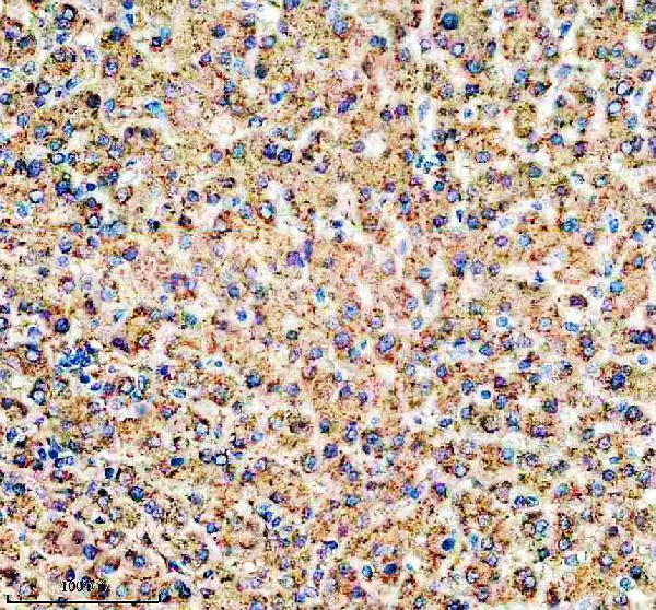
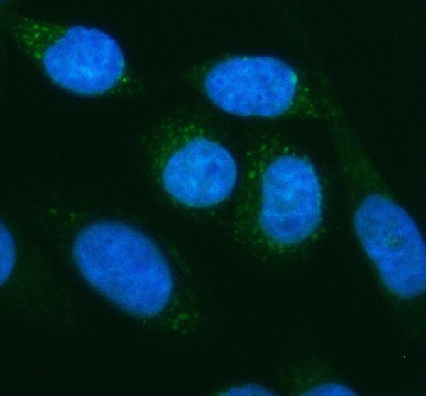
A01967-2 has IHC images from paraffin-embedded human liver and liver cancer sections, plus IF images from SIHA cells and human liver cancer sections (catalog image captions).
A01967-2 is listed for IHC, IF and ICC, with Human, Mouse and Rat reactivity (catalog applications and reactivity). Its IHC images show paraffin-embedded human liver and liver cancer sections; its IF images show SIHA cells and a paraffin-embedded human liver cancer section (catalog image captions).
Which to pick: For tissue IHC, choose A01967-2: its own IHC captions document paraffin-embedded human sections at 2 μg/ml after heat retrieval in EDTA, pH 8.0; the fixative is unreported (catalog IHC captions). For IF/ICC, A01967-2 is also listed for both applications, with IF images of SIHA cells and human liver cancer tissue at 5 μg/ml (catalog applications and IF captions). For cross-species work, A01967-2 is a polyclonal antibody listed as reactive with Human, Mouse and Rat, although its supplied IHC and IF images show human samples only (catalog dilution record, reactivity and image captions).