This website uses cookies to ensure you get the best experience on our website.


- Table of Contents
Plan MGP staining in paraffin sections using chondrocyte staining in cartilage as a positive reference (HPA tissue IHC). This guide covers fixation consistency, epitope coverage and interpretation of a secreted protein (UniProt).

Expected localisation, validated protocols, controls and antibodies — the at-a-glance facts below, then the full design guide.
| Expected localisation | Secreted; tissue compartment unresolved (UniProt) | |
| Staining pattern | High in chondrocytes; low in breast gland cells (HPA tissue IHC) | |
| Antigen retrieval | EDTA pH 8.0 HIER, heat-mediated (datasheet PB9954) | |
| Positive control | Cartilage+1 more · see all | |
| Negative control | Adipose tissue+4 more · see all |
| Fixation | Keep fixation consistent across sections. (standard IHC practice; not target-specific) | |
| Caveat | Secreted protein may stain beyond producing cells (UniProt) | |
| Regulation | Expression regulation not specified (UniProt) | |
| Isoform / epitope | 2 isoforms; mature chain 20–96; check epitope coverage (UniProt) |
The catalog antibody’s IHC-P protocol is paired with three published MGP immunohistochemistry protocols (PMC11178913; PMC11195983; PMC3731399).
| Sample | Paraffin-embedded human lung cancer tissue; fixative not specified (datasheet PB9954) |
| Fixation | Image fixative and duration unreported (datasheet PB9954); verify before use. |
| Sectioning | 4–5 µm sections on charged slides (standard) |
| Deparaffinisation | Xylene, graded ethanol series to water (standard) |
| Antigen retrieval | Heat retrieval: EDTA pH 8.0 (datasheet PB9954); 20 min, 95–100 °C (standard) |
| Peroxidase block | 3% H2O2, 10 min, room temperature (standard) |
| Blocking | 10% goat serum (datasheet PB9954) |
| Primary antibody | Rabbit anti-MGP, 0.5-1μg/ml (datasheet PB9954) |
| Primary incubation | Overnight at 4 °C (datasheet PB9954) |
| Detection | Streptavidin-biotin complex (SABC), DAB chromogen (datasheet PB9954) |
| Counterstain | Hematoxylin, blue, dehydrate and mount (standard) |
| Expected result | MGP-positive staining in chondrocytes of cartilage (HPA tissue IHC: High). HPA tissue profile: Expressed in chondrocytes and breast glands. No signal in the no-primary control. |
MGP is a secreted protein with no transmembrane segment and associates with the organic matrix of bone and cartilage (UniProt P08493). On IHC paraffin sections, expect the strongest reported staining in cartilage chondrocytes (HPA: High); prostate glandular cells show Medium staining and breast glandular cells Low staining (HPA tissue IHC). HPA rates its tissue staining profile Approved while cautioning that secreted protein and RNA locations may differ (HPA tissue IHC).
| Strong staining associated with cartilage chondrocytes, with possible nearby matrix staining. | This fits the reported High chondrocyte signal (HPA tissue IHC: cartilage). Nearby matrix signal is biologically plausible because MGP is secreted and associates with cartilage matrix (UniProt P08493). HPA does not specify a required intracellular or pericellular staining pattern, so assess the cell and surrounding tissue together. |
| Moderate prostate glandular staining or faint breast glandular staining. | These levels fit the reported Medium prostate and Low breast glandular signals (HPA tissue IHC). Compare each tissue with its own expected level; a weak breast result need not match the intensity of cartilage. Staining intensity alone does not establish which MGP form the antibody detects (UniProt P08493: processing and modified residues). |
| Predominantly nuclear staining, with little signal associated with the expected cells or surrounding tissue. | A nuclear-only pattern is discordant with MGP's Secreted annotation and lack of a transmembrane segment (UniProt P08493). Treat it as possible nonspecific staining and check controls and antibody performance (general IHC practice). Transient intracellular protein during secretion remains possible; the source record does not define a required intracellular compartment. |
| Strong staining in a cell population HPA reports as Not detected, such as adipocytes. | HPA reports MGP as Not detected in adipocytes (HPA tissue IHC: adipose tissue). Check whether staining follows tissue structures or appears across unrelated cells; cross-reactivity or endogenous detection activity are possible explanations (general IHC practice). One discrepant section alone does not identify which explanation applies. |
| Broad, similar chromogen deposition over positive and negative tissue areas. | A uniform pattern obscures the contrast expected between High chondrocytes and HPA Not detected cell populations (HPA tissue IHC). Consider background from detection chemistry, inadequate blocking, or excessive antibody concentration (general IHC practice). Reassess localisation only after controls show that the background is under control. |
| Secretion and processing | MGP has a signal peptide at residues 1–19 and a reported mature chain at 20–96 (UniProt P08493). Interpret cellular staining alongside possible deposited protein; HPA warns that secreted protein and tissue RNA locations may differ (HPA tissue IHC). The supplied record does not establish a required ratio of cellular to matrix staining. |
| Antibody recognition of MGP forms | UniProt lists two isoforms, a propeptide at 97–103, and modified residues including carboxyglutamate and phosphoserine (UniProt P08493). Without an epitope or form-specific validation in the supplied sources, do not infer which processed, modified, or isoform-specific species contributes to the IHC signal. |
| Strength of tissue and antibody evidence | HPA marks the tissue IHC profile Approved and lists antibody HPA013949 as IHC Approved (HPA tissue IHC; HPA antibodies). These labels support use of the reported pattern as a reference; the supplied antibody record does not say IHC Enhanced or document agreement between two independent antibodies. |
| Antigen retrieval in paraffin sections | Target-specific fixation effects are not established by the supplied assay evidence. Verify with a matched IHC source before attributing a result to fixation. |
| IF/ICC Q&A: Is a subcellular pattern established? | HPA calls MGP Secreted but provides no main ICC-IF location or cell-line images (HPA subcellular). The IHC tissue pattern therefore cannot define an IF punctate, cytoplasmic, or nuclear pattern. Assess IF/ICC localisation in its separate guide; no IF/ICC protocol is supplied here. |
| Situation | Likely cause | Next action |
|---|---|---|
| No staining in a cartilage section expected to be positive. | Chondrocytes are reported High (HPA tissue IHC: cartilage); a blank section could reflect assay performance or the sampled area (general IHC practice). The sources do not identify an MGP-specific fixation failure. | Confirm the section contains chondrocytes, then review the IHC-validated antibody, retrieval, primary incubation, and chromogenic detection with appropriate positive and omission controls (general IHC practice). |
| Prostate glandular cells appear negative while cartilage stains. | HPA reports Medium prostate glandular staining versus High cartilage chondrocyte staining (HPA tissue IHC). A weaker signal may be missed at the chosen detection threshold (general IHC practice). | Review glandular morphology and compare matched runs before changing interpretation; optimise detection against positive and negative controls without treating all tissues as equally intense (general IHC practice). |
| Breast glandular staining is weak or difficult to score. | HPA reports Low staining in breast glandular cells (HPA tissue IHC). Weak signal can be difficult to distinguish from chromogen background (general IHC practice). | Score the glandular cells against local background and control sections; use High cartilage staining as a run-level reference, not as the expected breast intensity (HPA tissue IHC; general IHC practice). |
| Nuclei dominate the stain, including in HPA Not detected cells. | A nuclear-only pattern conflicts with the Secreted annotation (UniProt P08493). Signal in HPA Not detected cells raises concern for nonspecific binding or detection activity (HPA tissue IHC; general IHC practice). | Inspect primary-omission and detection controls, confirm the expected cartilage pattern, and adjust blocking or antibody conditions if background persists (general IHC practice). |
| Diffuse chromogen covers both expected positive and negative areas. | Loss of tissue contrast can result from excessive primary or detection signal, inadequate blocking, or endogenous activity (general IHC practice). HPA's High and Not detected categories provide comparison points (HPA tissue IHC). | Check omission controls, review blocking and detection steps, and adjust staining conditions until morphology and tissue contrast are interpretable (general IHC practice). |
| Protein staining does not follow the tissue RNA enrichment signal. | HPA reports RNA as tissue enriched in blood vessel while its protein IHC profile highlights chondrocytes and breast glands; it warns that secreted protein and RNA locations may differ (HPA tissue IHC). | Interpret the stained section using its protein IHC cell pattern and controls. Do not label the RNA-to-protein mismatch an antibody failure without independent evidence (HPA tissue IHC; general IHC practice). |
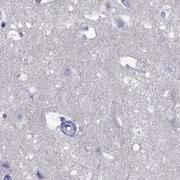


Comprehensive Human Protein Atlas IHC scoring per tissue (reliability: Approved — At least one protein variant secreted, tissue location of RNA and protein might differ and correlation is complex.). Rows are taken directly from the HPA tissue chart — click any row's HPA link to view the source.
| Tissue | Cell type | Level | Evidence | Source |
|---|---|---|---|---|
| Cartilage | Chondrocytes | High | Protein (IHC) | HPA → |
| Prostate | Glandular cells | Medium | Protein (IHC) | HPA → |
| Tissue | Cell type | Level | Evidence | Source |
|---|---|---|---|---|
| Adipose tissue | Adipocytes | Not detected | Protein (IHC) | HPA → |
| Adrenal gland | Glandular cells | Not detected | Protein (IHC) | HPA → |
| Appendix | Lymphoid tissue | Not detected | Protein (IHC) | HPA → |
| Bone marrow | Hematopoietic cells | Not detected | Protein (IHC) | HPA → |
| Bronchus | Respiratory epithelial cells | Not detected | Protein (IHC) | HPA → |
Troubleshoot MGP staining in paraffin sections by checking retrieval, compartment, controls and scoring before interpreting chromogenic signal.
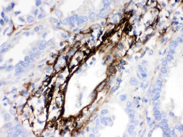
The IHC-validated anti-MGP antibody has real chromogenic IHC data from paraffin-embedded human lung cancer tissue (PB9954 image caption).
PB9954 is the sole card and is listed for human IHC (catalog applications/reactivity). Its figure shows MGP staining in a paraffin-embedded human lung cancer section with DAB detection (PB9954 image caption).
Which to pick: Choose PB9954 for human paraffin-section IHC: it is a rabbit antibody listed for human IHC, and its figure shows staining after EDTA retrieval at pH 8.0 (catalog host/applications/reactivity; PB9954 image caption). No listed SKU supports an IF/ICC or cross-species recommendation: PB9954 lists human IHC and WB, with no IF figure or nonhuman reactivity (catalog applications/reactivity; PB9954 image inventory). The figure caption reports paraffin embedding but does not identify the fixative, so fixation is unconfirmed (PB9954 image caption). The selected PB9954 tissue-IHC caption documents paraffin sections, but does not specify the fixative (selected-SKU IHC image PB9954).