MIA2 / Melanoma inhibitory activity protein 2 · IHC design guide

Design Immunohistochemistry for MIA2

Plan chromogenic MIA2 IHC in paraffin sections using the cytoplasmic staining seen in glandular and neuronal cells as a reference (HPA tissue IHC). This guide covers fixation, the catalog antibody’s 1:50 IHC dilution (datasheet A08319), and interpretation when the antibody may target more than one gene (HPA tissue IHC).

Evidence assembled Oct 2026 · For research use; verify linked source records and product datasheet before use
Immunohistochemistry protocol sheet for MIA2 (IHC for MIA2): expected localisation Cytoplasmic tissue staining (HPA tissue IHC); ER exit sites (UniProt), antibody A08319, validated IHC image, and IHC protocol steps
Printable MIA2 IHC protocol sheet — expected localisation Cytoplasmic tissue staining (HPA tissue IHC); ER exit sites (UniProt), antibody A08319, controls and protocol steps. Open the full MIA2 IHC guide →

MIA2 Immunohistochemistry Experimental Design Guide

Expected localisation, validated protocols, controls and antibodies — the at-a-glance facts below, then the full design guide.

Must know before staining
Expected localisation Cytoplasmic tissue staining (HPA tissue IHC); ER exit sites (UniProt)
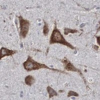
Staining pattern Cytoplasmic staining in glandular and neuronal cells (HPA tissue IHC)
Antigen retrieval Tris-EDTA pH 9.0 HIER, heat-mediated (datasheet A08319)
Positive control ⓘ Adrenal gland+4 more · see all
Negative control ⓘ Adipose tissue+2 more · see all
Important caveats
Reasons your staining may differ from the expected pattern.
Fixation Keep fixation consistent across sections (standard IHC practice; not target-specific)
Caveat Antibody targeting of more than one gene may confound staining (HPA tissue IHC)
Regulation Higher in severe fibrosis or inflammation (UniProt)
Isoform / epitope 12 isoforms; check epitopes against lumenal and cytoplasmic regions (UniProt)
Section 1

Recommended MIA2 IHC & IF Protocols

The catalog antibody’s IHC-P protocol (datasheet A08319) is accompanied by three published MIA2 IHC protocols (PMC8396825; PMC3629429; PMC5058745).

Recommended immunohistochemistry (IHC-P) protocol parameters
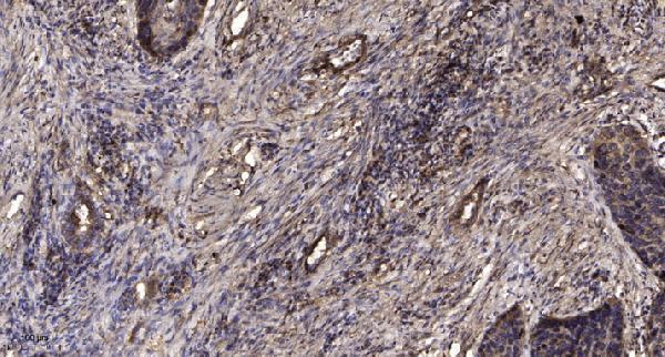
SampleParaffin-embedded human Squamous cell carcinoma of lung tissue; fixative not specified (datasheet A08319)
FixationImage fixative and duration unreported (datasheet A08319); verify before use.
Sectioning4–5 µm sections on charged slides (standard)
DeparaffinisationXylene, graded ethanol series to water (standard)
Antigen retrievalHeat retrieval: Tris-EDTA pH 9.0 (datasheet A08319); 20 min, 95–100 °C (standard)
Peroxidase block3% H2O2, 10 min, room temperature (standard)
Blocking10% normal serum of the secondary host, 30 min, room temperature (standard)
Primary antibodyRabbit anti-MIA2, 1:50 recommended; image 1:200 (datasheet A08319)
Primary incubationOvernight at 4 °C (standard)
DetectionHRP-polymer secondary, DAB chromogen 5–10 min (standard)
CounterstainHematoxylin, blue, dehydrate and mount (standard)
Expected resultMIA2-positive staining in glandular cells of adrenal gland (HPA tissue IHC: High). HPA tissue profile: Cytoplasmic expression in most tissues, most abundant in glandular and neuronal cells. No signal in the no-primary control.
💡Decision noteStart with heat retrieval in Tris-EDTA at pH 9.0 for the catalog antibody (datasheet A08319). Use each paper’s stated retrieval method when reproducing its protocol (PMC8396825; PMC3629429; PMC5058745).
Section 2

What Is the Expected MIA2 Staining Pattern?

MIA2 is an endoplasmic reticulum (ER) membrane protein concentrated at ER exit sites; its single transmembrane segment leaves a long cytoplasmic region (UniProt Q96PC5 topology). In tissue IHC, expect cytoplasmic staining chiefly in glandular and neuronal cells (HPA: tissue IHC). Interpret specificity cautiously: HPA rates the tissue profile Approved but reports low agreement with RNA and warns that staining may include protein from more than one gene (HPA: reliability).

What am I looking at on my slide?
Cytoplasmic signal in glandular cells or neurons, with little nuclear staining.This fits the observed tissue pattern and the ER membrane location (HPA: tissue IHC; UniProt Q96PC5: subcellular location). HPA reports High staining in adrenal, appendix and duodenal glandular cells and in caudate and cerebral cortex neurons (HPA: tissue IHC). Agreement in compartment and cell type supports interpretation, but HPA's cross-gene warning limits attribution to MIA2 alone (HPA: reliability).
Predominantly nuclear staining, with no convincing cytoplasmic signal.A nuclear-dominant pattern conflicts with the supported ER location and observed cytoplasmic tissue profile (UniProt Q96PC5: subcellular location; HPA: tissue IHC). Treat it as suspect staining; check controls and morphology before scoring it as MIA2 (general IHC practice).
Strong signal mainly in adipocytes, cardiomyocytes or skeletal myocytes.Those cells are listed as Not detected in the HPA tissue IHC profile (HPA: tissue IHC). Consider cross-reactivity or endogenous detection activity, especially if staining also appears in a negative control (general IHC practice). A different specimen result alone cannot establish which explanation applies; the HPA profile carries a specificity caution (HPA: reliability).
Diffuse colour across tissue structures, without a clear cellular pattern.This does not match HPA's cell-associated cytoplasmic profile (HPA: tissue IHC). Review the negative control, reagent background, blocking, washes and detection conditions before assigning cellular positivity (general IHC practice).
No signal in glandular or neuronal cells of a chosen positive-control section.First confirm that the section contains the cell type being judged. HPA reports High staining in specific glandular and neuronal populations, rather than every cell in those tissues (HPA: tissue IHC). If the relevant cells are present, check antibody, retrieval and detection controls before calling the specimen negative (general IHC practice).
💡Expected MIA2 appearanceCall a positive result when distinct cytoplasmic staining is present in the relevant glandular or neuronal cells, with stronger staining plausible in populations HPA rates High; nuclear-dominant or structure-wide diffuse colour is suspect (HPA: tissue IHC; UniProt Q96PC5: ER membrane location).
How each factor affects the staining
ER location and topology (UniProt Q96PC5: topology and subcellular location).MIA2 localizes to ER exit sites; residues 647–667 form a transmembrane segment, with lumenal and cytoplasmic regions on opposite sides (UniProt Q96PC5: topology). Interpret cytoplasmic compartment staining at tissue-section resolution; the record does not identify the catalog antibody's epitope, so it cannot predict that antibody's access or retrieval needs.
Tissue distribution and antibody confidence (HPA: tissue IHC and reliability).HPA reports High signal in several glandular and neuronal populations, but Not detected in adipocytes and muscle cells (HPA: tissue IHC). Its Approved tissue profile has low staining–RNA consistency and a cross-gene warning; a matching pattern is useful for judging a slide but is not independent proof of MIA2 specificity (HPA: reliability).
Isoforms and processing (UniProt Q96PC5: isoforms and processing).UniProt lists 12 isoforms and a 1–19 signal peptide; its annotated chain begins at residue 20 (UniProt Q96PC5: isoforms and processing). Because no antibody epitope is supplied, isoform coverage cannot be inferred. The record supplies no shedding annotation to explain extracellular staining.
IF/ICC Q: What pattern should be checked on its separate guide page?A: HPA supports ER localization in ICC-IF and lists images from A-549 and Hep-G2; HPA055459 has Supported ICC status (HPA: subcellular profile; HPA: antibodies). Those data support a compartment check, not an IF/ICC protocol or a claim that every IHC antibody has ICC support.
Why is my staining missing, weak or wrong?
SituationLikely causeNext action
No chromogenic signal in the selected positive-control section.The scored area may lack the relevant glandular or neuronal cells; reagent or detection failure is another possibility (HPA: tissue IHC; general IHC practice).Locate the expected cell population and verify that the antibody and detection controls worked. Review the catalog antibody's IHC-P instructions before adjusting retrieval or dilution; no MIA2-specific retrieval condition is established by this record (general IHC practice).
Strong nuclear staining dominates the section.A nuclear-dominant result is inconsistent with MIA2's ER membrane location and HPA's cytoplasmic tissue profile (UniProt Q96PC5: subcellular location; HPA: tissue IHC).Check the negative control, focus and cell boundaries, then score cytoplasmic and nuclear signal separately. Withhold an MIA2-positive call if the expected cytoplasmic pattern cannot be resolved (general IHC practice).
Adipocytes or muscle cells stain strongly.HPA reports Not detected staining in adipocytes, cardiomyocytes and skeletal myocytes; off-target binding or endogenous detection activity is possible (HPA: tissue IHC; general IHC practice).Compare with a negative control and with a tissue region containing an HPA-reported positive cell type. Check detection-related background before interpreting the unexpected signal; HPA also cautions that its tissue staining may reflect more than one gene (HPA: reliability; general IHC practice).
Colour is diffuse or obscures tissue architecture.Background from blocking, washing or detection can prevent assignment of signal to cells (general IHC practice).Inspect the negative control and repeat with the catalog antibody's IHC-P conditions, checking blocking, washes and detection timing. Score only signal that can be assigned to cells and a compartment (general IHC practice).
Two sections or antibodies give different staining distributions.HPA reports low consistency between tissue staining and RNA and cautions that its staining may include protein from more than one gene; antibody-specific recognition remains unresolved here (HPA: reliability).Compare the same cell types and controls across sections. Record which antibody produced each pattern; do not resolve the discrepancy from staining intensity alone. HPA lists four antibodies with Approved IHC status, which does not remove its profile-level caution (HPA: antibodies; HPA: reliability).

Sample controls for MIA2 IHC & IF

🧪Run duodenum first; its glandular cells should stain (HPA: High in duodenal glandular cells). Use skeletal muscle myocytes as the negative tissue (HPA: Not detected in skeletal muscle myocytes); on the duodenal slide, assess unstained non-glandular areas for background without assuming those cells are biologically negative (standard IHC practice).
Positive control tissue: Adrenal gland (Glandular cells, HPA High)
Negative control tissue: Adipose tissue (HPA Not detected)
ICC-IF cell lines (HPA subcellular resource): HPA ICC-IF images show MIA2 in A-549, Hep-G2, with annotated localisation: Endoplasmic reticulum (supported) (HPA subcellular).
Technical controls: Include a no-primary, secondary-only control, an isotype control matched to the primary antibody’s host species and clonality, and a MIA2 knockout or validated peptide-block control (standard IHC practice). For chromogenic staining, block endogenous peroxidase and check the duodenal slide for signal in inflammatory cells; block endogenous biotin if using avidin–biotin detection (standard IHC practice).
⚠️Feasibility: A target-specific fixation window and fixation effect are unreported in the supplied evidence; the fixative for the selected A08319 paraffin-section caption is also unreported (selected-SKU tissue-IHC caption). That caption used Tris–EDTA at pH 9.0 for retrieval and primary antibody at 1:200 overnight at 4 °C in lung squamous cell carcinoma, so retrieval dependence in duodenum remains unestablished (selected-SKU tissue-IHC caption). Whether frozen sections or IF are easier is unreported; IF has a supported endoplasmic reticulum localization reference, while endogenous peroxidase in inflammatory cells can complicate duodenal chromogenic scoring (HPA: endoplasmic reticulum supported; standard IHC practice).

HPA tissue IHC evidence for MIA2

Comprehensive Human Protein Atlas IHC scoring per tissue (reliability: Approved — Low consistency between antibody staining and RNA expression data. Caution, targets protein from more than one gene. Pending external verification.). Rows are taken directly from the HPA tissue chart — click any row's HPA link to view the source.

Positive expression · recommended positive controls

TissueCell typeLevelEvidenceSource
Adrenal gland Glandular cells High Protein (IHC) HPA →
Appendix Glandular cells High Protein (IHC) HPA →
Caudate Neuronal cells High Protein (IHC) HPA →
Cerebral cortex Neuronal cells High Protein (IHC) HPA →
Duodenum Glandular cells High Protein (IHC) HPA →

Undetected expression · recommended negative controls

TissueCell typeLevelEvidenceSource
Adipose tissue Adipocytes Not detected Protein (IHC) HPA →
Heart muscle Cardiomyocytes Not detected Protein (IHC) HPA →
Skeletal muscle Myocytes Not detected Protein (IHC) HPA →
Section 3

Advanced MIA2 IHC Tips

Use the catalog antibody’s paraffin-section result as a starting point, and interpret staining against MIA2’s reported ER localisation and tissue pattern.

Which retrieval conditions should I start with for MIA2 paraffin-section IHC?
Start with heat-mediated retrieval in Tris-EDTA at pH 9.0 for paraffin sections (datasheet A08319). The selected image used a 1:200 primary antibody dilution at 4°C overnight after that retrieval (caption A08319). If staining is weak, compare a gentler retrieval condition on adjacent sections while holding antibody dilution and detection constant; assess tissue preservation alongside signal (standard IHC practice). Score cytoplasmic staining in intact cells, since MIA2 is associated with ER membranes and ER exit sites (UniProt Q96PC5 localisation). Include a no-primary control to identify detection-derived staining before changing retrieval again (standard IHC practice).
How should I troubleshoot weak staining when fixation details are missing?
The selected paraffin-section caption does not report a fixative, so MIA2-specific sensitivity to fixation is unknown (caption A08319). Record each specimen’s fixative, fixation duration, processing history and section age before comparing staining intensity (standard IHC practice). Run sections from differently processed specimens in the same staining batch, using Tris-EDTA at pH 9.0 and the documented 1:200 primary dilution as starting conditions (datasheet A08319; caption A08319). If signal varies with processing, compare matched sections and tissue morphology before adjusting retrieval or antibody concentration (standard IHC practice). Do not assign the difference to a MIA2-specific fixation effect without direct evidence.
What staining pattern fits MIA2, and what pattern deserves scrutiny?
Expect intracellular cytoplasmic staining compatible with an ER-associated protein, potentially concentrated near ER exit sites (UniProt Q96PC5 localisation; HPA: cytoplasmic tissue profile). MIA2 spans the membrane at residues 647–667, with a lumenal N-terminal region and a cytoplasmic region from 668–1412 (UniProt Q96PC5 topology). Diffuse DAB throughout a section does not by itself establish that this distribution is specific; compare intact cells with a no-primary control (standard IHC practice). Check whether staining appears in glandular or neuronal cells, where HPA reports abundant signal, and review unexpected nuclear-only or extracellular staining separately (HPA: tissue profile; UniProt Q96PC5 localisation).
How can epitope position and MIA2 isoforms affect the IHC result?
Map the antibody’s disclosed immunogen to MIA2 before interpreting a negative section; this record lists 12 isoforms (UniProt Q96PC5 isoforms). An epitope in residues 20–605 is lumenal, whereas one in residues 668–1412 is cytoplasmic on the membrane-associated form (UniProt Q96PC5 topology). Glycosylation is reported at residues 59 and 368, but its effect on this antibody’s staining is not established (UniProt Q96PC5 glycosylation). If the immunogen is unavailable, do not infer isoform coverage from staining alone; compare sections with an independent antibody of known epitope where available (standard IHC validation practice).
How should I assess MIA2 localisation by IF alongside chromogenic IHC?
Treat IF as a separate assay: HPA reports supported ER localisation and lists A-549 and Hep-G2 among its ICC/IF image cell lines (HPA: subcellular). Multiplex MIA2 with a marker for the expected cell type, such as an appropriate glandular-cell marker in a glandular specimen, and check the channels separately (HPA: tissue profile; standard IF practice). Choose fluorophores after inspecting unstained tissue autofluorescence, favoring a channel with clear separation from that background (standard IF practice). Once the antibody epitope is mapped, optimize permeabilisation for access to its lumenal or cytoplasmic side; the membrane spans residues 647–667 (UniProt Q96PC5 topology; standard IF practice).
What should I change when MIA2 DAB staining is widespread or granular?
First compare the stained section with no-primary and detection-only controls to locate background from the detection system (standard IHC practice). Block endogenous peroxidase before DAB development and inspect pigment or precipitate in unstained sections; these are general chromogenic IHC checks (standard IHC practice). If background persists, titrate the primary antibody around the documented 1:200 condition and keep retrieval at Tris-EDTA pH 9.0 during that comparison (caption A08319; datasheet A08319). Assess staining in intact cells rather than section edges or damaged areas, and require a plausible cytoplasmic distribution for MIA2 (standard IHC practice; UniProt Q96PC5 localisation).
How should I score MIA2 staining across paraffin sections? ⚠ ANSWER MARKED FOR VERIFICATION
Define the cell population and region before scoring, then record the percentage of positive cells and an intensity-based H-score on matched sections (standard IHC quantification practice). Normalize positive-cell counts to all evaluable cells of that population, or report positive-cell density per mm² of viable tissue when counts are spatially sampled (standard IHC quantification practice). Keep retrieval, primary dilution, detection and image acquisition consistent across comparison groups; the documented starting dilution is 1:200 (caption A08319; standard IHC practice). Score cytoplasmic signal separately from background, and stratify cell types because HPA describes particularly abundant glandular and neuronal staining (HPA: tissue profile).
When is a MIA2-positive section convincing rather than an artefact?
Favor staining in intact cytoplasm with a plausible ER-associated distribution, consistent with MIA2 at ER membranes and exit sites (UniProt Q96PC5 localisation). Check cell identity: HPA reports abundant staining in glandular and neuronal cells, but its tissue IHC has low consistency with RNA data and may detect proteins from more than one gene (HPA: tissue profile; HPA: reliability). Discount signal confined to cut edges, necrosis, pigment or areas positive in no-primary controls; check endogenous peroxidase when DAB is used (standard IHC practice). A convincing result requires reproducible staining across comparable sections and independent specificity evidence before assigning the signal exclusively to MIA2 (standard IHC validation practice; HPA: reliability).
Boster reagents

Best MIA2 / Melanoma inhibitory activity protein 2 IHC Antibodies

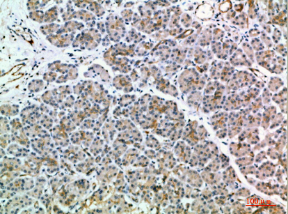
A08319 has IHC images of paraffin-embedded human lung squamous cell carcinoma and human pancreas (catalog image captions); listed reactivity covers human and mouse (catalog: reactivity).

Real IHC data Immunohistochemical analysis of paraffin-embedded human Squamous cell carcinoma of lung. 1, Antibody was diluted at 1:200 (4° overnight). 2, Tris-EDTA,pH9.0 was used for antigen retrieval. 3,Secondary antibody was diluted at 1:200 (room temperature, 45min).
Anti-MIA2 Antibody
Cat # A08319

A08319 is listed for IHC and WB in human and mouse (catalog: applications and reactivity). Its IHC images show paraffin-embedded human lung squamous cell carcinoma and human pancreas (catalog image captions).

Which to pick: For tissue IHC, choose A08319, a rabbit polyclonal antibody (catalog: host and dilution_raw); its lung image documents paraffin sections and Tris-EDTA retrieval at pH 9.0, while the fixative is unreported (catalog: lung image caption). No SKU in this payload lists IF/ICC validation, so there is no supported IF/ICC pick here (catalog: applications and IF image captions). For cross-species IHC, A08319 lists human and mouse reactivity, but its supplied IHC images show human tissues only (catalog: reactivity and IHC image captions).

Each figure is that product's own IHC / IF validation image from its datasheet.

References

  1. UniProt Consortium. UniProt entry Q96PC5 (MIA2_HUMAN, Melanoma inhibitory activity protein 2).
  2. Human Protein Atlas. MIA2 tissue IHC expression (reliability: Approved).
  3. Human Protein Atlas. MIA2 subcellular location (ICC-IF): Localized to the endoplasmic reticulum..
  4. Human Protein Atlas. MIA2 antibody validation summary (4 antibodies).
  5. Melanoma Inhibitory Activity and Melanoma Inhibitory Activity 2 as Novel Immunohistochemical Markers of Oral Epithelial Dysplasia. Journal of clinical medicine 2021 — PMC8396825.
  6. Protumoral roles of melanoma inhibitory activity 2 in oral squamous cell carcinoma. British journal of cancer 2013 — PMC3629429.
  7. A comprehensive expression analysis of the MIA gene family in malignancies: MIA gene family members are novel, useful markers of esophageal, lung, and cervical squamous cell carcinoma. Oncotarget 2016 — PMC5058745.
  8. Potential Immunohistochemical Biomarkers for Grading Oral Dysplasia: A Literature Review. Biomedicines 2024 — PMC10967812.
  9. PubMed PMID:9356211 — UniProt-cited evidence.
  10. PubMed PMID:12839582 — UniProt-cited evidence.
  11. PubMed PMID:27138255 — UniProt-cited evidence.