This website uses cookies to ensure you get the best experience on our website.


- Table of Contents
Plan chromogenic paraffin-section IHC for MRPL58 using the IHC-validated antibody at 1:25 (datasheet: A31846-1). Assess cytoplasmic tissue staining (HPA tissue IHC) while recognizing its mitochondrial localization (UniProt).

Expected localisation, validated protocols, controls and antibodies — the at-a-glance facts below, then the full design guide.
| Expected localisation | Observed cytoplasm (HPA tissue IHC); mitochondrial protein (UniProt) | |
| Staining pattern | Cytoplasmic staining in several tissues (HPA tissue IHC) | |
| Antigen retrieval | Citrate pH 6.0 HIER, 95–98 °C, 20 min (rule: cytoplasmic / membrane antigen) | |
| Positive control | Adipose tissue+4 more · see all | |
| Negative control | Bone marrow+4 more · see all |
| Fixation | Keep paraffin-section fixation consistent (standard IHC practice; not target-specific); Matched tissue-IHC evidence does not establish the fixation claim. Validate the specimen-specific method before use. (selected-SKU IHC image A31846-1) | |
| Caveat | Staining has low consistency with RNA expression (HPA tissue IHC) | |
| Regulation | Down-regulated during HT29-D4 differentiation (UniProt) | |
| Isoform / epitope | No annotated isoforms; mature chain spans 30–206 (UniProt) |
Compare the catalog antibody’s IHC-P protocol (datasheet) with the published human osteosarcoma tissue method (PMC12450695 methods).
| Sample | Paraffin-embedded H. colorectal carcinoma tissue; fixative not specified (datasheet A31846-1) |
| Fixation | Image fixative and duration unreported (datasheet A31846-1); verify before use. |
| Sectioning | 4–5 µm sections on charged slides (standard) |
| Deparaffinisation | Xylene, graded ethanol series to water (standard) |
| Antigen retrieval | Heat-induced epitope retrieval in citrate buffer, pH 6.0, 20 min at 95–98 °C (standard rule: cytoplasmic / membrane antigen) |
| Peroxidase block | 3% H2O2, 10 min, room temperature (standard) |
| Blocking | 10% normal serum of the secondary host, 30 min, room temperature (standard) |
| Primary antibody | Rabbit anti-MRPL58, 1:25 (datasheet A31846-1) |
| Primary incubation | Overnight at 4 °C (standard) |
| Detection | HRP-polymer secondary, DAB chromogen 5–10 min (standard) |
| Counterstain | Hematoxylin, blue, dehydrate and mount (standard) |
| Expected result | MRPL58-positive staining in adipocytes of adipose tissue (HPA tissue IHC: Medium). HPA tissue profile: Cytoplasmic expression in several tissues. No signal in the no-primary control. |
MRPL58 is a mitochondrial protein with no transmembrane segment (UniProt Q14197). In paraffin section IHC, expect cytoplasmic staining in selected cells, including colon glandular cells and cerebral cortex neurons, where HPA reports medium staining (HPA tissue IHC). HPA rates its IHC evidence “Approved” but reports low consistency between antibody staining and RNA expression; interpret tissue differences with that limitation (HPA tissue IHC).
| Cytoplasmic staining in colon glandular cells or cerebral cortex neurons. | This matches the cells and medium levels reported by HPA (HPA tissue IHC). A fine granular appearance can fit a mitochondrial protein, but paraffin section IHC alone does not establish that individual deposits are mitochondria (UniProt Q14197; general IHC practice). |
| Predominantly nuclear or cell-border staining, with little cytoplasmic signal. | Treat this as a compartment mismatch for tissue IHC: HPA describes cytoplasmic tissue expression, while UniProt places MRPL58 in mitochondria (HPA tissue IHC; UniProt Q14197). Review controls and detection before calling it MRPL58; HPA's additional nucleoplasmic and plasma-membrane IF locations are uncertain (HPA subcellular). |
| Strong staining in a cell population HPA lists as “Not detected,” such as bone marrow hematopoietic cells. | This conflicts with that reported tissue result and warrants checks for cross-reactivity or endogenous detection activity (HPA tissue IHC; general IHC practice). “Not detected” describes HPA's assay observation, not proof that every specimen or method must be negative (HPA tissue IHC). |
| Uniform color across tissue and empty spaces, with poor cell boundaries. | Interpret this as background rather than a resolved cellular pattern (general IHC practice). Check the no-primary control, blocking, washes and chromogen development; HPA's reported MRPL58 pattern is cytoplasmic expression in several tissues (HPA tissue IHC). |
| No staining in colon glandular cells in a run intended to show a positive result. | HPA reports medium staining there, so review tissue preservation, retrieval, primary-antibody application and detection controls before interpreting the absence biologically (HPA tissue IHC; general IHC practice). HPA's low staining-to-RNA consistency also limits certainty from a single specimen (HPA tissue IHC). |
| Cellular compartment | MRPL58 belongs to the mitochondrial large ribosomal subunit and has no transmembrane segment (UniProt Q14197). HPA describes tissue IHC as cytoplasmic; do not require a distinct mitochondrial outline in chromogenic sections (HPA tissue IHC; general IHC practice). |
| Tissue and cell choice | HPA reports medium staining in colon glandular cells and adipocytes, but “Not detected” in salivary glandular cells and skeletal myocytes (HPA tissue IHC). Compare the named cell populations, since a whole-section average can obscure their different reported results (general IHC practice). |
| Evidence strength | The tissue IHC reliability label is “Approved,” with low consistency between staining and RNA expression; the listed IHC antibody is also “Approved,” not “Enhanced” (HPA tissue IHC; HPA antibodies). Use the pattern as a reference for interpretation, with controls for each run (general IHC practice). |
| IF/ICC Q&A: Should nuclear or membrane fluorescence be expected? | Mitochondria are the supported main location; nucleoplasm and plasma membrane are additional uncertain locations (HPA subcellular). Treat those extra signals cautiously. This IF/ICC observation does not establish an IHC-P staining pattern (HPA subcellular; HPA tissue IHC). |
| Situation | Likely cause | Next action |
|---|---|---|
| The expected cytoplasmic signal is absent in colon glands. | A staining-run failure is possible; HPA reports medium staining in these cells (HPA tissue IHC; general IHC practice). | Check the run's positive control, primary-antibody application, retrieval and detection steps, then repeat if a control failed (general IHC practice). No MRPL58-specific retrieval condition is established by the supplied evidence. |
| Color is strongest in nuclei or along cell borders. | The distribution conflicts with HPA's cytoplasmic tissue profile and UniProt's mitochondrial location (HPA tissue IHC; UniProt Q14197). | Compare with a no-primary control and reassess antibody specificity and detection background before assigning the signal to MRPL58 (general IHC practice). |
| Bone marrow hematopoietic cells stain strongly. | HPA lists these cells as “Not detected”; cross-reactivity or endogenous detection activity may explain unexpected color (HPA tissue IHC; general IHC practice). | Inspect the no-primary control and the detection and blocking steps; report the discrepancy by cell type rather than calling the specimen globally positive (general IHC practice). |
| Diffuse chromogen obscures cell boundaries. | Background from the staining workflow may overwhelm a cellular signal (general IHC practice). | Check blocking, wash quality and chromogen development against the no-primary control; score MRPL58 only where a cellular cytoplasmic pattern remains distinguishable (general IHC practice; HPA tissue IHC). |
| A reportedly negative cell type shows faint color. | “Not detected” is an HPA observation, and faint color alone cannot separate weak signal from background (HPA tissue IHC; general IHC practice). | Compare intensity with nearby background and run controls, document the named cell type, and avoid converting the HPA observation into an absolute biological negative (HPA tissue IHC; general IHC practice). |
| The slide pattern differs from a mitochondrial IF image. | HPA's supported mitochondrial localization comes from ICC-IF, while its tissue IHC profile is reported as cytoplasmic (HPA subcellular; HPA tissue IHC). | Judge chromogenic sections by cell-resolved cytoplasmic staining and IHC controls; use IF localization as context rather than requiring identical visual detail (HPA tissue IHC; HPA subcellular; general IHC practice). |


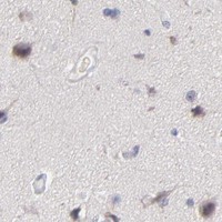
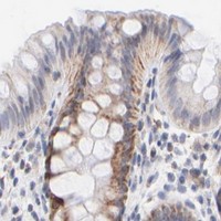
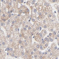


Comprehensive Human Protein Atlas IHC scoring per tissue (reliability: Approved — Low consistency between antibody staining and RNA expression data.). Rows are taken directly from the HPA tissue chart — click any row's HPA link to view the source.
| Tissue | Cell type | Level | Evidence | Source |
|---|---|---|---|---|
| Adipose tissue | Adipocytes | Medium | Protein (IHC) | HPA → |
| Adrenal gland | Glandular cells | Medium | Protein (IHC) | HPA → |
| Appendix | Glandular cells | Medium | Protein (IHC) | HPA → |
| Bronchus | Respiratory epithelial cells | Medium | Protein (IHC) | HPA → |
| Cerebellum | Cells in molecular layer | Medium | Protein (IHC) | HPA → |
| Tissue | Cell type | Level | Evidence | Source |
|---|---|---|---|---|
| Bone marrow | Hematopoietic cells | Not detected | Protein (IHC) | HPA → |
| Hippocampus | Glial cells | Not detected | Protein (IHC) | HPA → |
| Ovary | Ovarian stroma cells | Not detected | Protein (IHC) | HPA → |
| Salivary gland | Glandular cells | Not detected | Protein (IHC) | HPA → |
| Skeletal muscle | Myocytes | Not detected | Protein (IHC) | HPA → |
Troubleshooting MRPL58 chromogenic IHC in paraffin sections, with one entry for secondary IF/ICC experiments.
A31846-1 has IHC images from human paraffin-embedded colorectal carcinoma and skin sections (catalog image captions). No IF/ICC images are provided (catalog payload).
A31846-1 is the sole SKU with a rendered card, showing human paraffin-embedded colorectal carcinoma IHC at 1:25 (card caption). Its catalog also shows human paraffin-embedded skin IHC at 1:25 (catalog image caption).
Which to pick: Choose polyclonal A31846-1 for human paraffin-section IHC because its own captions show staining in colorectal carcinoma and skin sections; the fixative is unreported (A31846-1 catalog and image captions). For cross-species IHC, A31846 lists human, mouse and rat reactivity and an IHC application, but provides no IHC image (A31846 catalog). Neither SKU lists IF/ICC or provides an IF image, so there is no catalog-supported IF/ICC pick (catalog payload).