This website uses cookies to ensure you get the best experience on our website.


- Table of Contents
Plan MTNR1B staining in paraffin sections using cerebral cortex neurons, retinal nerve fibers and hippocampal glia as high-staining reference sites (HPA tissue IHC). Interpret the observed cytoplasmic pattern (HPA tissue IHC) alongside its cell-membrane topology (UniProt) and the reported off-target staining concern (HPA tissue IHC).

Expected localisation, validated protocols, controls and antibodies — the at-a-glance facts below, then the full design guide.
| Expected localisation | Cytoplasmic tissue signal (HPA tissue IHC); cell membrane (UniProt) | |
| Staining pattern | Cytoplasmic neuronal processes and placental decidual cells (HPA tissue IHC) | |
| Antigen retrieval | Tris-EDTA pH 9.0 HIER, heat-mediated (datasheet A30844) | |
| Positive control | Caudate+4 more · see all | |
| Negative control | Adipose tissue+4 more · see all |
| Fixation | Matched tissue-IHC evidence does not establish the fixation claim. Validate the specimen-specific method before use. (selected-SKU IHC image A30844) | |
| Caveat | Presumed off-target staining warrants careful interpretation (HPA tissue IHC) | |
| Regulation | No expression regulator specified (UniProt) | |
| Isoform / epitope | No isoforms annotated; epitope side matters across 7 transmembrane segments (UniProt) |
The catalog antibody’s IHC-P protocol uses heat-mediated Tris-EDTA retrieval at pH 9.0 (datasheet A30844). The published options below cover human glioma and sheep CNS tissue (PMC13263238; PMC3608070).
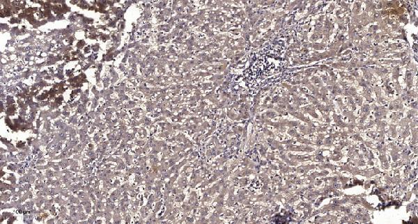
| Sample | Paraffin-embedded human liver cancer tissue; fixative not specified (datasheet A30844) |
| Fixation | Image fixative and duration unreported (datasheet A30844); verify before use. |
| Sectioning | 4–5 µm sections on charged slides (standard) |
| Deparaffinisation | Xylene, graded ethanol series to water (standard) |
| Antigen retrieval | Heat retrieval: Tris-EDTA pH 9.0 (datasheet A30844); 20 min, 95–100 °C (standard) |
| Peroxidase block | 3% H2O2, 10 min, room temperature (standard) |
| Blocking | 10% normal serum of the secondary host, 30 min, room temperature (standard) |
| Primary antibody | Rabbit anti-MTNR1B, 1:50 recommended; image 1:200 (datasheet A30844) |
| Primary incubation | Overnight at 4 °C (standard) |
| Detection | HRP-polymer secondary, DAB chromogen 5–10 min (standard) |
| Counterstain | Hematoxylin, blue, dehydrate and mount (standard) |
| Expected result | MTNR1B-positive staining in glial cells of caudate (HPA tissue IHC: High). HPA tissue profile: Cytoplasmic expression in neuronal processes in CNS and retina, and decidual cells in placenta. No signal in the no-primary control. |
MTNR1B is a 7-pass cell-membrane receptor (UniProt P49286 topology). In paraffin-section IHC, expect staining in neuronal processes in CNS and retina, and decidual cells in placenta (HPA: tissue IHC, Supported). HPA also reports high staining in specified neuronal and glial cells; its Supported assessment notes presumed off-target staining was disregarded and that RNA data do not corroborate the antibody pattern (HPA: tissue IHC).
| Staining follows neuronal processes or the retinal nerve fiber layer, with little nuclear signal (HPA: tissue IHC; UniProt P49286 topology). | This fits the reported cytoplasmic-appearing process pattern for a membrane receptor (HPA: tissue IHC; UniProt P49286 subcellular location). Compare the named cell compartment and tissue; a continuous cell outline is not required to recognize process staining in a section (general IHC practice). |
| Signal is mainly nuclear in cells expected to stain (HPA: tissue IHC). | Nuclear dominance conflicts with the cell-membrane assignment and reported process or decidual-cell staining (UniProt P49286 subcellular location; HPA: tissue IHC). Treat it as suspect until controls exclude nonspecific staining or a detection artefact (general IHC practice). |
| Strong staining appears in adipocytes or adrenal glandular cells (HPA: Not detected in those cells). | These are poor matches to the HPA tissue pattern, so review cell identification and background before calling them MTNR1B-positive (HPA: tissue IHC). Cross-reactivity or endogenous detection activity can mimic a positive chromogenic signal (general IHC practice). |
| Color is diffuse across cells and surrounding tissue, including areas without a recognizable cell pattern (general IHC practice). | Diffuse staining cannot establish the reported neuronal-process, retinal nerve-fiber, or decidual-cell pattern (HPA: tissue IHC). Compare a no-primary control and inspect blocking, washes, and detection chemistry for background (general IHC practice). |
| No signal appears in a section containing retinal nerve fiber layer or cortical neuronal cells (HPA: High in those locations). | First check that the expected cells are present and the detection run worked (general IHC practice). An absent signal conflicts with the HPA IHC observations, but it cannot by itself prove absent MTNR1B: antibody and section conditions still need review (HPA: tissue IHC; general IHC practice). |
| Compartment and topology (UniProt P49286 topology; HPA: tissue IHC) | Seven membrane-spanning segments support a membrane-associated interpretation; the HPA section profile describes cytoplasmic-appearing neuronal processes (UniProt P49286 topology; HPA: tissue IHC). Interpret section morphology in that context rather than requiring a sharp membrane rim (general IHC practice). |
| Tissue and cell context (HPA: tissue IHC) | High staining is reported in cortical neuronal cells, caudate and hippocampal glial cells, and retinal nerve fiber layer; placenta decidual cells show medium staining (HPA: tissue IHC). Score the named cells, since a whole-section average can obscure their pattern (general IHC practice). |
| Evidence strength (HPA: IHC Supported; HPA: tissue IHC) | The listed antibody has Supported IHC status, with external characterization backing the observed pattern; HPA also says RNA is not detected and presumed off-target staining was disregarded (HPA: tissue IHC; HPA074024 IHC Supported). Use morphology and controls when judging an unexpected positive (general IHC practice). |
| Extracellular terminus and epitope uncertainty (UniProt P49286 topology and glycosylation) | Residues 1–42 are extracellular and residue 4 is annotated as glycosylated (UniProt P49286 topology and glycosylation). The supplied sources do not map this antibody's epitope or report MTNR1B-specific retrieval or fixation sensitivity; do not infer a retrieval effect from topology alone. |
| Situation | Likely cause | Next action |
|---|---|---|
| Expected positive tissue is blank (HPA: High in retinal nerve fiber layer and cortical neuronal cells). | The section may lack the relevant cells, or the IHC run may have failed (HPA: tissue IHC; general IHC practice). | Confirm the cell compartment on the counterstain and check a working positive control, primary-antibody step, retrieval record, and chromogenic detection (general IHC practice). No MTNR1B-specific retrieval setting is established by these sources. |
| Signal is predominantly nuclear (UniProt P49286: cell membrane). | A dominant nuclear pattern is inconsistent with the reported localization; nonspecific staining or detection background is possible (UniProt P49286 subcellular location; HPA: tissue IHC; general IHC practice). | Compare a no-primary control, inspect background distribution, and reassess the cellular compartment before scoring positive (general IHC practice). |
| Adipocytes or adrenal glandular cells stain strongly (HPA: Not detected in those cells). | Cell misidentification, cross-reactivity, or endogenous detection activity may explain staining outside the reported pattern (HPA: tissue IHC; general IHC practice). | Verify morphology and compare a no-primary control; apply suitable endogenous-activity blocking for the detection system if control staining warrants it (general IHC practice). |
| The whole section has diffuse chromogen (general IHC practice). | Background from primary or detection reagents, inadequate blocking, or insufficient washing can obscure a cell-specific pattern (general IHC practice). | Review no-primary control, blocking and wash steps, then reassess whether staining resolves into HPA-reported cells and processes (general IHC practice; HPA: tissue IHC). |
| Placental decidual staining looks weaker than CNS staining (HPA: Medium in decidual cells; High in listed CNS cells). | Different reported HPA levels can produce this comparison without indicating assay failure (HPA: tissue IHC). | Judge decidual cells against their own expected medium pattern and local background; avoid applying a CNS intensity threshold to placenta (HPA: tissue IHC; general IHC practice). |
| Can ICC-IF confirm the IHC pattern? (HPA: subcellular summary Membrane). | HPA lists membrane as its ICC-IF summary but provides no ICC-IF images or ICC validation status for the listed antibody (HPA: subcellular; HPA074024 antibody record). | Treat the ICC-IF summary as limited context. Base this section's positive call on the Supported tissue-IHC pattern and appropriate IHC controls (HPA: tissue IHC; HPA074024 IHC Supported; general IHC practice). |


Comprehensive Human Protein Atlas IHC scoring per tissue (reliability: Supported — Presumed off target binding observed and disregarded. External characterization data supports antibody staining pattern while RNA data does not.). Rows are taken directly from the HPA tissue chart — click any row's HPA link to view the source.
| Tissue | Cell type | Level | Evidence | Source |
|---|---|---|---|---|
| Caudate | Glial cells | High | Protein (IHC) | HPA → |
| Cerebral cortex | Neuronal cells | High | Protein (IHC) | HPA → |
| Hippocampus | Glial cells | High | Protein (IHC) | HPA → |
| Retina | Nerve fiber layer | High | Protein (IHC) | HPA → |
| Cerebellum | Cells in molecular layer | Medium | Protein (IHC) | HPA → |
| Tissue | Cell type | Level | Evidence | Source |
|---|---|---|---|---|
| Adipose tissue | Adipocytes | Not detected | Protein (IHC) | HPA → |
| Adrenal gland | Glandular cells | Not detected | Protein (IHC) | HPA → |
| Appendix | Glandular cells | Not detected | Protein (IHC) | HPA → |
| Bone marrow | Hematopoietic cells | Not detected | Protein (IHC) | HPA → |
| Breast | Adipocytes | Not detected | Protein (IHC) | HPA → |
Troubleshoot MTNR1B staining in paraffin sections by checking retrieval, compartment, cell type and controls before assigning biological meaning.
A30844 is a human-reactive anti-MTNR1B antibody with IHC data from paraffin-embedded human liver cancer (catalog: A30844; image caption: A30844).
A30844 lists IHC among its applications and human reactivity (catalog: A30844). Its IHC image shows paraffin-embedded human liver cancer (image caption: A30844).
Which to pick: For tissue IHC, choose A30844: it is rabbit polyclonal, lists human reactivity, and has an IHC image from paraffin-embedded human liver cancer (catalog: A30844; image caption: A30844). The caption reports Tris-EDTA pH 9.0 retrieval and a 1:200 primary dilution overnight at 4°C; the fixative is unreported (image caption: A30844). No SKU in this payload lists IF/ICC or nonhuman reactivity, so there is no supported IF/ICC or cross-species pick (catalog: A30844 applications/reactivity).