MZF1 / Myeloid zinc finger 1 · IHC design guide

Design Immunohistochemistry for MZF1

Plan MZF1 IHC in paraffin sections around the nuclear staining reported across several tissues (HPA tissue IHC). This guide covers fixation consistency, controls and interpretation of cytoplasmic staining, which HPA disregards as off target (HPA tissue IHC).

Evidence assembled Oct 2026 · For research use; verify linked source records and product datasheet before use
Immunohistochemistry protocol sheet for MZF1 (IHC for MZF1): expected localisation Nuclear staining in several tissues (HPA tissue IHC), antibody A04865-2, validated IHC image, and IHC protocol steps
Printable MZF1 IHC protocol sheet — expected localisation Nuclear staining in several tissues (HPA tissue IHC), antibody A04865-2, controls and protocol steps. Open the full MZF1 IHC guide →

MZF1 Immunohistochemistry Experimental Design Guide

Expected localisation, validated protocols, controls and antibodies — the at-a-glance facts below, then the full design guide.

Must know before staining
Expected localisation Nuclear staining in several tissues (HPA tissue IHC)
Staining pattern Nuclear staining across several tissue cell types (HPA tissue IHC)
Antigen retrieval Tris-EDTA pH 9.0 HIER, 95–98 °C, 20 min (rule: nuclear antigen)
Positive control ⓘ Caudate+4 more · see all
Negative control ⓘ Adipose tissue+4 more · see all
Important caveats
Reasons your staining may differ from the expected pattern.
Fixation Keep fixation consistent across sections. (standard IHC practice; not target-specific)
Caveat Disregard cytoplasmic staining as off target (HPA tissue IHC)
Regulation Preferential in differentiating myeloid cells (UniProt)
Isoform / epitope 3 isoforms; epitope coverage is unreported (UniProt)
Section 1

Recommended MZF1 IHC & IF Protocols

The catalog antibody’s IHC-P protocol is paired with two published MZF1 chromogenic IHC methods (PMC10701130; PMC5130010).

Recommended immunohistochemistry (IHC-P) protocol parameters
SampleParaffin-embedded human stomach carcinoma tissue; fixative not specified (datasheet A04865-2)
FixationImage fixative and duration unreported (datasheet A04865-2); verify before use.
Sectioning4–5 µm sections on charged slides (standard)
DeparaffinisationXylene, graded ethanol series to water (standard)
Antigen retrievalHeat-induced epitope retrieval in Tris-EDTA buffer, pH 9.0, 20 min at 95–98 °C (standard rule: nuclear antigen)
Peroxidase block3% H2O2, 10 min, room temperature (standard)
Blocking10% normal serum of the secondary host, 30 min, room temperature (standard)
Primary antibodyRabbit anti-MZF1, 1:50-1:200 (datasheet A04865-2)
Primary incubationOvernight at 4 °C (standard)
DetectionHRP-polymer secondary, DAB chromogen 5–10 min (standard)
CounterstainHematoxylin, blue, dehydrate and mount (standard)
Expected resultMZF1-positive staining in glial cells of caudate (HPA tissue IHC: High). HPA tissue profile: Nuclear expression in several different tissue types. No signal in the no-primary control.
💡Decision noteStart with Tris-EDTA pH 9.0 retrieval at 95–98 °C for 20 min (page retrieval rule; UniProt P28698: nucleus).
Section 2

What Is the Expected MZF1 Staining Pattern?

MZF1 should be scored as nuclear staining in paraffin sections: UniProt places it in the nucleus and reports no transmembrane segment (UniProt P28698 subcellular location; topology). HPA reports nuclear expression in several tissue types and rates its tissue IHC pattern Approved (HPA: tissue IHC). Strong reference examples are glial cells in caudate, cerebral cortex and hippocampus, and thyroid glandular cells (HPA: High in those cells).

What am I looking at on my slide?
Distinct nuclear chromogen in glial cells of caudate, cerebral cortex or hippocampus, or in thyroid glandular cells.This matches the reported compartment and High cell-specific staining (HPA: nuclear tissue profile; High in the named cells). Judge nuclei against the counterstain and nearby cells; a High HPA rating is a reference pattern, not a required intensity for every section (standard IHC practice).
Predominantly cytoplasmic color, with little or no convincing nuclear signal.Do not score this as MZF1-positive staining: HPA explicitly disregarded observed cytoplasmic staining as off-target binding (HPA: Approved reliability description). Recheck the nuclear pattern in a reported positive cell population before interpreting the sample (HPA: tissue IHC; standard IHC practice).
Signal appears in a cell population reported as Not detected, such as adipocytes or cardiomyocytes.This conflicts with those cell-specific HPA observations, but does not establish that an entire tissue must be negative (HPA: Not detected in adipocytes and cardiomyocytes). Compare nuclear localization and control sections; unexpected chromogen may reflect cross-reactivity or endogenous detection activity (standard IHC practice).
Weak, diffuse color covers nuclei, cytoplasm and spaces between cells.The broad pattern cannot reliably identify nuclear MZF1 (UniProt P28698 subcellular location; HPA: nuclear tissue profile). Assess a no-primary control and review blocking, washing and chromogen development to distinguish assay background from localized staining (standard IHC practice).
No nuclear signal in a reported High reference population, while the section remains interpretable.The run has not demonstrated detection in its positive control (HPA: High in glial cells of caudate, cortex and hippocampus, and thyroid glandular cells). First verify tissue identity, cell type and detection controls; absent signal alone cannot establish that the tested sample lacks MZF1 (standard IHC practice).
💡Expected MZF1 appearanceCall the result positive when chromogen is centered on nuclei in the relevant cells, with strong reference staining possible in HPA High glial or thyroid glandular populations; predominantly cytoplasmic color is a false-positive pattern to disregard (UniProt P28698 subcellular location; HPA: High tissue IHC; HPA: reliability description).
How each factor affects the staining
Cell-specific reference patternUse named cells, not whole organs, as comparators: HPA reports High glial staining in three brain regions and High thyroid glandular staining, but Not detected in adipocytes, cardiomyocytes and several other specified populations (HPA: tissue IHC).
Molecular location and processingMZF1 is nuclear, has no transmembrane segment and is listed as one 1–734 chain without a signal peptide or propeptide (UniProt P28698 subcellular location; topology; processing). These annotations support nuclear scoring; they do not establish a fixation or retrieval requirement.
Isoforms and antibody evidenceUniProt lists three isoforms (UniProt P28698 isoforms). HPA lists HPA001757 as Approved for IHC and Supported for ICC; the supplied antibody record does not specify which isoforms its IHC staining detects (HPA: antibody validation).
IF/ICC Q: Where should MZF1 fluorescence appear?A: In the nucleoplasm (HPA: supported ICC-IF localization). This supports a compartment check only; the ICC-IF record does not provide a paraffin IHC protocol or establish identical intensities across applications (HPA: subcellular summary; antibody validation).
Why is my staining missing, weak or wrong?
SituationLikely causeNext action
Only cytoplasmic staining is visible in a putative positive section.The observed compartment matches staining HPA classified as off target (HPA: reliability description).Withhold a positive MZF1 call. Compare a reported High cell population and inspect a no-primary control; review detection and background before repeating the IHC run (HPA: tissue IHC; standard IHC practice).
A reported High reference population has no nuclear chromogen.The positive reference has not demonstrated assay detection; the supplied sources do not identify a MZF1-specific fixation or retrieval failure (HPA: High tissue IHC).Confirm the named cell type and section quality, then check retrieval, primary incubation and detection against run controls as general IHC troubleshooting (standard IHC practice).
Brown color appears broadly in a reported Not detected cell population.That observation conflicts with its HPA cell-specific pattern; nonspecific antibody binding or endogenous detection activity may contribute (HPA: tissue IHC; standard IHC practice).Compare nuclear localization with a High reference and use a no-primary control. If chromogen persists without primary antibody, investigate the detection chemistry (standard IHC practice).
Diffuse haze makes nuclear scoring uncertain.Background can obscure the nuclear pattern expected for MZF1 (UniProt P28698 subcellular location; HPA: tissue IHC).Use the counterstain to identify nuclei, inspect a no-primary control, and review blocking, washes and chromogen development before scoring (standard IHC practice).
Some cells stain within a tissue that also contains negative cells.HPA assigns levels to specified cell populations, and reports nuclear expression across several tissue types (HPA: tissue IHC).Score the identified cell population and its nuclear compartment separately; do not transfer a Not detected rating from one cell type to every cell in its organ (HPA: tissue IHC; standard IHC practice).
An unexpected cell type shows convincing nuclear staining.Nuclear location alone is insufficient to establish antibody specificity, particularly outside the listed HPA reference populations (HPA: tissue IHC; antibody validation).Document the cell type, compare positive and negative reference populations, and repeat with appropriate controls before treating the unexpected pattern as MZF1 expression (HPA: tissue IHC; standard IHC practice).

Sample controls for MZF1 IHC & IF

🧪Run thyroid gland first and expect nuclear staining in glandular cells (High; HPA: thyroid gland glandular cells; UniProt P28698: nucleus). Use adipose tissue adipocytes as the negative tissue (HPA: adipocytes not detected); on the thyroid slide, cells lacking specific nuclear staining should show counterstain only, but the supplied HPA rows do not identify a confirmed negative cell type within thyroid.
Positive control tissue: Caudate (Glial cells, HPA High)
Negative control tissue: Adipose tissue (HPA Not detected)
ICC-IF cell lines (HPA subcellular resource): HPA ICC-IF images show MZF1 in A-431, HEK293, U2OS, with annotated localisation: Nucleoplasm (supported) (HPA subcellular).
Technical controls: Include a no-primary, secondary-only control, a rabbit polyclonal IgG isotype control matched to the catalog antibody, and MZF1-knockout tissue or cells as a biological negative (selected-SKU caption: ZNF42 pAb and anti-rabbit secondary). Block endogenous peroxidase and assess endogenous biotin background when using the captioned avidin–peroxidase detection (selected-SKU caption: avidin–peroxidase).
⚠️Feasibility: A target-specific fixation window and the effect of fixation are unreported, and the exact A04865-2 paraffin-section caption does not report a fixative (selected-SKU caption: fixative not stated). Retrieval dependence is unreported, so compare retrieved and unretrieved sections in a pilot; the supplied evidence does not establish whether frozen sections or IF/ICC would be easier. For thyroid sections, check glandular luminal material for nonspecific chromogen background; assess autofluorescence if adapting the assay to IF (standard IHC/IF practice).

HPA tissue IHC evidence for MZF1

Comprehensive Human Protein Atlas IHC scoring per tissue (reliability: Approved — Cytoplasmic staining viewed as off target binding observed and disregarded.). Rows are taken directly from the HPA tissue chart — click any row's HPA link to view the source.

Positive expression · recommended positive controls

TissueCell typeLevelEvidenceSource
Caudate Glial cells High Protein (IHC) HPA →
Cerebral cortex Glial cells High Protein (IHC) HPA →
Hippocampus Glial cells High Protein (IHC) HPA →
Thyroid gland Glandular cells High Protein (IHC) HPA →
Cerebellum Cells in granular layer Medium Protein (IHC) HPA →

Undetected expression · recommended negative controls

TissueCell typeLevelEvidenceSource
Adipose tissue Adipocytes Not detected Protein (IHC) HPA →
Cervix Glandular cells Not detected Protein (IHC) HPA →
Duodenum Glandular cells Not detected Protein (IHC) HPA →
Esophagus Squamous epithelial cells Not detected Protein (IHC) HPA →
Fallopian tube Glandular cells Not detected Protein (IHC) HPA →
Section 3

Advanced MZF1 IHC Tips

Troubleshoot MZF1 staining in paraffin sections by checking nuclear localisation, controls, and tissue context before interpreting chromogenic signal.

How should I retrieve MZF1 when nuclear staining is weak?
Start with Tris-EDTA pH 9.0 heat-induced retrieval at 95–98 °C for 20 min (page retrieval setting: nuclear antigen). After cooling and washing, compare nuclear staining with a matched no-primary control while keeping antibody dilution and detection conditions constant (standard IHC practice). Use a section containing thyroid glandular cells as a reference for detectable nuclear signal, while recognising that expression varies by tissue and cell type (HPA: High in thyroid glandular cells; HPA: nuclear expression in several tissue types). If signal remains weak, adjust retrieval duration on adjacent sections and assess tissue integrity alongside staining (standard IHC practice).
Could fixation explain inconsistent MZF1 staining between paraffin blocks?
Target-specific fixation sensitivity is unknown because the supplied MZF1 evidence gives no fixation comparison, and the selected tissue caption does not state its fixative (selected caption: A04865-2). Record each block’s fixative, fixation interval, processing history, and section age before comparing staining across blocks (standard IHC practice). Run blocks together with the same retrieval, antibody dilution, detection reagents, and development time to limit technical variation (standard IHC practice). If nuclear signal differs, compare morphology and an appropriate positive reference section before attributing the difference to fixation or biology (HPA: nuclear expression; standard IHC practice).
Should I score cytoplasmic staining as MZF1 expression?
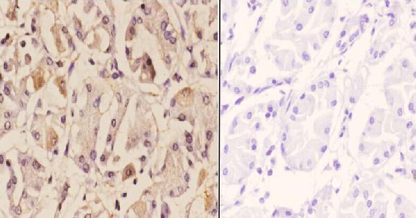
Score nuclear staining as the principal MZF1 signal because the protein is assigned to the nucleus and supported in the nucleoplasm (UniProt P28698: nucleus; HPA subcellular: nucleoplasm). The selected paraffin-section image reports both cytoplasmic and nuclear staining, but its no-primary image controls only for signal arising without the primary antibody (selected caption: A04865-2). HPA explicitly disregards cytoplasmic tissue staining as off-target binding, so keep it separate from nuclear scores (HPA tissue IHC: cytoplasmic staining viewed as off target). Check nuclear boundaries with the counterstain and review any widespread cytoplasmic colour against a matched no-primary section (standard IHC practice).
How can isoforms and epitope accessibility affect MZF1 staining?
MZF1 has 3 listed isoforms, and the supplied evidence does not identify which epitopes the selected antibody recognises (UniProt P28698: isoforms; selected caption: A04865-2). Interpret a negative paraffin-section result cautiously until the antibody’s immunogen or epitope information establishes which isoforms it can detect (UniProt P28698: alternative splicing; standard IHC practice). The SCAN box occupies residues 44–125, and a phosphoserine is reported at residue 111; neither establishes an antibody-specific staining effect here (UniProt P28698: domains and modified residues). Compare adjacent sections under the same retrieval conditions and use a documented positive reference before assigning differences to isoform expression (standard IHC practice).
How would I investigate MZF1 by IF alongside a cell-type marker?
On this IHC page, treat IF as a separate assay requiring its own antibody and staining validation (standard IF practice). Pair MZF1 with a marker for the expected cell type, and check nuclear overlap with a DNA counterstain; HPA supports nucleoplasmic localisation (HPA subcellular: nucleoplasm). Choose fluorophores after inspecting unstained tissue for autofluorescence, and place the weaker signal in a channel with lower background (standard IF practice). Because MZF1 has no transmembrane segment and is nuclear, permeabilise fixed cells to allow antibody access to the nuclear epitope, then verify that permeabilisation preserves morphology (UniProt P28698: topology and nucleus; standard IF practice).
What should I check when MZF1 chromogenic staining looks diffuse?
First compare the stained section with a no-primary control; the selected paraffin-section image includes a PBS-instead-of-primary control (selected caption: A04865-2). If diffuse colour appears in both, review endogenous peroxidase blocking, secondary-reagent binding, and chromogen development time (standard chromogenic IHC practice). If only the primary-treated section has cytoplasmic colour, avoid counting it as MZF1 without independent support because HPA disregards cytoplasmic staining as off-target binding (HPA tissue IHC: reliability description). Compare nuclear signal at the working antibody dilution with the selected image’s 1:50 condition, while accounting for differences in processing and detection (selected caption: A04865-2; standard IHC practice).
What is a defensible way to quantify MZF1 in paraffin sections? ⚠ ANSWER MARKED FOR VERIFICATION
Define the cell population and tissue area before scoring, and count nuclear staining separately from cytoplasmic colour (HPA tissue IHC: nuclear expression; cytoplasmic staining disregarded). Report an H-score using the percentage of nuclei at each intensity, or report the percentage of positive nuclei when intensity cannot be graded reliably (standard IHC scoring practice). For spatial comparisons, express positive nuclei per mm² of viable tissue and also report total eligible nuclei or area assessed (standard histology quantification practice). Apply one threshold and sampling plan across slides, excluding folds, edges, and necrotic regions from the denominator (standard IHC practice).
How can I distinguish convincing MZF1 staining from artefact?
A convincing result is reproducible nuclear staining in intact cells, consistent with MZF1’s nuclear assignment and HPA’s supported nucleoplasmic location (UniProt P28698: nucleus; HPA subcellular: nucleoplasm). Check the labelled cell type against tissue context: HPA reports High nuclear expression in thyroid glandular cells and no detection in adipocytes (HPA tissue IHC: thyroid gland and adipose tissue). Treat cytoplasmic colour cautiously because HPA identifies it as off-target, even though the selected paraffin image shows cytoplasmic and nuclear staining (HPA tissue IHC: reliability description; selected caption: A04865-2). Discount staining concentrated at section edges, necrotic areas, or endogenous-enzyme-rich regions when matched controls or morphology indicate artefact (standard chromogenic IHC practice).
Boster reagents

Best MZF1 / Myeloid zinc finger 1 IHC Antibodies

A04865-2 has real IHC data from a human paraffin-embedded stomach carcinoma section (catalog image caption). Human, mouse and rat reactivity is listed; ICC is listed without an IF image (catalog applications and reactivity).

Real IHC data Immunohistochemistry (IHC) analyzes of ZNF42 pAb in paraffin-embedded human stomach carcinoma tissue at 1:50.showing cytoplasmic and nucleus staining. Negative control (the right)Using PBS instead of primary antibody, secondary antibody is Goat Anti-Rabbit IgG-biotin followed by avidin-peroxidase.
Anti-ZNF42 MZF1 Antibody
Cat # A04865-2

A04865-2 is shown staining a human paraffin-embedded stomach carcinoma section at 1:50, with nuclear and cytoplasmic staining (catalog image caption). The catalog also lists ICC and human, mouse and rat reactivity for A04865-2, but supplies no IF image (catalog applications, reactivity and image records).

Which to pick: Choose A04865-2 for paraffin-section IHC because its own image documents that sample and dilution; the fixative is unreported (catalog image caption). For IF/ICC, A04865-2 lists ICC and a 1:50–1:200 dilution, but has no IF image (catalog applications, dilution and image records). It is also the listed option for human, mouse and rat reactivity, though the supplied IHC image documents human tissue only (catalog reactivity and image caption).

Each figure is that product's own IHC / IF validation image from its datasheet.

References

  1. UniProt Consortium. UniProt entry P28698 (MZF1_HUMAN, Myeloid zinc finger 1).
  2. Human Protein Atlas. MZF1 tissue IHC expression (reliability: Approved).
  3. Human Protein Atlas. MZF1 subcellular location (ICC-IF): Localized to the nucleoplasm..
  4. Human Protein Atlas. MZF1 antibody validation summary (1 antibodies).
  5. MZF1 promotes tumour progression and resistance to anti-PD-L1 antibody treatment in hepatocellular carcinoma. JHEP reports : innovation in hepatology 2024 — PMC10701130.
  6. miRNA-337-3p inhibits gastric cancer progression through repressing myeloid zinc finger 1-facilitated expression of matrix metalloproteinase 14. Oncotarget 2016 — PMC5130010.
  7. Therapeutic targeting of YY1/MZF1 axis by MZF1-uPEP inhibits aerobic glycolysis and neuroblastoma progression. Theranostics 2020 — PMC6993229.
  8. Coordinated DNA 5-mC and RNA m(5)C methylation epigenetically regulates MZF1 splice variants to drive EGFR-TKI resistance. Experimental & molecular medicine 2026 — PMC13434211.
  9. PubMed PMID:1860835 — UniProt-cited evidence.
  10. PubMed PMID:10974541 — UniProt-cited evidence.
  11. PubMed PMID:15057824 — UniProt-cited evidence.